 |
 |
|
|
|
||||
|
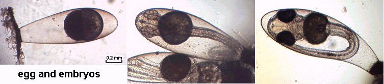
As we are talking about fishes, sometimes when catching plankton, we can find some fish eggs. At other times, eggs are found deposited in the open shells of dead molluscs, or on submerged pieces of wood and you can see thousands of eggs. In this case, sample only some of them and avoid destroying others. The chances for an egg to become an adult fish are probably 1 in 100 or 1 in 1000. (It's perhaps a good thing, otherwise it would be possible to cross the Channel, walking on dried sea fish!). Some of these eggs are particularly interesting because fish embryo can be seen inside and all the structures are still transparent. It's always impressive to see a beating heart and blood circulation when the fish is close to hatching. The pictures below show some stages of egg development. |
|
|
 |
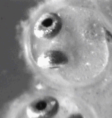
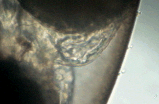
These embryos are probably one-two days old. The picture on the upper left shows the underside view of an embryo through the spherical yolk. The head is located on the left. Often round fat globules can be seen within an egg. In the picture left, note the developing eye: its crystalline form is noticeable. |
| I have found many eggs of a coastal fish species laid in one half of a mussel shell. By keeping them in the refrigerator I can observe embryo growth inside the eggs and the hatching of young fish. Low temperatures slow down the process and you must wait for two or three weeks before they hatch. In the video sequence below you can see eye movement, the beating heart and circulating blood; direct observation is more impressive because blood already has its red color. It's difficult to identify this fish species, (its probably a blenny), but these observations are really fascinating; maybe because fish are vertebrates and a little closer to us in evolutionary history than copepods or other crustaceans! | ||
 |
||
| Image above:
Note spots on skin used as a camouflage.
Black and white animated gif sequence taken under a stereo microscope using reflected light. You can see the beating heart slightly right of the upper eye. |
 |
|
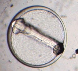
| Last summer I found a submerged piece of wood ca. 12 x 8 cm in size, covered by oval eggs and each was individually fixed by a sort of stalk. They were probably goby eggs. Each one was 0,5 x 1 mm in size or 0,5 mm². 120 x 80 = 9600 mm² / 0,5: thus there were approx. 19 000 eggs on the piece of wood! |
 |
|
 |
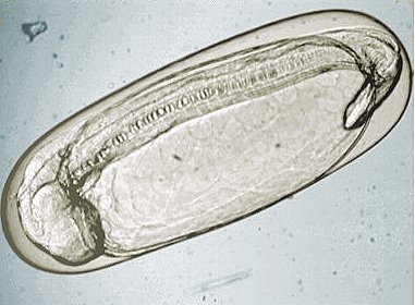
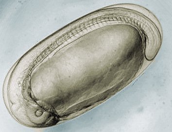
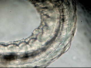
In the pictures
above you can see some stages of egg development. The left hand picture
shows a stage near hatching.
Darkfield works well too, with this kind of subject. The yellow spot in the center is the remains of the yolk. |
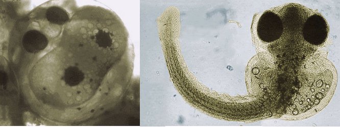
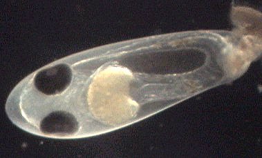
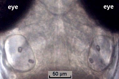
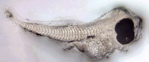
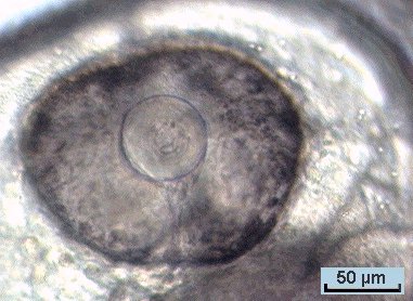
| A closer look at an eye: the crystalline form is clearly seen. Behind the eyes are two sorts of pouch forming the internal ears, in which are two otoliths used as equilibrium organs (right hand picture below). | ||
|
|
 |
|
| In the right hand picture above, vertebrae are also clearly visible and blood circulation in the caudal artery. When the eyes are formed, embryos become sensitive to light and they move within the egg during observation. Heart rate (left hand animation below) is increased showing a form of 'stress' induced by light. | ||
 |
 |
|
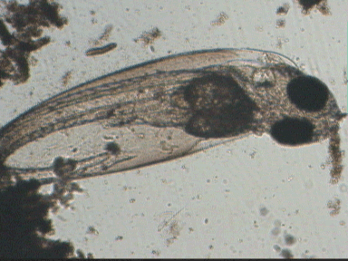
| When young fish hatch, observation becomes more difficult because they swim rapidly and in particular because the skin darkens and internal organs are hidden. Click HERE for a video sequence (lapsed time 5 min duration.) Don't forget to release baby fishes because it's quite impossible to feed them: they are only attracted by moving prey but the usual artemia nauplii are too large for their mouths! Some fish breeders use rotifers (Brachionus species) as food. | |
 |
Eyes, spines
and digestive gut are still clearly visible in a hatched young.
(Preserved specimen.) |
| To learn more about fish embryology go to: http://biol1.bio.nagoya-u.ac.jp:8000/stage-map.html. The web page has a striking animation of the stages in embryo development. |
|
|
|
Microscopy
UK Front Page
All drawings and photographs © Jean-Marie Cavanihac 2001 Published in the January 2002 edition of Micscape Magazine. Please report any Web problems or offer general comments
to the Micscape
Editor,
Micscape is the on-line monthly magazine of the Microscopy
UK web
|