Editor's note: I'm sure we've all acquired tips, techniques, 'tricks of the trade' or perhaps made simple gadgets while pursuing the fascinating hobby of amateur microscopy. Why not share some of yours with our readers, who may not have come across them. Just send us a short note in an e-mail (contact in footer), enclosing a scanned picture or drawing if you wish and we would be pleased to compile, upload and acknowledge your contributions.
Use of the Abbe Condenser in Viewing Certain Specimens with 4X Objectives
It is fairly common knowledge that when viewing specimens using low-power objectives, such as the 4X, the top section of the condenser should be removed and the condenser racked all the way down or the condenser removed entirely (see footnote). When viewing occurs using objectives of 10X and higher, the condenser is usually racked all the way up and the iris stopped down to about two-thirds. This is said to provide the maximum resolution for a given objective.
Generally, when working at low power, I simply rack the condenser down and observe without removing the top section. To me, the image seems the same whether the top section is removed or the whole condenser is simply racked all the way down.
I have found, however, that when viewing certain pond specimens, I can increase contrast (and apparent sharpness) with the 4X objective by racking the condenser all the way up and stopping down the iris to its minimum opening. This also calls for a slight increase in illumination. Although the procedure defies the laws of resolution, I find that at such low powers, it does not seem to matter. I use the technique only on a rare occasion, such as when viewing an especially transparent protist at 40X. Higher powers, of course, dictate the proper use of the condenser and iris diaphragm, and I believe no deviation should be allowed here in order to achieve maximum resolution.
I would be interested in hearing what other microscopists have discovered using the low-power contrast technique.
Contribution by Chuck Huck, comments welcomed.Editor's footnote: Depending on the condenser design, the optics supplied for use at low powers can vary. E.g. with the Russian Abbe condenser a swing in bulls-eye lens is used for low powers, with the Russian aplanatic condenser a screw-on NA 0.3 optic is supplied.
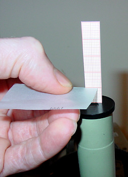
Measuring the eyepoint (exit pupil) of a microscope eyepiece for use with a consumer digital camera.
One of the popular ways of using a consumer digital camera on a microscope is by supporting the camera close to the eyepiece. In which case, an eyepiece design with a high eyepoint often works best to minimise vignetting (light cut off), as the image will be projected further into the camera's optics.
Modern eyepieces usually have a spectacle symbol if a high eyepoint design, but older ones don't and the eyepoint can vary widely. So it's a useful exercise to quickly measure and tabulate the eyepoints for each older eyepiece possessed and then the higher eyepoint designs would be the first choice for trying with a digicam.
One way to measure the eyepoint, is to support a small strip of 1 mm graph paper vertically on the eyepiece with plasticine, then with microscope focussed with lamp on, move a small piece of translucent paper over the eyepiece until the illuminated circle is smallest; note this position against the paper ruler. For the author's Russian and CTS eyepieces, the eyepoint varied from only 5 mm (12x CTS compensating eyepiece) to 14 mm (Russian 15x compensating eyepiece).
In my own case this was a valuable exercise, because the 15x eyepiece with the highest eyepoint was in a spares box, and wouldn't have been a first choice to use with the digicam. This eyepiece did prove to be the most successful with a digicam whereas the eyepieces I use for viewing (7x and 10x) had eyepoints of 5 - 9 mm and caused more vignetting. (An eyepoint of at least 10 mm is recommended for digicam use, e.g. see reference 1.)
A high eyepoint eyepiece alone may not entirely eliminate vignetting, there are other considerations such as the digicam's lens design and whether it has zoom optics.
Contribution by Dave Walker, comments welcomed.
Reference
1) The New Photomicrography by Peter Evenett. Proceedings RMS, Vol. 35 (4), December 2000.
This article is available online (in pdf Acrobat format) on the Royal Microscopical Society's web site. Follow the 'Publications' and 'Proceedings' links on the RMS web site at http://www.rms.org.uk. The article is an excellent introduction to the considerations when using consumer digicams on a microscope, with particular reference to the Nikon Coolpix 950 and 990 models.