|
Of a Fern By Wan Yu, China
|
 |
|
Of a Fern By Wan Yu, China
|
|
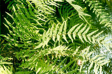
When I was walking near a pool which was not far from my house on a fine afternoon, I suddenly discovered some brown lines on the back of holly fern leaves. I observed them carefully and I was so interested at the structure of the leaves that I brought some of them home.I made a slide of the leaves and used my microscope to observe it. I discovered there were some interesting structures and I took some images of them.
Images
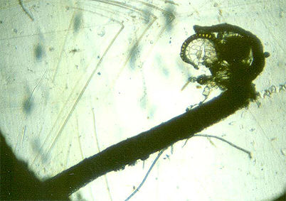 |
Image
1 (75X):
It
shows the whole structure of the leaf. The leaf is curled at its tip. Within
the curl of the leaf tip there are some sporangia which were made up of
special thin cells.
|
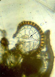 |
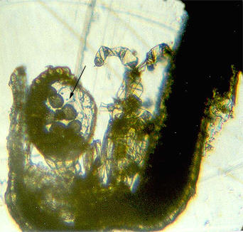
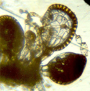
Image 2 (200X):
It shows the detailed
structure
of a sporangium. It has dehisced and the spores have gone, so you
cannot see them, but you can see the large and thin cells which made up
the wall of the sporangium.
|
 |
 |
Images 3 and 4 (200X):The sporangia which have not dehisced are shown in these images. So you can see some spores (arrowed in the left hand image). There are some additional structures around the sporangia.
All comments to the author Wan Yu are welcomed.
Editor's note: Wan Yu has started his own web site to promote microscopy as a hobby in China.
Microscopy UK Front Page
Micscape Magazine
Article Library
© Microscopy UK or their contributors.
Published in the January 2002 edition of Micscape Magazine. WIDTH=1Please report any Web problems or offer general comments to the Micscape Editor,
via the contact on current Micscape Index.Micscape is the on-line monthly magazine of the Microscopy UK web
site at Microscopy-UK