|
|
|
|
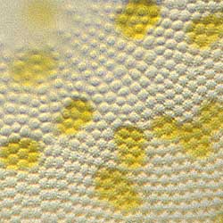
| With high power objectives you can get a glimpse of the finer details of the cell wall. This picture, cropped from an image made with a 40X lens, shows that the cell structure is not a simple net but that there are two layers with different texture. The finest details of diatoms are on the edge of what is visible with the light microscope. Scanning electron microscopy is needed to see the actual structure. |
All comments to the author Wim van Egmond are welcomed.
Visit Wim's home page for links to his many web pages on microscopy
Please report any Web problems or
offer general comments to the Micscape
Editor,
via the contact on current Micscape
Index.
Micscape is the on-line monthly magazine
of the Microscopy UK web
site at Microscopy-UK