Folliculina a marine Ciliate

by Wim van Egmond, The Netherlands


Folliculina in dark-field illumination.
Not only is marine plankton a rich source of subjects for a microscopist. Sessile organisms are at least as interesting. Organisms that are attached to floating platforms in harbors are always worth examining. Colonies of Hydrozoan polyps, red algae, Ectoprocts (moss animalcules) are very interesting study objects to study the many basic principles of life. And between these larger animals many interesting protozoa find shelter.

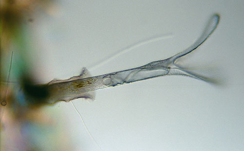
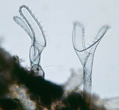
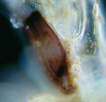
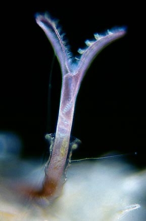
One such micro-organism I only knew from text books is Folliculina, a ciliate that somewhat resembles the well known Stentor. I found it living in its protective tube attached to a colony of Ectoprocts. I thought it would be interesting to photograph the organism. (A quick search on the Internet did not show many images). This was not easy since it lived on the rather thick 'branching' Ectoprocts.

I tried to cut off small parts of the Ectoproct 'stems' and carefully placed these on a slide. Then it was a matter of patience before the ciliate would come out of its house. 

It worked. I could make a series of images that was, although not perfect, good enough to see several of the main features of the organism. It was impossible to show the whole organism since in this species the tube-like house (lorica) had a bend in it.


The different illumination methods each give another view of the organism. This image was taken with differential interference contrast. The thickness of the slide diminishes the D.I.C. effect a bit. The colours on the left are of the out of focus houses of the Ectoprocts. The crystalline structure of the houses show different colours in the polarised light of the interference contrast.
 
In bright-field illumination you can close the diaphragm of the condenser a bit to enhance the depth of field. Although this gives less resolution it gives a better idea of the actual shape of the organism.

Folliculina is a Spirotrich ciliate. Ciliates belonging to this class all have membranelles (series of short rows of cilia) running clockwise towards the mouth. Other members of this class are Stentor and Spirostomum.

The order of Coliphorida consists of several families. The organism depicted in this article is a rather large species with a length of almost half a millimeter.

 
Retracted to the base of its house the surface of the cell of Folliculina shows the rows of cilia that resemble Stentor.

Although not easy to find it is one of the most spectacular ciliates I have found so far. The peristome (modified region that leads toward the mouth) is very spectacular and shaped like two long flaps. Along these run the fast moving membranelles that work small food particles towards the mouth. In the dark-field image (top) it is easy to see how small bundles of cilia form the membranelle.


Visit John Walsh's Micrographia web site for an interesting feature 'HomeBush Bay.
A microscopical investigation.' where a study of Folliculina is described.

All comments to the author Wim van Egmond are welcomed.

Visit Wims home page for links to his many web pages on microscopy

An Introduction to Microscopy

Microscopy UK Front Page
Micscape Magazine
Article Library


© Microscopy UK or their contributors.

Published in the January 2003 edition of Micscape Magazine.

Please report any Web problems or offer general comments to the Micscape Editor,
via the contact on current Micscape Index.

Micscape is the on-line monthly magazine of the Microscopy UK web
site at Microscopy-UK


© Onview.net Ltd, Microscopy-UK, and all contributors 1995 onwards. All rights reserved. Main site is at www.microscopy-uk.org.uk with full mirror at www.microscopy-uk.net.