Vaginicola |
Vaginicola |
Cothurnia fixed on algae |
Thuricola |
|
|
|
||||
|
Microscopy beginners often ask the question: "Where can I find subjects to observe?". Here is a true short story which may help them.... Last summer, my wife and I were on our holidays in the country of her parents in south west France. On a sunny day, at the end of our stay, we were visiting a town a few kilometers from their house. It was around 4pm and we decided to take a rest in a public garden to eat a cake. Sat down on a bench, we observed tall chestnut trees in the garden, flowers groves, a nice little wooden bridge crossing over an artificial pond .... POND. Have I said POND? My microscopy instinct appeared immediately: pond = fresh water microscopic life! Unfortunately I didn't have an adequate container with me to take a pond water sample. A frustrating situation for me... But I remembered I had in my pocket an empty 35 mm film container for my camera and I reused the container as a sampling jar. |
| Safety note: It's important to state that in such a case, eat food FIRST and THEN take a sample and not vice versa, because your fingers may be contaminated when sampling pond water and you can ingest some unfriendly amoebas! BE CAREFUL WHEN SAMPLING!! |
| The pond water was not very clear with extensive algal growths and a lot of large carp struggling to eat pieces of bread launched into the water by children, but I bravely plunged the container into a 30 cm depth of water, scraping a little the stones with the container edge and also took a bit of broken algae floating near the shore. |
| Well, I stop here my fascinating holiday memories, I think they are without interest for you! More interesting is what was in the film container; I came back home and put some drops of the water into a Petri dish to take pictures of critters which were present in this less than 25 ml of water sample. |
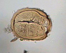
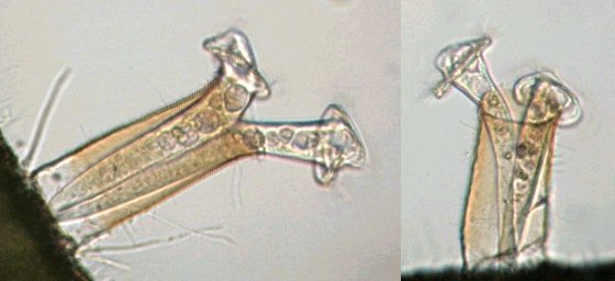
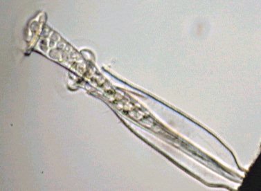
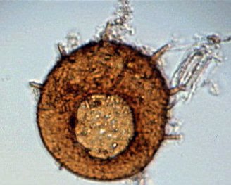
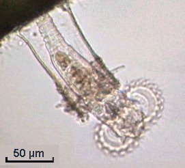
| The first inhabitants I found were the well known protozoa like Euplotes and Vorticella, as well as species with elegant shapes: Vaginicola, Thuricola and Cothurnia. Commonly, two individuals share the same lorica and retract themselves simultaneously. I found some yellow loricas which may be remnants of a cyst? (Image below left.) The animated GIF shows a flow of green particles inside the cytoplasm. |
|
Vaginicola |
|
Vaginicola |
|
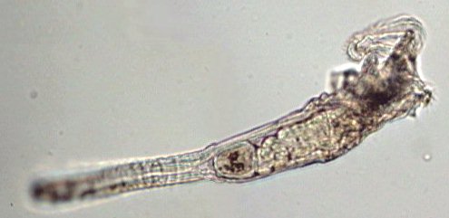
Cothurnia fixed on algae |
|
Thuricola |
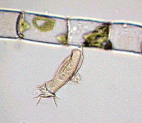
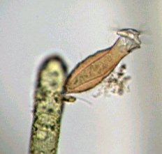
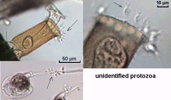
| Strange very small protozoans were found which were almost invisible, with a round body surrounded by some sort of tentacles (shown below (arrowed) on the top of a lorica and on a Vorticella stalk). Even with a x40 objective it's difficult to see their very transparent body. |
 |
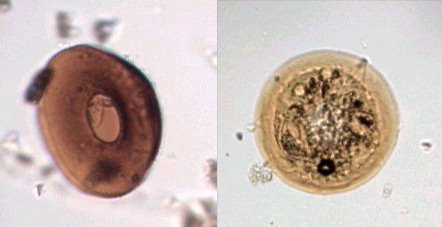
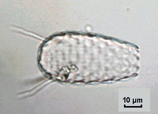
| Of course, the surface deposit I had scraped from the stones contained Arcella (side and top view shown below), also Euglypha acanthophora with spines and Centropyxis, another shelled amoeba: | |
 |
 |
 |
|
| Above left image pair Arcella; above right Centropyxis; image left Euglypha acanthophora. | |
| Some diatoms like Gomphonema and Pinnularia were also found (but you know these species well so I don't illustrate them). |
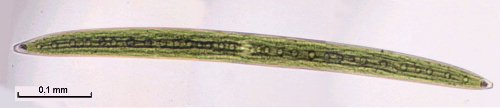
| Desmids were seen like this long Closterium (below left) and Pediastrum (below right). |
 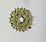 |
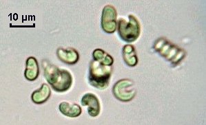
| Other chlorophyticalgae found included the well known Spirogyra, Eudorina (flagella are well seen), and also groups of small croissant-shaped algae (image below right). | |
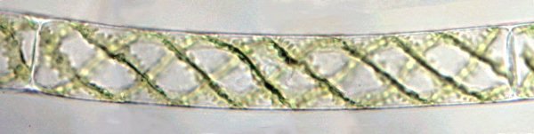
Spirogyra |
|
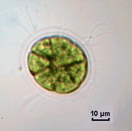 |
Left - Eudorina |
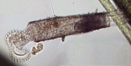
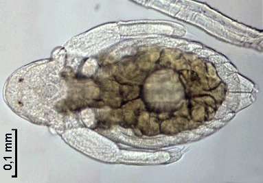
| Pursuing my observations amongst the algae, I found some loricas containing a rotifer - probably Limnias melicerta: when it is disturbed, the rotifer leaves the lorica and swims using its ciliated crown. (The last picture in the three below showing this rotifer). Other small rotifers were present too, such as Lecanes. | |
 |
 |
 |
|
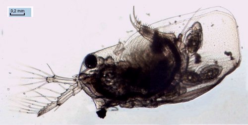
| Suddenly a strange critter appeared: | |
 |
It seemed to
be still a baby, moving very slowly some appendages, then another appeared
some millimeters away and finally the 'mother' (picture below) showed up
which was a large water flea, probably Daphnia Obtusa: the picture
below shows some babies hatching from the incubation chamber of the adult.
Note: The pictures of these large water fleas were taken with x2,5 objective, and were made from five original images manually assembled using PSP software. (The pictures are reduced by a factor of two.) |
 |
|
| It was the end of my holidays and coming back to my ordinary job the following day was a little frustrating, but these pictures helped me to have a little more enthusiasm! Observations of this tiny jungle, although made without premeditation (I had not taken with me any accessories to sample or concentrate water...) are among the most exciting I've made. Conclusion: you don't need anything more than your interest in microscopy (plus may be just a bit of opportunity...), or need to travel through exotic countries to make many fascinating discoveries! |
| PS: Some months ago in the fall, the algae had disappeared in the park's pond, water fleas and rotifers were no longer visible.. they may be under dead leaves covering the bottom of the pond, waiting for the next summer. The carp though were still present and always hungry! |
|
Microscopy
UK Front Page
All drawings and photographs © Jean-Marie Cavanihac 2003 WIDTH=1 Published in the January 2003 edition of Micscape Magazine. Please report any Web problems or offer general comments to the Micscape Editor. Micscape is the on-line monthly magazine of the Microscopy
UK web
|