|
An Overview of Bacteria by Matt Emery (UK), an A level sciences student, and amateur microbiologist |
Essentially,
there are 3 general types of bacteria, each being specialised
to different environments and having specific advantages in
various organisms in relation to other forms. They exist all
around us, in the air we breathe, on the surfaces we touch, on
our skin, and within us. Many bacteria, contrary to popular
misconception do not actually do any harm to mankind, and in
fact, can be helpful to us, such as the bacteria responsible
for turning milk to yoghurt, breaking down detritus into soil,
and those which live symbiotically in our gut.
Cocci:-
These
bacteria are generally some of the smallest, and simplest,
being small and spherical, hence
Cocci
(berry shaped). There are a
number of bacteria in this category which are pathogenic
(disease causing) such as
Staphlycoccus
aureus
, which causes a type of food poisoning, and is rapidly
becoming known as the hospital superbug, a variant of this
species called MRSA (Methicillin Resistant
Staphylococcus
aureus
),
N.
meningitidis,
which causes the often deadly diseases,
meningitis,
Staphylococcus
epidermis
which inhabits the skin and can cause spots and boils, and
finally
Moraxella
catarrhalis, which generally tends to cause infections in the lower
respiratory tract in humans.
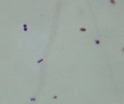
Fig 1 - Micrograph of a pair of Cocci (Stained with Fuschin, 100x objective (oil)).
Bacillus :-
The
second and slightly more complex type of bacteria are the
genus Bacillus. These bacteria are rod shaped, and can be very
short or very long, approx 5 - 30 microns long, although there
are some larger species. Most bacilli are capable of forming
spores, which can protect the bacteria from harsh conditions
such as dryness etc, and the bacteria within can remain viable
for up to 100 years. Bacilli are also capable of causing many
diseases, such as
Yersinia
pestis, which causes bubonic, pneumonic and sceptacaemic plague,
and
Bacillus
anthracis, which causes the, by now probably very famous disease, due
to the post 9/11 attacks, anthrax. Common types of bacillus
are
Lactobacillus
spp which generally cause milk
to spoil, and a number of various soil dwelling
species.
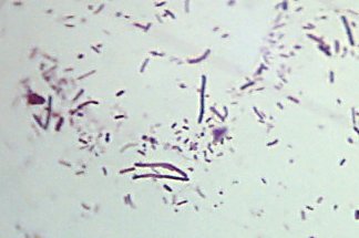
Fig 2 - Micrograph of Bacilli (Stained with Fuschin, 100x objective (oil)).
Spirochetea:-
A variation of the bacillus genus is the vibrio, which is part of the spirochaete family, but looks like a curved rod. This type of bacteria is generally responsible for diseases such as cholera ( Vibrio Cholerae ), and are generally found in stagnant water, and water which has been contaminated with sewage.
The
final type of bacteria, which I generally regard to be the
most complexly shaped is the spirochetea, which is basically a
corkscrew shaped bacterium. It causes diseases such as lyme
disease
(
Borrelia
burgdorferi
).
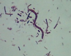
Fig 3 - Micrograph of a Spirochaete (Stained with Fuschin, 100x objective (oil)).
After
all of this incriminating talk about bacteria, I feel that I
should also mention the fact that many bacteria do no harm to
man, such as those found in probiotic yoghurt, the wide
spectrum of bacterial flora naturally found in the mouth, and
bacteria found in root nodules of plants such as clover which
naturally produce fertiliser for the plant they infect, along
with many types of bacteria that exist around us everyday and
still do no harm to us.
Microscopic examination of bacteria
The easiest way to study bacteria without any specialised equipment such as nutrient agars and petri dishes is to let a layer of plaque accumulate on the teeth for a day (make sure to brush your teeth at bedtime!) and mix a small amount of the plaque with a single drop of water on a cleaned, grease free slide (wash slide and pass through a bunsen flame twice). Then add a drop of a simple stain to the preparation on the slide such as crystal violet or methylene blue, and lower a coverslip onto this preparation. Don't bother wasting time with the 4x objective, skip to the 10x objective and find a space with a high concentration of small stained specs, then switch to the 40x objective.
You should be able to observe a number of various types of bacteria such as bacilli and cocci, however it is unlikely that there will be any spirochetea in the normal flora of the mouth. These bacteria, provided that the stain concentration is not so great as to kill the cells, should be observed to be moving around in the water, this is accomplished by numerous flagellae around the surface of the bacteria (small "hairs" which enable the bacteria to "swim").
Ideally, bacteria should be observed as a dry, heat fixed, stained smear without a coverslip, under the 100x oil immersion objective to give the best image, however, many bacteria can be seen at 400x for those who are unfortunate enough to not possess a microscope with a 100x objective.
The procedure for this is to prepare a smear as in the example above, and instead of placing a coverslip on the sample, spread it over a portion of the slide and leave to dry, then pass the slide through a flame smear side up twice to heat fix the cells to the slide.
Then
stain with a simple stain such as methylene blue. Rinse the
slide, then allow it to dry. After this, place a single drop
of immersion oil on the slide and lower the 100x objective
onto the oil and focus. A fairly high amount of information
can be obtained about the bacteria via this method, especially
if the specimen has been gram stained (a method of
differentiating between types of bacteria, used in medical
diagnostics and bacteriology laboratories)
My humble work environment and
equipment
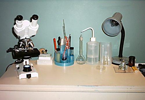
Here is my workbench, cultures and all. My microscope, a Bresser Biolam BLK, by all means not the best microscope in the world, but it has excellent build quality, and very good optics for a microscope costing £280. Note the desk tidy to keep inoculating loops, spreaders, pippettes etc in to keep mess to a minimum, along with the grey trays on the side of the bench (just visible on the right). Overall, I find the study of bacteria very fascinating, and once I finish my A level courses, I aim to study microbiology for my degree.
Micrographs were all taken by myself, using a Polaroid PDC 2070 2.1 Mpixel digital camera held to the eyepiece of the microscope.
Safety note :- after working with any kind of bacteria, once finished, immediately place the slides into a disinfectant solution and wash, wipe work surfaces down with an appropriate disinfectant / antibacterial agent such as 70 % ethanol, sodium hypochlorite (aqueous, 10% sol), or a household disinfectant made up to the manufacturer's directions, and wash hands with an antiseptic soap. Avoid hand contact with the eyes or mouth whilst working with bacteria, and always handle cultures in the correct manner, assume everything is a pathogen.
I hope this article has provided some useful information on bacteria.
Any constructive comments welcome to the author, Matt Emery
Please report any Web problems or offer general comments to the Micscape Editor.
Micscape is the on-line monthly magazine of the Microscopy UK web site at Microscopy-UK.