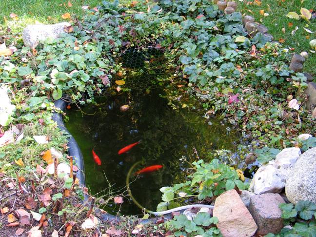
Figure 1: Goldfishes in the garden pond
|
Green Algae from a Garden Pond: Tetrabaena socialis Bernd Laber, Idstein, Germany |
The motile volvocid green algae Tetrabaena socialis (= Gonium sociale) forms square colonies of four ovoid cells, each with two equal flagella, two contractile vacuoles, and a pyrenoid and a red eyespot within a single green chloroplast. Colonial cells are attached to each other by the protuberances of their cellular sheaths and are also held together by a gelatinous capsule surrounding the entire colony. To accomplish asexual reproduction, each cell of a colony divides twice to form a daughter colony. Photomicrographs illustrating these features are presented.
Figure 1: Goldfishes in the garden pond
Our garden pond consists of a preformed plastic pond liner frequently offered in garden centres and DIY stores. It is oval, roughly 1.3 x 0.7 m in size, 0.5 m deep and contains about 400 litres of water. A small pump ensures mixing of the pond water. The pond is inhabited by three goldfishes (Fig. 1), and due to their waste the water is always quite rich in nutrients. Since I avoid pond care for algae control, microscopic pond life is usually rich and the pond water therefore is a rewarding source of material for microscopic investigations all over the year.
One of the smaller inhabitants of the garden pond is the flagellate green algae Tetrabaena socialis. Taxonomically, T. socialis, also known as Gonium sociale, is a member of the Chlorophyta (green algae). It belongs to the class Chlorophyceae and the order Volvocales. The order Volvocales consists of unicellular and colonial flagellates, most of which are frequently found in water bodies with elevated levels of nutrients. The most famous colonial member of the order Volvocales is Volvox globator, whose spherical colonies are formed by up to several thousand cells. But even the smaller and less well known members of this order, like T. socialis, which I would like to introduce here in more detail, may reveal many details of the morphology of the colony members and the construction of the colony, as well as an interesting lifestyle, when studied under the microscope.
Methods
Water samples were collected from the garden pond in November and December 2005 and immediately studied under the microscope. Photomicrographs were captured with a Canon PowerShot G5 digital camera attached with a Zeiss Universal Digital Camera Adapter to a Zeiss Axioskop equipped with a Plan-Neofluar 40x/0.75 objective, using brightfield illumination. Clipping of photographs as well as some histogram stretching and mild contrast enhancement was performed with Adobe Photoshop Elements 2.0.
Morphology of Tetrabaena socialis
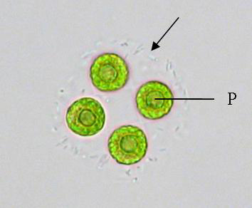
Figure 2: Above view of a colony of T. socialis. The border of the gelatinous capsule (arrow)
is clearly visible. P = pyrenoid.
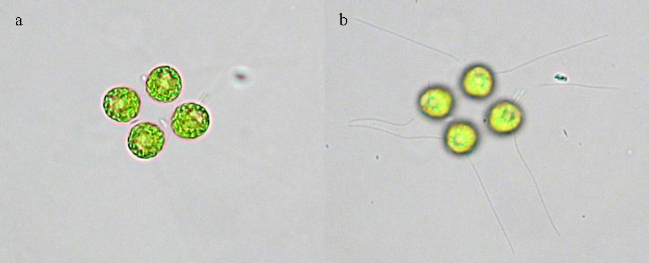
Figure 3: Above
view of T. socialis. (a) In this colony the border of the
gelatinous capsule is invisible.
(b) High focus of the same colony shows two equal flagella per colony
member.
When T. socialis colonies are viewed from above or below, which is the typical sight when there is only a small amount of water under the coverslip, the four colony members form a square (Fig. 2). Each cell is nearly circular and a single bright green chloroplast fills the entire cell. A single circular pyrenoid, which is the centre for starch production, is located within the chloroplast and is clearly visible in the middle of each cell. Typically, colonies are about 30 - 35 µm in diameter, and the diameter of a single cell is about 10 µm. The colonial members are attached to each other by the protuberances of their cellular sheaths (Figs. 4b and 5b), but this feature may be difficult to discern using brightfield illumination. The colonial cells are also held together by a gelatinous capsule surrounding the entire colony. In many cases, the border of this capsule is easily visible under the microscope, but in the same sample there are usually also many colonies whose gelatinous capsules are invisible (Fig. 3a). A possible explanation for this difference is given below. Two equal flagella of about 40 µm length arise from the front (apex) of each cell and project out from the mucilage. Since all colony members are orientated in the same direction, all flagella may become visible at the same time when the focus is raised to a point slightly above the colony (Fig. 3b).
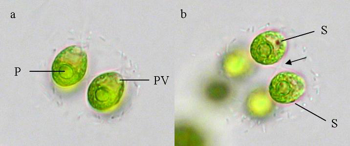
Figure 4: Side view of T. socialis. The front ends of the colony members point to the upper right. (a) The two cells in focus cover the other two colony members. (b) Two cells of the colony are in focus, the other two are visible as bright yellow-green patches to the lower left. Note the protuberances of the cellular sheaths that connect both cells (arrow). P = pyrenoid, PV = contractile (pulsing) vacuole, S = stigma.
If there is plenty of water under the coverslip, free-swimming colonies of T. socialis may be observed. When these are viewed with low magnification, i.e. 100-fold, it becomes obvious that the colonies actually are not spheroid but rather disc-shaped. The flagella of the algal cells can sometimes be discerned when the iris diaphragm of the condenser is closed very much to raise contrast and increase the depth of field. Switching to higher magnifications, such as 400-fold, reveals that the colony members are egg-shaped (ovoid) when viewed from the side (Fig. 4). They are 12 - 15 µm long, about 10 µm wide and their longitudinal axes are oriented almost parallel to each other. Prominent features visible inside the cell are the circular pyrenoid at the posterior (back) end and a median red eyespot (stigma) located inside the cup-shaped chloroplast. Two contractile or pulsing vacuoles may be discerned at the front end of the cell near the base of the flagella. The flagella themselves usually stay invisible due to their rapid movement above and below the actual plane of focus.
Asexual Reproduction
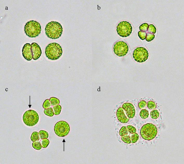
Figure 5: Top views of colonies of T. socialis undergoing asexual reproduction. (a) The bottom left cell has divided for the first time. (b) The top right cell has divided twice to form four daughter cells which are not yet fully separated. Note the protuberances of the cellular sheaths which attach all colony members to each other. (c) Intermediary stage of reproduction. The stigmas of the two cells which have not divided yet are marked by arrows. (d) All intermediary stages of asexual reproduction are visible in this single colony: An undivided cell (bottom right), cells that divided for the first (top left) and second time (bottom left) and a daughter colony (top right).
According to my experience, asexual reproduction preferentially occurs after nightfall, since I regularly spotted many colonies undergoing reproduction when I investigated pond water sampled in darkness late in the evening. On the other hand, I only very rarely found propagating colonies in pond water sampled during daylight hours. However, I was unable to observe and photograph the asexual reproduction of one single colony up to now, most likely due to the fact that colony reproduction apparently proceeds rather slowly. For example, it took a cell which had just started its first division 41 minutes to complete this process. In a different colony, a cell which had already completed its first division required another 17 minutes to divide for the second time. Nevertheless, all the various stages of asexual reproduction can easily be observed by looking at several different colonies. Fig. 5 summarizes these observations.
Asexual reproduction starts when a mother cell divides her body longitudinally to produce two daughter cells (Fig. 5a). Subsequently, both daughter cells undergo another longitudinal division perpendicular to the first one. The four resulting daughter cells are initially contained inside the cellular sheath of their mother cell (Fig. 5b), but produce their own cellular sheaths and separate from each other later on.
Asexual reproduction is not synchronized since the colony members divide independent from each other. Consequently, different stages of reproduction can typically be observed in a single colony (Fig. 5c). With a little luck it is even possible to observe all intermediary stages in a single colony (Fig. 5d).
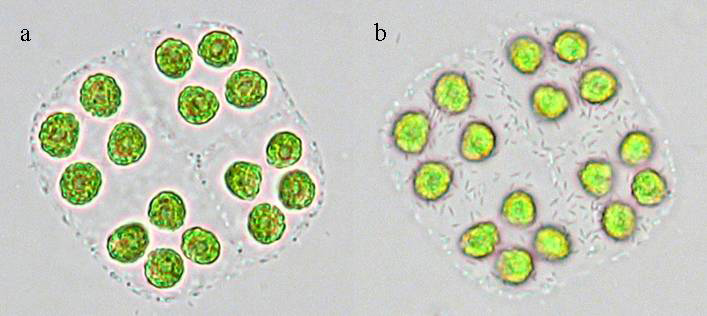
Figure 6: (a) Top view of four daughter colonies of T. socialis prior to their separation. (b) High focus of the colony reveals several rod-like structures on top of or inside the gelatinous capsule.
After all colony members have divided twice, the daughter cells soon become daughter colonies, but their gelatinous capsules initially remain attached to each, thus pretending to be a 16-membered colony (Fig. 6a). High focus of this colony (Fig. 6b) revealed the presence of many rod-like structures covering the gelatinous capsule, perhaps bacteria living on top of or inside the mucilage. However, these rod-like structures highlight the borders of the gelatinous capsule, which cannot be discerned in their absence (Fig 3a, Fig. 5a - c).
Finally, after reaching maturity, the daughter colonies separate from each other to continue their life as independent colonies.
Comments to the author, Bernd Laber, are welcomed.
References
Nozaki, H. (2003) Flagellated Green Algae. In: Freshwater Algae of North America. Ecology and Classification (J. D. Wehr and R. G. Sheath, eds.), pp. 225 - 252, Academic Press.
http://microscope.mbl.edu/scripts/microscope.php?func=imgDetail&imageID=12549
Microscopy
UK Front Page
Micscape
Magazine
Article
Library
Published in the January 2006 edition of Micscape Magazine.
Please report any Web problems or offer general comments to the Micscape Editor.
Micscape is the on-line monthly magazine of the Microscopy UK website at Microscopy-UK
©
Onview.net Ltd, Microscopy-UK, and all contributors 1995 onwards. All
rights
reserved.
Main site is at www.microscopy-uk.org.uk with full
mirror at www.microscopy-uk.net.