|
THE GRIFFITH CLUB MICROSCOPE, A “QUEEN OF GRACE AND UTILITY AMONGST MICROSCOPES.” Manuel del Cerro, Pittsford, NY. |
"This is a full-sized, first-class monocular, made of brass, steady when in position, free from tremor, unique in design, and beautiful in appearance." E. Griffith, 1890.
INTRODUCTION.
With the proud words reproduced above the maker described his creation, the Club Microscope. Here I will discuss this microscope as well as information on Mr. Griffith, and the village where his most important work developed. I will comment on the Club Microscope used by the physician who may have treated Griffith’s wife. This article is an updated and shortened version of an article published elsewhere (del Cerro, 2001).
EZRA HOLLACE GRIFFITH
Padgitt (1975) and Watson (1995) are the key sources of information on the life and work of Ezra Hollace Griffith, of Fairport, New York. Padgitt describes him as “an active microscopist, and designer of microscopes, objectives, and accessories. He is mainly remembered by his creation of what he (Griffith) called the “Griffith Club Microscope.” According to Padgitt, Griffith may have created the original design for “the Club” during the 1870s. Griffith designed the stands and the optics but initially relied on Bausch & Lomb for manufacturing the stands and objectives. As we will see later, Griffith’s relationship with Bausch & Lomb ended on an unhappy note; thereafter his stands were made by John Field of Birmingham, England, and by Gundlach of Rochester, New York, and New York City. Unfortunately, no Gundlach-made Griffith stand is known exist currently. Eventually, Griffith moved from Fairport to Rochester (presently a 20 minute drive, but back then a trip taking hours). He left a person by the name of Arthur B. Newman as his Fairport sales agent. Padgitt comments that “sometime after 1892, Griffith moved to Chicago and nothing more is known of him thereafter.” However, according to Watson, Griffith received considerable recognition in Chicago, where he arrived in 1893. Particularly significant was the award received for one of his microscopes at Chicago’s “World’s Columbian Exhibition” of 1893. The official declaration states that the award was given as a “substantial recognition for this queen of grace and utility amongst microscopes.” (Watson, 1995; emphasis added).
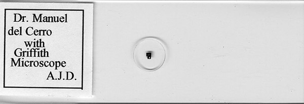
Figure 1. Scan of a 1980s microphotograph by master microphotographer Anthony J. DiDonato, Norwood, PA.
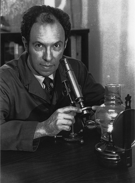
Figure 2. The microphotograph showed in fig. 1 was photographed on film using a 10X Olympus plano-apochromat objective and a 3.3X photographic ocular. The illustration depicts the Griffith Club microscope serial # 933 (MdC #210) being tested by the author in the mid-1980s. A kerosene lamp provided illumination and ambiance. Question: what is the correct manner of referring to a photomicrograph of a microphotograph?
FAIRPORT, NEW YORK
Today, Fairport is a charming and prosperous suburb of Rochester, New York. According to the U.S. Census Bureau, the population of Fairport for the year 2000 was 5740 persons. Although he was the designer of one of the most distinctive American microscopes, Mr. Ezra Griffith has left no lasting memory in the town where his main creation took place. His name was not recognized by the staff of the Fairport Historical Museum. Search of historical records did show his name in the 1875 town census. Griffith was at that time living with his wife, Jenny (31 years old) and daughter Josephine (9 years old). The 1880 Village census shows that a son, Charles, was by then age four. After that point there are no other records of Ezra Griffith in Fairport; he is not listed in the 1900 census, although an article by him published in June 1890 still gives Fairport as his place of residence. This fact agrees with Padgitt’s assertion that eventually Griffith moved from Fairport to Rochester, leaving Arthur B. Newman as his Fairport sales agent. In fact, the 1900 Fairport census lists Arthur Newman as a village resident.
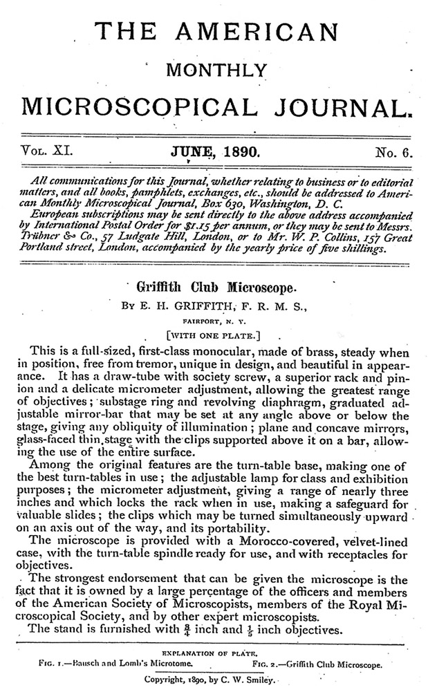
Figure 3. The Griffith Club microscope in the words of his creator.
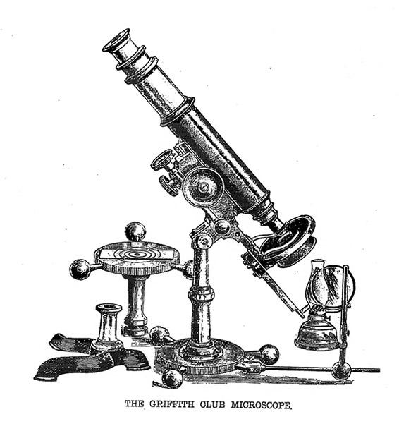
Figure 4. The first illustration of the Club microscope. Drawing attached to Griffith’s 1890 article.
A highly original feature of the Club microscope was its fine adjustment mechanism. Griffith had described it in the December 1889 issue of The American Monthly Microscopical Journal (Fig. 5), several months in advance of his description of the full instrument.
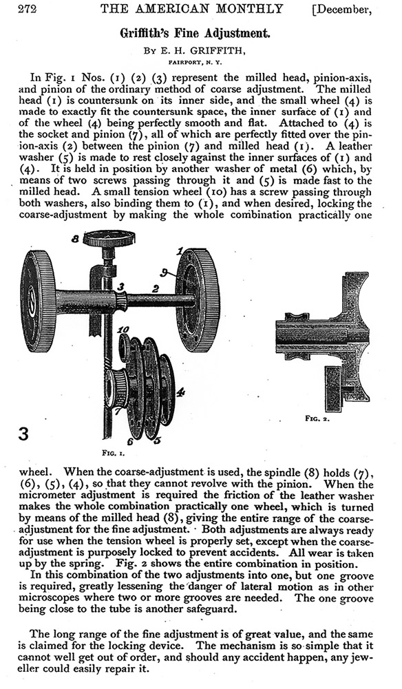
Figure 5. Griffith’s illustrated description of the fine focus mechanism used in his Club model. As noted in the text, this description was published in advance of the description of the microscope itself.
MR. GRIFFITH AND DR. BRIGGS
Charles Menzies Briggs, M.D., (1855-1933) (fig.4), was a gynecologist practicing for many years in the village of Fairport., New York. He was a contemporary and neighbor of Ezra Griffith. Historical record shows that Dr. Briggs was a beloved physician with an extensive practice in a small community. When a local newspaper published a laudatory article describing his services to the community, pointing out that he had attended the birth of “two thousand babies,” Dr. Briggs modestly commented, “It is a little exaggerated, my record is only about 1,800 children.” Dr. Briggs deserves our attention for a different reason than his clinical work; he was an avid microscopist. He was a member of the Rochester Pathological Society, the American Society of Microscopists, and others. More to the point yet, he had and used for his studies a Griffith Club microscope. Dr. Briggs’ microscope, his slide collection, and documents have been preserved as the Briggs Legate in the Miner Library at the University of Rochester Medical School, in Rochester, New York. I have had the pleasure of examining them while preparing this article.
The slide collection is housed in a small but elegant wooden box holding a total of 59 slides in eleven trays. Dr. Briggs made a number of slides himself, others were obtained form a variety of makers. The two slides more relevant for our discussion are the one labeled “Diatomacea, Aranoidiscus erenbergii” by A. B. Newman of Fairport, New York (Griffith’s agent), and particularly the one that carries a handwritten label reading: “Foraminifera, 1884. Presented by E. H. Griffith, Fairport, New York.”
Dr. Briggs’ Club Club microscope model is signed on the circular base “E. H. Griffith, Fairport, N.Y.” and carries as a serial number the inscription “C27.” The ocular carries no indication of its power. The objective is engraved “3/4”, indicating a focal length of 3/4 of an inch, or about 18 mm, a focal length that corresponds to an approximate magnification of 8X. A second objective is inscribed 1/5, which is approximately 5 mm and about 40X. The instrument has no condenser or wheel of fixed diaphragms. Instead, under the stage there is a short cylindrical component made of brass with a threaded hole. An attempt to screw an RMS objective into it was successful. In other words, this particular microscope was designed to use an objective as a condenser, a practice not uncommon in British microscopes made during the late 19th century. This microscope is still fully functional, optically and mechanically.
GRIFFITH AND THE ROYAL MICROSCOPICAL SOCIETY OF LONDON
In 1883, Griffith was elected as a Fellow of the prestigious Royal Microscopical Society. The fact suggests that Griffith had reached wide recognition as an active microscopist and designer of microscopes and accessories. It should be noted that First Lady Frances Folsom Cleveland, is known to have had a Griffith microscope. Not a small achievement for someone who made a living as a traveling salesman for a soda manufacturer (Watson, 1995). Griffith may have appreciated the distinction bestowed upon him, since he presented one of his microscopes to the Society, a nickel-plated stand. The instrument is still in the Society’s collection (catalog # 98) but no record of the date of accession into the collection exists (Turner, 1989). Produced before the introduction of the (Improved) Club model, this microscope already shows some of the design elements that give the Club its distinctive identity.
GRIFFITH’S MICROSCOPES AT THE BILLINGS COLLECTION, WASHINGTON, DC
Two microscopes by Griffith comprise part of the Billing’s Collection of microscopes at the National Museum of Health and Medicine, in Washigton, DC. Inexplicably, neither of these instruments is ascribed to Griffith! The oldest, Armed Forces Institute of Pathology (AFIP) 71789, is a Griffith microscope with the stand made in England. In view of the information contained in the original accession note (fig. 6), it is incomprehensive that John Field is identified in the catalog (Purtle, 1974) as the maker. Ezra Griffith, who designed, patented, and commissioned the making of the instrument, is not mentioned.
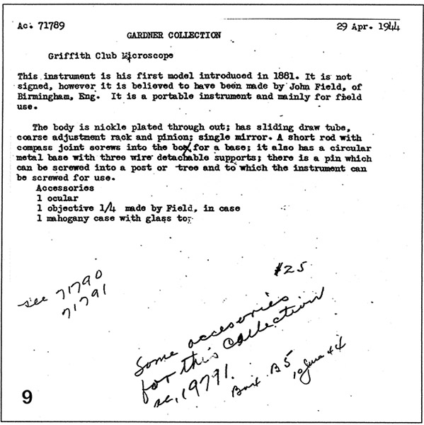
Fig. 6. Original accession note for the oldest Griffith microscope in the Billings Collection (reproduced by kind permission of the National Museum of Health and Medicine, Washington, DC).
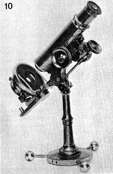
The reference to item 71790 (fig. 7), handwritten in the accession note reproduced above is to the second Griffith microscope in the collection (Purtle, 1974, p. 90). Alas, that one too is attributed to another maker, as we will see below. The corresponding entry reads:
“The circular base of this instrument is 3-5/16 inches in diameter and 1/4 inch thick. Three screw-in ball-end supports, 1-1/2 inches long, form a flat tripod, that supports the 5-1/4-inch-high pillar with screw division and cradle joint; there is a Jackson-type limb and arm with a double milled-head pinion. The fine adjustment is an endless screw micrometer.
The swinging tailpiece has a 1-3/8-inch-diameter double mirror in a dovetail slide. The 2-5/8-inch circular stage with a glass top is fixed to the limb, and has a 1-inch aperture.
The swing-out substage has an Abbe condenser and filter holder. The body tube is 5 x 1/4 inches, has a rack, graduated drawtube, and cone nose. The circular base may also be used as a turntable. The instrument is all brass with nickel trim and is 10-3/4 inches high. It is signed, “E.H. Griffith, Pat. Dec. 14, 1866 [sic], Rochester, N.Y., 1069.”
The writer of this entry gave a suitable description of the stand (fig. 7). The two most characteristic features of the second Griffith model are noted. These are the circular base with its very distinctive ball-ended arms, and the capability for adapting it for use as a turntable. So too is the other, even more unique feature of this model, the endless-screw fine focus devise. The regrettable omission of any reference to the ocular(s) and objective(s) is not unique to this entry, but rather, an unfortunate feature of the Billings Catalog. Two other points arise questions. One is the date given to the Griffith patent, 1866. This is just a typographical error, since the microscope is identified as “C. 1886.” More unsettling is the fact that the legend for the picture reproduced to the right (fig. 10) reads:
“Fig. 167. Bausch & Lomb Optical Co., Rochester, N. Y.; compound monocular; C. 1886; (AFIP 192 – 604713-339).”
The written description clearly states that “It is signed E. H. Griffith, Pat. Dec. 14, 1866 [see above], Rochester, N.Y., 1069.” Nonetheless, the microscope is attributed to Bausch & Lomb. This time Goliath won over David!
OBSERVATIONS ON AN EVOLVING STAND
Randy Watson (1995) has written an interesting article on the evolution of the Griffith microscope. Watson recognizes two forms of Griffith stands, the Club Microscope and the Improved Club Microscope. Unfortunately, the nomenclature for these instruments is not straightforward. Griffith called his early stands, ca. 1880, “Club Microscope” emphasizing the fact that these were highly portable instruments designed to assist members of microscopic clubs during their outings and meetings. The microscope he gave to the RMS around 1883 is engraved “Griffith Club Microscope.” Griffith later developed a new model, one with the circular base and the unique fine focus arrangement; it was produced from 1886 on. He called that too “Improved Club”, although he admitted that the two models had little in common besides their names. Modern writers have in general, failed to follow Griffith’s usage. In current practice, the designation “Club microscope” is reserved for the second model. This model features a circular base with three ball-ended arms; another characteristic is the distinctive fine focus mechanism driven by a worn wheel (figs. 5). The practice of referring to this model as the “Club microscope” is so widespread that I doubt it can be changed.
Confusion also arises from the fact that after the rift with Bausch & Lomb (see below), Griffith started using the expression “Improved Club.” This might have been an attempt to imply that the stands he was selling directly were an advanced version of those previously sold by Bausch & Lomb. Eventually Griffith did improve his second model sometime after 1890, by adding an Abbe condenser and double nosepiece to it. Paradoxically, he didn’t attach a new designation to that truly improved design.
An alternative to the circular base was offered; it was a “claw” base (fig. 4, far left) similar to that used in contemporary Bausch & Lomb microscopes. Judging by the surviving examples the unique and more functional circular base was by far the most popular.
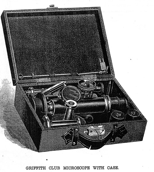
Fig. 8. A plate facing p. 221 in the October 1890 issue of The American Monthly Microscopical Journal shows a Griffith microscope disassembled and stored in its carrying case. The small tear in the leather handle may be an accident of printing. If so, it was quite appropriate, as apparently no Club microscope case has survived with an intact handle. Notice that this 1890 microscope is still identified as “Club,” not “Improved Club” although it is an example of the second Griffith model.
THE BREAKEUP
The end of the Griffith – B&L relationship was abrupt, bitter, and public. I have had the good fortune of receiving copies of some of the original documents describing the separation of the parties. They show how commercial disagreements were vented at the closing of the 19th century.
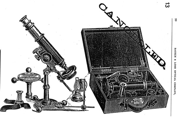
Figure 9 from a contemporary B&L catalog present that Company’s way of terminating their relationship with Griffith.
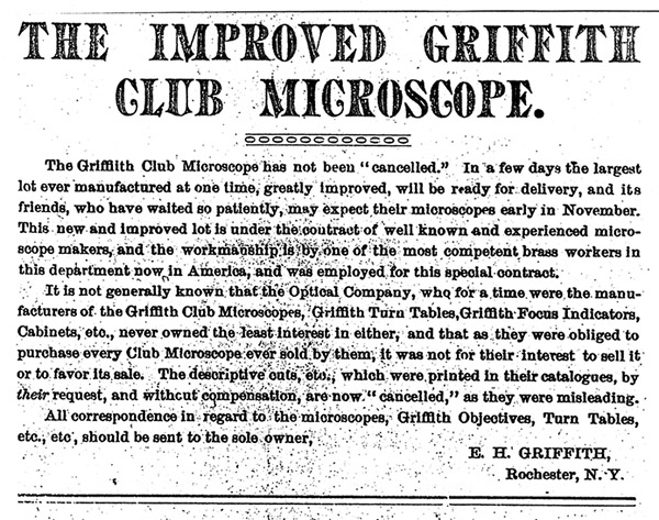
Figure 10 illustrates Griffith’s angry rebuke as published in a Public Notice / Advertisement in the January 1892 issue of The Microscope (A. Stokes, Ed). Griffith’s public notice appeared next to an advertisement for the Gundlach Optical Company. Gundlach was to replace B&L in producing Griffith’s stands. As Griffith, Gundlach had severed his association with B&L on an unhappy note.
MODERN EVALUATION OF PERFORMANCE
How does a Griffith microscope perform when examined using slides of different types and ages? To answer this question I tested two Club microscopes, the Briggs microscope discussed earlier, and microscope #210 in the MdC collection (serial #933).
I tested the Briggs microscope using slides from Dr. Charles M. Briggs own slide collection (discussed above). In particular I choose slides of human pathology and used overhead illumination reflected from the microscope’s concave mirror. The images obtained with either of the objectives were quite acceptable for uncoated optics. Had the slides not been labeled, it would not have been difficult to make a diagnosis of the specimens using this one hundred and ten year-old, amateur microscope. The only minor difficulty was with in fine focusing the image with the higher power, 1/5 objective; it was caused by partially dry lubrication rather than by any mechanical deficiency.
In testing the Club microscope MdC 210, serial #933 (figure 11), I used the concave mirror and natural daylight from a window. This lighting approximated what 19th century microscopists considered optimal daylight conditions. The unmarked drawtube was pushed in completely at the start of the observation and later extended as required in order to obtain the best possible image. Three biological slides, different in nature and preparation date (early and late 20th century) were used for subjective evaluation of image quality; a modern stage micrometer was also used to obtain quantitative data. The first slide made ca. 1900 by B&L, was a transverse paraffin section of the spinal cord, stained with hematoxylin-eosin; the second is plastic section of huan testicle made ca. 1980 by the Carolina Biological Supply Co.; it is also stained with hematoxylin-eosin. The last of the biological slides, is a cross-section of horsetail, made by the noted plant histologist John H. Nichols from Norfolk, England, in 1992. It was stained with a carmine-light green combination. The stage micrometer is an advanced design made in the early 1990s by Graticules Limited of Kent, England.
In all cases the clarity and contrast of the image was highly dependent on the proper positioning of the mirror. This is understandable, as while working without an understage condenser, the concave mirror fulfills two functions, that of a light reflecting surface and that of a low power condenser. Using the 2/3 objective (approximately 8X), the central image was crisp and with reasonable contrast. As expected, of the three biological slides, the 1.5 µm-thick section of human testis was the most challenging for the optics of this instrument. It was affected by the field curvature of the objective. Only in the central half of the field was in focus, around it extended a circle of badly out of focus image. The heads of the spermatozoa could be seen (4.5 µm long, 3 µm wide, 1 µm thick) but not the tails (1 µm thick). Neural cells and fibers could be seen with acceptable clarity in the spinal cord. The field of view of this objective was a little more than 2 mm in diameter (2,100 µm). Testing the 1/5 objective (approximately 40X and field of view of 180µm) gave disappointing results. Some features could be seen in the botanical section and the spinal cord, but only an observer aware of what the structures were and how they should look could have interpreted those images. No detail of any significance could be seen in the 1.5 µm-thick plastic section. The lines and numbers on the stage micrometer did not appear black against a colorless background, but rather gray and blurry. It was not obvious what caused this poor performance. The surfaces of the lenses were clean, and a few pits on the outer surface of the objective lens facing the object were far too minute to justify such a deterioration of the image. Extending the drawtube had the effect of improving the image but only to a very limited extent. It should be tentatively concluded that this was originally a not very good objective. Its performance was poorer than that of contemporary examples by Bausch & Lomb or Gundlach tested. Today we would call it a case of poor quality control.
THE CLUB MICROSCOPE IN THE MARKETPLACE
Griffith Club Microscopes are offered for sale only infrequently. The asking prices for those that come to the market vary considerably. We will give a few examples here. A Club carrying the serial number 915 was offered for $1295 (Perceptions Scientifica, 1987/88), another was auctioned in London, UK (Sotheby’s, 1994) with a estimated value of 8,000 British pounds. The Tesseract Catalogue of Early Scientific Instruments offered Griffith Club Microscopes on two occasions. The winter 1994/1995 issue offered a Griffith serial #1070. This was a late instrument that was different from every other example known to me; it had a two-objective revolving nosepiece and centrable condenser. Without case or lamp-holding bracket the instrument was offered at $6,500. The base of this instrument was engraved "Rochester, NY" instead of "Fairport, NY" as were the instruments in the 900s series. Tesseract Catalogue of Early Scientific Instruments (#51 winter 1995/1996), offered a Griffith fitted with a Gundlach objective. This was a presentation piece, said to be the second of the three known versions of the “Club.” Tesseract offered it at $8,200. The Tesseract commentary describes this as an important example of an uniquely American instrument. Finally, a Griffith microscope, serial # 1007, complete with case and (possibly) original lamp, was auctioned at ebay on 2001 07 28. The winning bid was at $9,000.
CONCLUDING REMARKS
The Perceptions Scientifica Catalog 1987/88 makes the following comment regarding the Club Microscope: “Few American Microscope manufacturers had singularly unique designs, most being heavily influenced by English examples. Griffith’s microscope, developed in the 1870s and 80s, is uniquely American.” Those words offer an appropriate ending for this article.
ACKNOWLEDGEMENTS
A number of persons helped me in the preparation of this article. Paul Ferraglio’s knowledge of the Griffith microscope was most helpful. Alan Hawk, Curator of Scientific Instruments at the National Museum of Health & Medicine, Washington, was most generous with his time and knowledge. He allowed me to examine the early Griffith microscope in the Billings collection and provided me with valuable documentation. Christopher Hoolihan, Librarian, History of Medicine Section, Edward G. Miner Library, University of Rochester Medical School, gave me access to information on Dr. Briggs and the opportunity to examine Dr. Briggs’ microscope. The staff of the Fairport Historical Museum kindly assisted me in searching for records pertaining to Ezra Griffith.
Comments to the author is most welcome.
BIBLIOGRAPHIC REFERENCES
• del Cerro, Manuel (2001) The Griffith Club Microscope, an American Original. Journal of the Microscope Historical Society, 9: 39-57.
• Griffith, Ezra (1890) Griffith’s Fine Adjustment. The American Monthly Microscopical Journal, 5: 272-273.
• Griffith, E. H. (1890) Griffith Club Microscope. The American Monthly Microscopical Journal, 6: 121.
• Padgitt, Donald L. (1975) A Short History of The Early American Microscopes. Microscope Publications Ltd., London, pp. 116-118.
• Purtle, Helen R. (Ed.) (1974) The Billings Microscope Collection of the Medical Museum Armed Forces Institute of Pathology. Armed Forces Institute of Pathology, 2nd Edition, p. 83-84 & 90.
• Stevenson, Betty Briggs Satterwhite (1992) The Story of my Grandfather, Charles Menzies Briggs, M.D., 1855-1933. Privately printed.
• Turner, Gerard L’E. (1989) The Great Age of the Microscope. A Hilger, Bristol, pp. 106 & 283.
• Watson, Randy D. (1995) E. H. Griffith and Griffith Club Microscopes. Rittenhouse, 9: 25-31.
___________________________________________________
Published in the January 2007 edition of Micscape.
Please report any Web problems or offer general comments to the Micscape Editor .
Micscape is the on-line monthly magazine of the Microscopy UK web site at Microscopy-UK
© Onview.net Ltd, Microscopy-UK, and all contributors 1995
onwards. All rights reserved.
Main site is
at www.microscopy-uk.org.uk
with full mirror
at www.microscopy-uk.net
.