|
Oddities and Entities by Richard L. Howey, Wyoming, USA |
From time to time, odd little notions about bits of technique, cost-saving substitutes for lab implements or captivating items to look at with the microscope occur to me, but are rarely such that they would serve as the basis for an article. So, I decided to ramble on a bit about some of these disconnected bits and pieces and not pay much, if any, attention to continuity–which may say something about the character of my mind. In fact, one of the reasons that I greatly enjoy browsing in copies of the late 19 th and early 20th Century microscopical journals, proceedings, and transactions is that they cover wide ranges of topics with some longer articles on specialized topics along with many short notes, observations, suggestions, descriptions of techniques or organisms thus providing a full evening entertainment for a microscopical eccentric such as myself. So, on to the rambles.
1) Recently I have been making up slides of various chemical crystals to examine under polarized light. Some of these have involved mixtures of the salts or solutions of more than a single substance and, of course, one needs to be careful what one mixes together, since you don’t want to generate toxic gases, caustic sprays, or explosive reactions. So, my rule is to be cautious to the point of timidity.
The preparation of a large number of slides can involve the use of a large number of pipets and even if you are using plastic or glass ones of the disposable variety, this can get rather expensive. As a consequence, I have taken to using small, plastic straws about 5 inches long. Slide the straw into the tube of solution with your finger over the outer end, remove your finger letting the straw take up some of the liquid, replace your finger over the end, put the straw over the center of a clean slide, remove your finger again and deposit the required amount of solution on the slide. Discard the straw. I buy these straws in packages of 500 for one dollar and recently found a store offering them for 50 cents!–real bargain-priced pipets.
2) Again on the subject of crystals. If you are mixing two solutions, after adding the second one, you may want to stir them a bit. A box of flat toothpicks–usually less than a dollar, unless you buy the rosewood ones–will provide you with a plentiful source of stirring sticks. In addition, if you are making crystal preparations directly on the slide, rather than preparing solutions in tubes, you can place a drop of water (or other solvent) on the slide, and using a flat toothpick (moistened if necessary) transfer some crystals to the drop of water and stir until dissolved.
3) Sometimes one needs a fine, sharp needle to sort or arrange forams, radiolaria, diatoms butterfly scales, etc. For good-sized sturdy forams, one can use a standard dissecting needle, but such a needle is too large for smaller specimens and also may crush fragile forms. So, at a local craft shop, I buy a packet of about 100 porcupine quills for 3 or 4 dollars. (You can also find them on the Internet.) My very first encounter with these fiendish hairs from Hades made me very cautious in handling them–they are painfully SHARP! About 35 years ago, when walking in the mountains, my wife and I came across a very large, dead, male porcupine. I didn’t attempt to harvest any quills–a lost opportunity, as I could have provided many microscopists a lifetime supply.
If I am unpacking, sorting, manipulating the quills, I usually use a pair of forceps to spare my fingers. The other day, I thought that perhaps now that I am retired as a philosophy professor, who dwelt on the mental–in fact many of my colleagues and students thought I was mental–that I might now focus on the physical and learn acupuncture employing quills and call it porcupunture. If I had the patience to pull it off, I could probably make a fortune in this world full of incredibly gullible people.
The quills are quite flexible and fine-tipped and so are admirably suited to sorting and arranging minute specimens, but are not utilizable for dissection. I have been using the quills without a handle, but it would be an easy matter to fashion a handle, if one wanted great length. For that matter, one could place a quill in a pin vise.
4) Have you noticed how, when a relatively simply and fairly ordinary device is modified and becomes a “scientific” implement, the price escalates? Consider a slide warming tray. You can order one from a scientific supply house for a mere $200! I guess these people either had wealthy parents or they don’t like amateur microscopists. So, I went on eBay and found a number of retro buffet warming trays with “thermostatic” control from “Lo” to “Hi”. These can be had at prices of 20 or 30 dollars and can serve essentially the same function as a slide warming tray. Don’t however try to find a new one at your local appliance store. The chances are that the clerk will look at you quizzically as you mumble and stammer trying to explain what you want and if they do have one, it will likely cost about $200.
I ended up getting such a tray for $20 plus $10 shipping. It can be used for accelerating the drying of slides mounted in balsam or synthetic resins and is also very handy for preparing slides of crystals. A variety of substances crystallize in very different patterns at different temperatures. Since my model doesn’t have a calibrated control, I plan to do a rough calibration by measuring the temperature in a beaker of water at various dial settings to provide myself with an eccentric, but repeatable standard.
5) Another example of a case where the description “scientific” means “expensive”, is the water bath. This device is, for the amateur, especially handy for melting glycerine jelly when preparing slides of delicate specimens that do not well withstand the rigors of dehydration for mounting in resins. Check a scientific supply house catalog and you will discover that water baths cost an outrageous amount. I looked at the catalog of one popular company and the price ranges from $427 to $635 depending on the size! Clearly manufactured and distributed by a bunch of amateur-microscopist-hating-wealthy spoiled brats. The solution is another retro device known as a crock pot or slow cooker (sounds like a description of some of my colleagues). Put some water in it and let it heat up to a temperature which will just keep the glycerine jelly liquid. Put the jelly in a heat-resistant beaker and use only a small amount so that you aren’t wasting it. It can be reheated, but it is best to simply use very small quantities to avoid doing so. If you go to a yard sale, you can probably find a working crock pot for less than 5 dollars.
6) Micro-forceps. You can get a nice pair of Swiss fine point forceps for $35 or $40 or buy from a specialty manufacturer of fine scientific tools and pay $600 or more. I buy 10 pair at a time of inexpensive micro-forceps made in Pakistan for about $15 total with shipping. Fine surgical tools these are not and the quality seems to vary with the batch. However, with a bit of work, you can produce forceps that are useful and adequate for many purposes. I bought a small fisherman’s diamond pocket hone at a discount store for about 5 dollars and it has a groove down the center of the long surface for sharpening hooks. With a bit of practice, you can sharpen the tips to make inexpensive and very serviceable micro-forceps. In the process of producing these handy tools, you make the tips thinner and more susceptible to bending. This is readily fixed by using the hone to touch up the tips as needed. Furthermore, this hone is excellent for putting very sharp tips on dissecting needles or single piece scalpels. (Don’t bother trying to sharpen disposable scalpels, since it’s unlikely that you can really get them suitably sharp again and the blades are brittle and can easily break. The flying pieces can pose a threat to hands, face, and eyes. Dispose of scalpels, pipets, glassware and all other laboratory materials in a proper manner. Pack things for disposal in such a way as to minimize risk. Just imagine yourself as your own garbage collector and consider how you would want such items packaged if you had to haul them off. Sorry–end of lecture. I just sometimes fear that we are too casual and forgetful of others and in the process have become seriously inconsiderate.)
7) Special laboratory dishes can also get expensive, so whenever I’m at a large discount store, I poke around in the kitchen accessories. Plastic condiment dishes can be purchased for less than a dollar in either clear or white. I purchased a few of each and find them very handy for sorting dry specimens such as foram samples. Recently I also bought two 4 oz. measuring glasses for under two dollars each. They are calibrated for tablespoons, teaspoons, ounces, and milliliters and this is very handy indeed when dealing with 19th and early 20th Century formularies which may employ all four forms of measurement. I bought one with black markings for measuring water and other harmless solutions and one with red markings for measuring chemical solutions. I also found a packet of four 2 ounce metal sauce dishes for less than a dollar a packet. so, when you’re out shopping, keep your eyes peeled for items useful in your lab. The hardware section has useful gizmos as well.
8) There are some types of specimens that constitute my M.U.S.T. list (M.U.S.T. is my acronym for M icroscopical U ltimate S pecimen T axon–how can we be modern if we don’t use acronyms?). I keep finding items to add to my list, but let me share some of my top contenders with you. I realize that other people’s lists will likely be quite different and I hope that some fellow microscopists will submit their Top Ten lists, so that we can all enjoy and share the fun of discovering some new treasures.
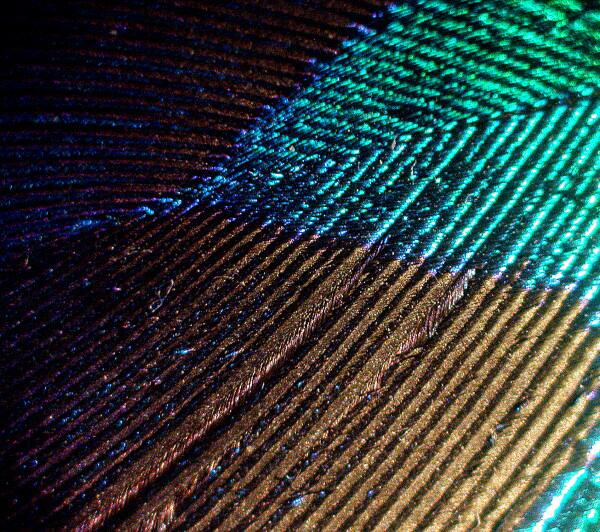
a) Peacock feathers. Fortunately you don’t even need to own a gun to obtain these; once again your local craft shop can very probably come to the rescue. If not, you can find them for sale on the Internet and, believe me, they are worth the trouble of tracking down. The “eyes” in the tail feathers provide especially stunning spectacles. The most intricately woven human fabrics pale by comparison. Here are blues, greens, golds, purples, and yellows with metallic sheens that defy description.
Particularly intriguing to me is the fact that some of these colors are due to pigments and others are a result of refraction due to structural characteristics. These are best observed with a stereo-dissecting microscope. Move your light source around to achieve a variety of angles of illumination and you will have a marvelous display. This is an experience not to be missed.
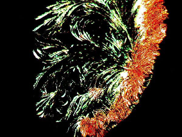
b) Orange G. This is a common biological stain usually used as a contrast stain. If possible, purchase it in an alcoholic solution or buy some of the crystals and dissolve some of them in 70% isopropyl alcohol. [Always exercise great care in handling the crystals or powders of stains. Many are very dangerous if the powder is inhaled and a number are known or suspected cancer-causing agents.] You can use an aqueous solution but you’ll have to be very patient and persistent to see the display, so I strongly recommend the alcoholic solution. (So would W.C. Fields.) Place a drop of the solution on a slide and quickly place it on the stage of your compound microscope which should be prepared in advance for polarization with the polars crossed. After a few moments, you will have a dazzling light show. After the crystals form, rotate the polarizer for an additional delight. There are many substances that produce striking crystals, but Orange G is a special treat to show your friends.
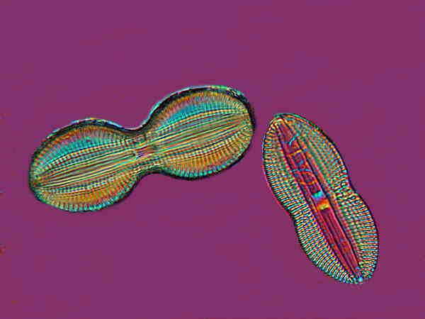
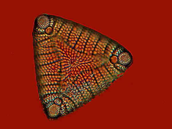
c) Diatoms and Radiolaria. These are difficult to prepare and mount properly unless you are a specialist but, at minimum, a few good prepared slides of these should be in virtually every microscopist’s collection. Diatoms used to be regarded as plants and radiolaria as animals, but these days they are classified as protists. The sculpting of some diatom shells is a challenge for even the very best lenses which microscope manufacturers can produce. The elegance and variety of these tiny bits of silica have seduced significant numbers of microscopists into a lifetime study of these remarkable organisms.
Radiolaria and their close relatives, the Acantharians, produce some of the most intricate and aesthetically pleasing structures in all of nature. In relation to diatoms they are generally significantly “thicker” and thus more of a challenge when one is trying to acquire an overall picture of their morphology.
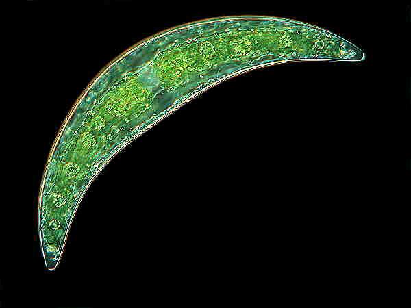
d) Desmids . Every microscopist with a bit of imagination makes associations with certain types or groups of organisms or even non-living specimens. I have always had a special fondness for desmids and somehow they always seem to make me rather cheerful. Here the protective and enclosing structure is not silica, but an organic membrane of considerable complexity. Their chloroplasts are frequently of an especially rich, deep green color and in terms of shape, desmids range from crescents, such as, Closterium to spiny stellate forms.
e) Volvox . Neither my wife nor Wim van Egmond ('Volvox, one of the seven wonders of the micro world' ) would forgive me if I were to omit this incredible creature from my list. In many respects, Volvox is an enigma; it is a colony that can produce subcolonies within itself, sometimes exclusively female ones, sometimes exclusively male ones, and sometimes a mixture. Its appearance is that of a delicately-tinted green, glass sphere. It is composed of hundreds to thousands of tiny flagellates each containing chloroplasts and an eyespot and these “cells” reside in a gelatinous surface matrix and are interconnected by a lattice of fibrils. Here in the mountain ponds, early spring and late fall are the best times to collect Volvox . It is difficult to imagine ever forgetting one’s first experience of observing these extraordinary organisms rolling through the water like miniatures space stations.
f) Termite flagellates. ('You want to bring those into the house?') Trust me! I’m not joking and, just think, your experimentation could save someone’s house from being devoured by voracious termites. Living termites can be obtained from biological supply houses. Prepare a solution of insect saline–0.6% non-iodized salt in distilled water. Decapitate a termite, place the body in a small amount of the insect saline in a small Petri dish and open up the stomach and intestines with dissecting needles. Talk about a need for population control! You will, I think, be overwhelmed by the sheer number of organisms in such a small volume but, even better, organisms that are very high on the scale of oddity. Trichonympha campanula, one of the largest and most common, immediately catches one’s attention.
It is a hypermastigote, which means, among other things, that it has an enormous number of very long flagella. There are a significant number of species in both termites and woodroaches that are still unidentified. I suspect that there is a wide variety of symbiotic and parasitic organisms in the guts of a great variety of insects, so keep your bottle of insect saline handy.
g) Butterfly and Moth Wings. These structures are wonderful both for the larger patterns in the wings and also for the variety of scales that make up those patterns. Examine the wings with a stereo-dissecting microscope and then scrape some scales onto a slide containing a drop of immersion oil, place a cover glass on the oil drop and examine with a compound microscope. As with feathers, some colors are a consequence of pigments and others of refraction. It is also rewarding to examine the elytra or wing cases of beetles, especially of iridescent ones.
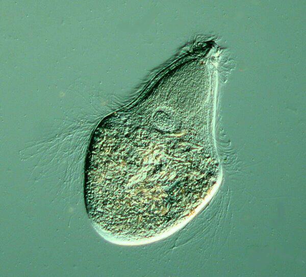
h) Hydra . A stunning aquatic creature is named for the mythological beast, Hydra. It is an unforgettable experience to be examining a dish of pond water and suddenly encounter a white, brown, or green stalk with tentacles waving or drifting slowly in the water. The stalk may have bumps on it which can be ovaries or spermaries. At higher magnifications, one can also see much smaller bumps on the tentacles. These are nematocysts or “stinging cells” of which there are nearly a dozen types. If a hapless Daphnia (water flea) chances to brush against the tentacles, it can easily become a meal for the Hydra . This is a remarkable contest, because Daphnia is a strong swimmer, but the tentacles and the toxins in the nematocysts of the Hydra often win out.
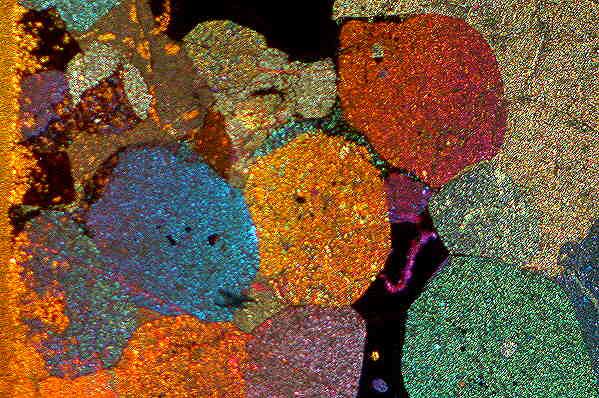
i) Thin Rock Sections. Unless you have an inordinate amount of time and patience or are willing to invest a large sum in specialized rock cutting and grinding equipment, it is best to purchase the occasional prepared slide which starts at about $20. These are especially interesting under polarized light.
Well, the list could go on and on, but I have a few other random items I would like to mention before concluding.
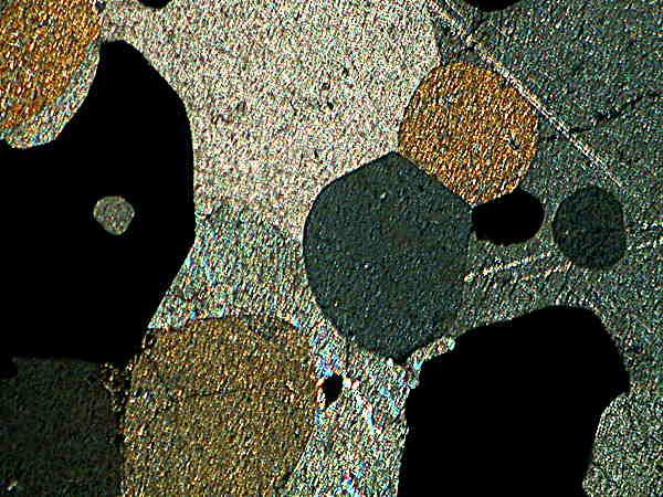
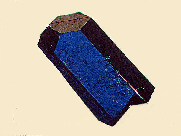
9) Some mineral collectors are specialists in micro-mounts; small sections, often less than a cubic inch which have been selected for nearly perfect small crystals. Large crystals can, of course, be extraordinarily impressive, but the larger they are, the more likely they are to have imperfections. Many of the small crystals have a stunning beauty that a stereo microscope can reveal. I have taken small chunks of rock, which to the naked eye appeared rather dull, but which under the microscope revealed marvelous and intricate crystal growths hidden away in little nooks and crannies. If you wish to attempt photomicrography with such specimens, you will likely experience a number of frustrations and soon discover that you require a significant amount of space, equipment, and patience for such an enterprise. Lighting becomes a major issue and inevitably the Koch-I-Nor of micro-crystals which you are trying to photograph will be lodged in a niche that defies all of your attempts at illumination as though it were a black hole.
WARNING ! Micro-mount enthusiasts should stop reading at this point. What I am going to propose may induce cardiac arrest. Well, it’s not quite as bad as that, because, after all, there are superb micro-mounts ranging on down to very good, mediocre, and junk. What I’m suggesting is taking a piece—one which is not superb, but has some very nice crystals and removing some individual crystals or small clusters to place them in a context such that they can more readily be photographed. I wrote earlier about producing fine-pointed, sharp needles using dissecting needles and a diamond hone. Using the same implements, you can produce micro-chisels which are sharp, relatively sturdy, and easily resharpened. With your micro-needles, micro-chisels and persistence, you can sometimes extract lovely clusters which can be photographed readily once they are no longer embedded in a matrix.
With a bit of ingenuity, one can make a variety of useful, inexpensive micro-tools and by letting your imagination run rampant, you can find endless odd items in both usual and unusual places to expand your microscopical investigations.
All comments to the author Richard Howey are welcomed.
Editor's note: Visit Richard Howey's new website at http://rhowey.googlepages.com/home where he plans to share aspects of his wide interests.
Microscopy UK Front
Page
Micscape
Magazine
Article
Library
Please report any Web problems or offer general comments to the Micscape Editor .
Micscape is the on-line monthly magazine of the Microscopy UK website at Microscopy-UK .