(White card under slide for photo.)
|
Enjoying a M—ller 80 form diatom type-slide with a microphotograph setting by David Walker, UK |
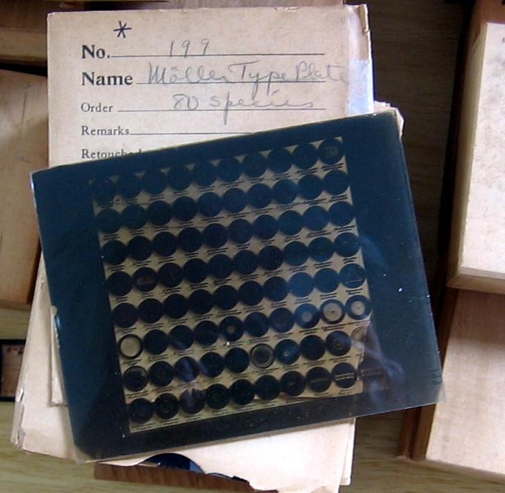
The arranged diatom type-slides by some of the 19th century mounters are very collectable; one of the more noted mounters was J D M—ller. His more elaborate mounts are not usually within the budget of my brother and I, but we decided to treat ourselves to a more modest example using part of some unexpected prize money. Aspects of the slide are shared below as an example of his remarkable skills both as a diatom mounter and microphotographer. The author's long term project is to photograph each diatom on the slide to create an interactive online tour of the slide.
The author is neither a historian nor diatom expert, so presents the article mainly as an owner's 'hands-on tour' of the slide with references. There is a wide range of resources on the life and work of Johann Diedrich M—ller and a selection are mentioned in the text and provided in the references with additional notes. Matthias Burba was very helpful in answering questions on aspects of the slide. Comparison of this slide's features with the maker's equivalent 335 form with microphotograph setting was possible using Little Imp Publication's extensive CD-ROM resource on this larger type-slide. (See Acknowledgements and References.)
Slide summary
(White card under slide for photo.)
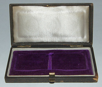
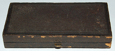
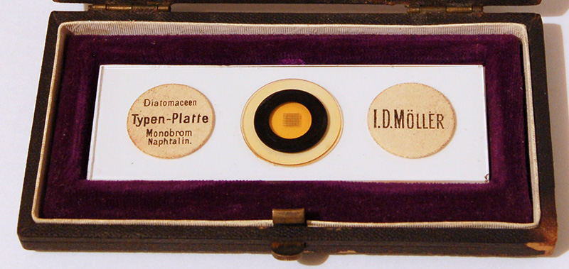
The box (ca. 10x5x1.5
cm)
supplied with the 3x1 inch slide seems to be of the period but uncertain if the
original. Although its style, apart from lack of any maker or dealer markings
for this example, looks very similar to that described for the equivalent 335
type-plate (ref. 9).
There is an outer 19.5 mm diameter coverslip with smaller raised black ring where the mountant is noticeably darker; suggesting
a coverslip was mounted on another (see ref. 11.) When studying the diatoms under the microscope,
the microphoto' is at a slightly higher plane of focus than each diatom,
presumably consistent with the microphoto' being made on the underside of smaller
coverslip to which diatoms were added.
The slide is quite simply labelled as shown above (the label looks identical to that on the 335 form, ref. 9). The righthand label apparently states 'I. D. MøLLER', but in a footnote in Horrocks's paper, he remarks that 'there has never been an I. D. M—ller', also see ref. 10. The seller stated the slide was made ca. 1880 (see ref. 12).
The slide subject shows 80 species of diatoms displayed in a 10x8 microphotographed array, with the species name below each. See below for more details. Both Horrocks and Bracegirdle note that apparently only two types of slides with this particular named species microphotograph were made, an 80 form as this example and a 335 form (see ref. 9 which describes and illustrates how this larger type plate with microphotograph settings was arranged). The references below give details on M—ller's other arranged diatom slides, both commercial and specials, which include the 'Universum Diatomacearum M—llerianum' with 4026 species (see ref. 7)!
Monobromo-naphthalene appears to be a quite common diatom mount of the period; its RI of 1.658 being higher than Canada Balsam (1.526), a little lower than Sirax / Hyrax (1.7) and much lower than the exotic Realgar (2.4). (RIs are quoted from ref. 4.)
Slide detail
To
the present author the striking aspects of this slide is the professionalism,
elegance and practicality
of the mount. Elegant because it looks a beautifully balanced composition. The photo-reduced
setting is exactly 3.0 x 3.0 mm in size and each diatom circle is 0.30 mm in diameter and the
microphoto' is crisply executed.
The use of microphotography to present each species with its name works very
well, avoiding the need to refer to an index. Many of the other microphotographs
of this era by other mounters, although undoubtedly technically accomplished
aren't used in a practical way but show photo-reduced artwork, famous people
etc.
The author has never owned microphotographs by mounters such as Dancer to compare the quality; Tither (ref. 3) remarked on the comparative quality of some M—ller and Dancer microphotographs.
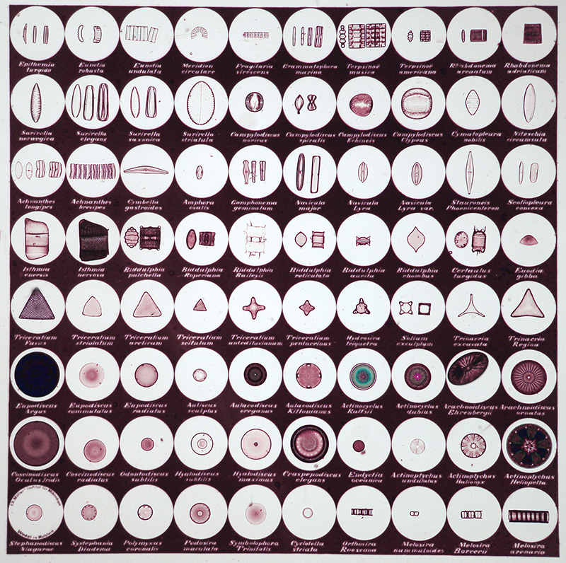
The microphotograph setting is 3 x 3 mm square with 0.30 mm diameter species circles. The array sits comfortably in the field of view of the Zeiss 2.5X objective on a Zeiss stand with 1.25x Optovar and fills field of a 4x Zeiss planapo on a 160 mm stand with 1x trino head (10x widefield eyepieces). It was photographed with the 2.5x objective. The size chosen by M—ller for the microphoto array and number of species shown gives a well balanced, uncluttered composition. (The setting is perfectly square visually, there's a slight distortion in photography.) See ref. 7a for a M—ller photographic negative of what appears to be a similar type-slide.
Although the mount to the eye is a pale brown, it's not very noticeable visually under the microscope and a colour cast correction easily corrects it in digital photomicrographs.
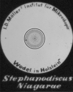
Left: The diatom
at bottom lefthand corner of the array has an inscription incorporated into the
microphotograph which reads: 'I. D. M—llers Institut f■r Mikroskopie'
and 'Wedel in Holstein'. The microphoto' still looks essentially grain free with
the 16x objective used here.
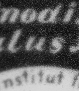
Right: To measure the silver grain
size to any accuracy would require an oiled 100x objective and decided not to
use oil on this slide. With the dry 40x objective used here, the grain is now
visible.
Species
Given
the species names are stated on the slide, I'm uncertain whether it would have
been originally supplied with a paper index. The author has catalogued
the species below in alphabetical order. There are 45 genera represented. The
presentation of a number of the species with multiple views from 2 - 4,
is very useful to understand a given diatom's structure.
By comparing this species list with that given for the equivalent 335 form (ref. 9), all the species on the 80 form are also represented on the larger plate except two, ('Hyalodiscus maximus' and 'Navicula lyra var.') The species which M—ller chose to present as multiple views also seem the same on the 335 form for where comparisons were possible.
A selection of diatoms are shown below to illustrate typical diatom presentations. The photos were taken with a Zeiss 16x objective to show both the diatom and name, but the plane of focus of text can be slightly higher than the typical focus plane of diatom (e.g. the Cymbella below). The name next to a species is a useful aid when studying them but for detailed visual and photographic studies of a given species, higher mag objectives would be needed.
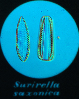
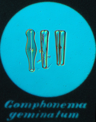
Surirella
saxonica and Gomphonema geminatum. Circle diameter 300 çm.
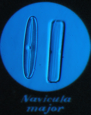

Navicula
major and Cymbella gastroides (phase).
Comments
My
brother and I don't regard ourselves as owners of this slide but more 'custodians'.
The
slide is remarkably well preserved and hopefully can be enjoyed by future microscopy
enthusiasts if looked after with care. Micscape would be delighted to hear
from owners of any interesting slides if they wish to share aspects
of them.
Comments to the author David Walker are welcomed.
Acknowledgements
Any inaccuracies
in this article are the present author's (and would welcome
any corrections).
I'd
like to thank Matthias Burba who is an authority on the life and work
of J D M—ller and has prepared both public exhibitions of M—ller's work and
also written on many aspects of his life and work, with access to the M—ller
archives (see refs.). Matthias patiently answered questions on aspects of my
own slide as well as kindly providing copies of his own articles and additional
references.
Raymond Hummelink who presented his photos of the M—ller Hamburg exhibition online in the Yahoo microscopehobby forum, gave permission to link to his folder and put me in contact with Matthias Burba.
Keith Shaw for permission to link to, and host a copy locally, of his image of the 4x5 inch negative of a M—ller 80 form type slide in the 'Chappell collection' (ref. 7a).
Little Imp Publications
(Steve Gill and Mike
Samworth ) have prepared a CD-ROM on M—ller's 335 form
type-plate with microphotographed setting and species names, which is a
'virtual tour' as well as providing historical background material (ref.
9). The CD is a superb example of how rare slides can be presented in an interactive digital resource
for others to enjoy.
I acquired this resource after completing the draft
article,
but this has been invaluable in allowing fascinating comparisons between the
80 form slide owned and the 335 form. Some comments in the above text have been
added based on this comparison.
References, additional
notes and resources
1)
F T Horrocks, 'Johannn Diedrich M—ller', Quekett J. Microscopy, 1969, 31,
210 - 214.
Includes references to German biographies.
2)
B Bracegirdle, 'Microscopical mounts and mounters', Quekett Microscopical Club,
London, 1998. (Available from Savona
Books).
'M—ller J. D.' biographical entry p.68, illustrated slides
include Plate 45 which shows a wide selection.
3) R Tither, 'Unrecorded
Dancer slides and other microphotographic matters', Quekett J. Microscopy, 1969,
31, 123.
4) F C Wise, 'The President's address. A short contribution to the history of diatom mounting', Quekett J. Microscopy, 1959, 28, 180 - 194.
5) 'Novelties', Quekett
J. Microscopy, 1868, 1, 141 - 142.
Report of a Quekett Club meeting
where a J D M—ller ca. 400 diatom 'type-slide' was exhibited.
6) H van Heurck, 'A
Treatise
on the Diatomaceae', English translation by W E Baxter, 1896. Book free to download
at www.archive.org.
In Chapter I, page 78 in the section titled
'Preparation of diatoms. Systematic preparations of Type Slides and selected
diatoms',
the author remarks:
'Mr
M—ller employs a specially constructed microscope to make his type slides
and he adopts his own particular methods, which he has hitherto disclosed
to but three or four persons, of which I am fortunately one.
I have now
known it for a long time but am under an obligation not to reveal anything
in connection with the subject.'
The above entry is followed by van Heurck quoting an extended description of methods for arranging and preparing diatom type slides provided by 'Mr H Peragallo, the eminent French diatomist'.
7) A Yahoo 'microscopehobby' Forum member Raymond Hummelink shares a note on a M—ller exhibition in Hamburg with a selection of his photos in the Forum's Photos section (the photos are accessible only by members). An image is included of the 4026 species 'Universum Diatomacearum M—llerianum'.
 7a)
Photographic
negative (4x5 inch) of a M—ller 80 form type-plate, image by Keith Shaw.
It forms part of the 'Chappell collection', of which Keith Shaw is
custodian. Hosted in the Yahoo
'microscopehobby' forum Photos section (the photos are accessible only by members)
but shown right with permission of Keith Shaw (click thumbnail for larger).
Not to be used without his permission.
7a)
Photographic
negative (4x5 inch) of a M—ller 80 form type-plate, image by Keith Shaw.
It forms part of the 'Chappell collection', of which Keith Shaw is
custodian. Hosted in the Yahoo
'microscopehobby' forum Photos section (the photos are accessible only by members)
but shown right with permission of Keith Shaw (click thumbnail for larger).
Not to be used without his permission.
8) Nikon Small World 2008 'Image of distinction'. A beautiful photomicrograph by Matthias Burba of a striking '1891 J D M—ller arrangement' of diatoms.
9) Little Imp Publications (Steven Gill and Michael Samworth, with some contents supplied by Dr J Lubran) CD-ROM. 'J D M—ller's Diatomaceen Typen Platte. 335 Form'. (Available from Savona Books ref. M81).
10) Matthias Burba, in a personal communication, confirmed and remarked that 'there was nobody with the name I. D. M—ller'. He comments that in the first 15 years of M—ller's slide making he used labels where the 'J' looked like an 'I'.
11) Matthias Burba, in a personal communication, confirmed that M—ller used two cover glasses for his arrangements. Matthias discusses this in the ref. 16 he supplied.
12) Matthias Burba, in a personal communication, confirmed that the date of 1880 will be correct within a 10 year range. Matthias notes that after 1891 J D M—ller stopped making arranged slides.
The following references were kindly supplied by Matthias Burba.
13) 'Joann Diedrich M—ller, 1844 - 1907, Die Kunst Diatomeen zur Legen, Ausstellung im Zoologischen und Botanischen Museum der Universitðt Hamburg 15. November 2007 - 15. April 2008.' An illustrated article (downloadable pdf) on the exhibit at the Hamburg museum. Matthias remarks 'the Exhibition will be closing in March 2009, if somebody wishes a guided tour, please ask.'
14) Zeit Online 'PartyspaÔ Alge' - 14 page gallery with commentary showing a variety of M—ller's arranged slides and historical background, including his working area.
15) Matthias Burba, 'Die gr—sste Typenplatte der Welt und ihre Herstellung', Mikrokosmos 2008, 321-327.
16) Matthias Burba, 'Johann Diedrich M—ller (1844 - 1907) Éber die Kunst, Diatomeen zu legen', Mikrokosmos 2007, 7-17.
Published in the January 2009 edition of Micscape.
Please report any Web problems or offer general comments to the Micscape Editor .
Micscape is the on-line monthly magazine of the Microscopy UK web site at Microscopy-UK
ˋ
Onview.net Ltd, Microscopy-UK, and all contributors 1995
onwards. All rights reserved.
Main site is at
www.microscopy-uk.org.uk
with full mirror
at
www.microscopy-uk.net
.