|
In 2009, I was intrigued by Olympus objectives marked 'RP' on
eBay
and I wanted to know more about them. I) Material I am equipped with Olympus microscopes and for a long time, my main model is the BHS. With the help of internet auction sites, I have added accessories and also bought other second-hand microscopes. |
|
I bought in particular in the United States a few years ago, an Olympus CK2 inverted microscope. It is 110V but I added a 110-220V transformer for use in France! 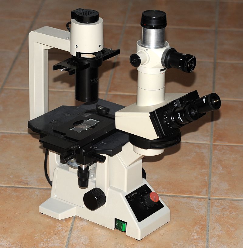 I used it first with objectives A4Pl, A10PL with the phase rings supplied and a LWD20PL (this last one has a collar for regulation of thickness of object cover 0 - 2mm). |
|
As remarked at the start of this article, last year I 10x
achro
RP NA 0,25 and Note that
the 40x is designed for a thickness of object cover of 1mm! | 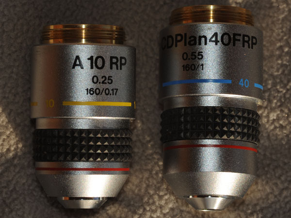
|
|
I made rings for phase corresponding to those in the original condenser of the CK2 but this one had an insufficient opening for the 40X. | |
|
Now at the same time, I bought second hand two partial Hoffman Modulation Contrast sets for the Nikon Diaphot. So I have a whole set of lenses and two Nikon condensers of NA 0,5. | |
|
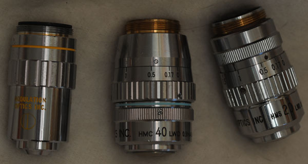
One was
associated with the objectives for contrast of Hoffman:
| 
|
|
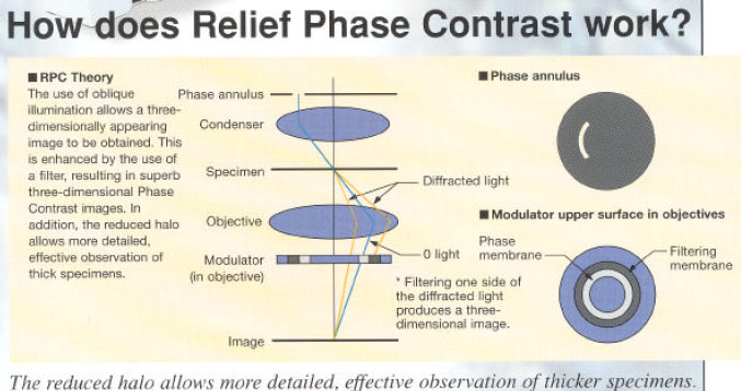
The other condenser with four openings in turret, serves for do-it-yourself work and in particular for this relief contrast. I made phase rings with cardboard. 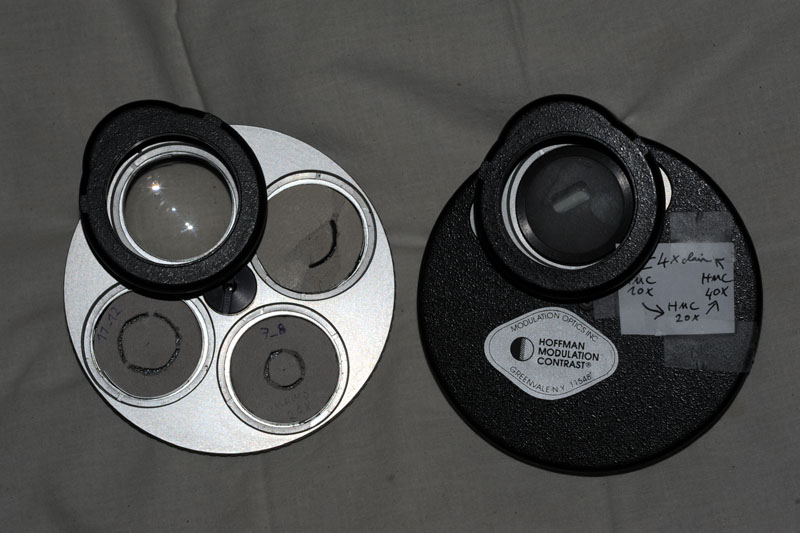
A brochure associated with the Olympus CK microscope (ref 1) gave me a model to make the phase rings adapted to two objectives for this "Relief Phase Contrast ( RPC)". It is necessary to note that the brochure
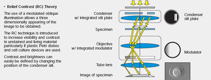
dedicated to the more recent microscopes CKX (ref 2) describes a
different technique
"Relief Contrast" which seems very close to the contrast of
Hoffman ( HMC). Did the patents of this technique become exploitable by Olympus? I have no details of the story, but my tests by setting up the two techniques cover two generations of equipment. |
|
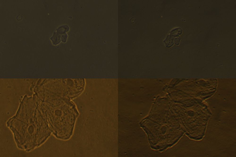
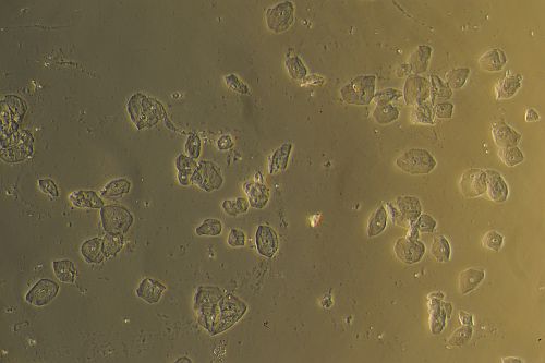
2) Results of the tests To study the techniques images were taken with a digital Nikon camera that show. the classical oral epithelium cells. Pictures are all resampled for 500 pixels length. |
| 1st series (scale 0,5 = 2 photosites for 1 pixel in the image): | |
| Phase Contrast "Relief phase contrast" Olympus | |
| 10x 40x |
|
|
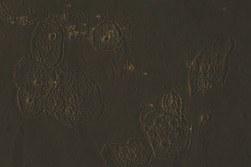
2nd series (this time the full field with the projection NFK2,5x eyepiece which was used, is kept): | |
| Phase | "Relief phase contrast" Olympus |
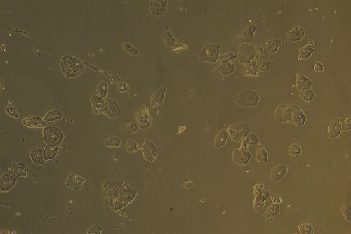 10x 10x
| 
|
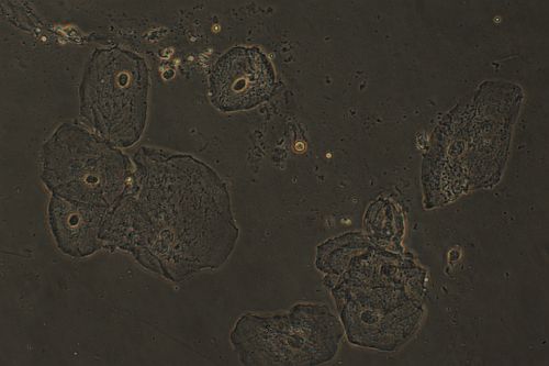 40x 40x
| 
|
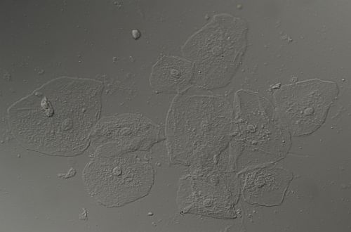
| At 40x, I also compared the images with Hoffman modulation and differential interference contrast, but I did not retain the same field after change of microscope. | |
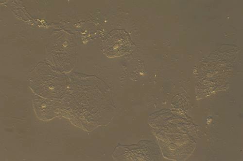 40x 40x
| 
|
| Hoffman modulation contrast | Differential interference contrast |
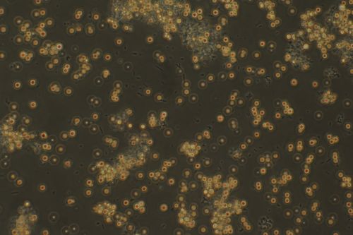
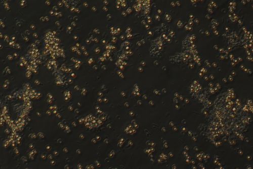
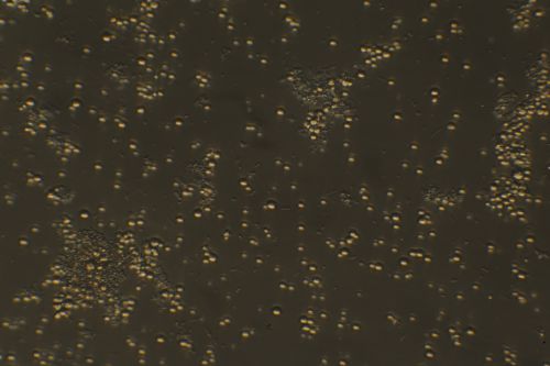
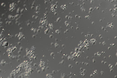
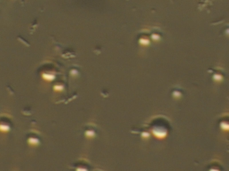
2nd test subject: only using the 40x objectives, were observations of a drop of liquid floating of natural yogurt to see bacteria responsible for the processing of the milk: long lactobacteria and chains of streptococci, among globules of fat
| Phase Contrast | Relief phase contrast |
| 
|
| 
|
| Hoffman modulation contrast | Differential interference contrast |
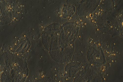
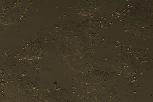
| Now, three examples on the same line for easier comparison: | ||
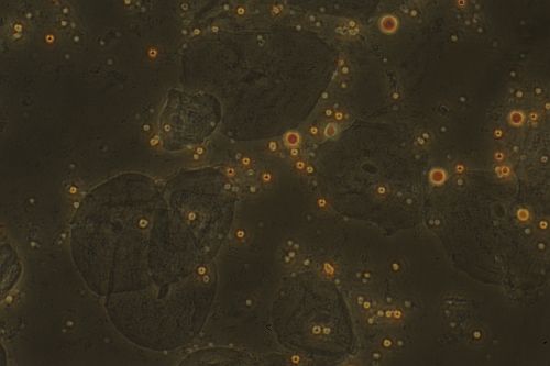
| 
| 
|
| Phase Contrast | Relief phase contrast | Hoffman modulation contrast |
 | ||
| Phase Contrast | Relief phase contrast | Hoffman modulation contrast |
|
3) Discussion The images in RPC have an overall similar look to phase contrast, but the halo is present only with the latter one. It gives a light impression of relief but which has nothing to do with that seen with DIC. This technique RPC seems to me to bring little benefit. It was nevertheless proposed recently (2007) by a working doctor in Romania Joerg Piper (ref. 3). It seems that Zeiss (ref. 4) also used a similar idea for a while (Variable relief contrast = Varel) before developing in 2003 an interference contrast method suitable for plastic dishes i.e. PlasDIC. (Ref. 5) Differential interference contrast ( DIC) is much preferred every time we can use it. The Hoffman contrast works with the plastic material dishes incompatible with DIC. It is the current technique with my Olympus microscope, named "relief contrast" by Olympus. The
techniques based on an oblique illumination are however interesting
because
they have only a low cost and can be implemented by amateurs. The PlasDIC would certainly be interesting to try but it is reserved for Zeiss equipment. I wonder however if it would not be possible to realize it with a prism IC for reflected light? Perhaps it is a new aspect to explore by do-it-yourself. Daniel Nardin. Please feel free to e-mail me if you have any questions or comments. |
|
Bibliography and Internet references : (1) Olympus relief phase contrast Brochure CK : http://www.iolympus.cz/mikroskopy/prospekty/Relief%20phase%20contrast.pdf (2)
Olympus
relief contrast Brochure CKX:
http://www.iolympus.cz/mikroskopy/prospekty/CKX2.pdf
(4) Zeiss
Varel |
Microscopy UK Front Page
Micscape Magazine
Article Library
© Microscopy UK or their contributors.
Published in the January 2010 edition of Micscape.
Please report any Web problems or offer general comments to theMicscape Editor .
Micscape is the on-line monthly magazine of the Microscopy UK web site atMicroscopy-UK
© Onview.net Ltd, Microscopy-UK, and all contributors 1995 onwards. All rights reserved.
Main site is at www.microscopy-uk.org.uk with full mirror at www.microscopy-uk.net .

