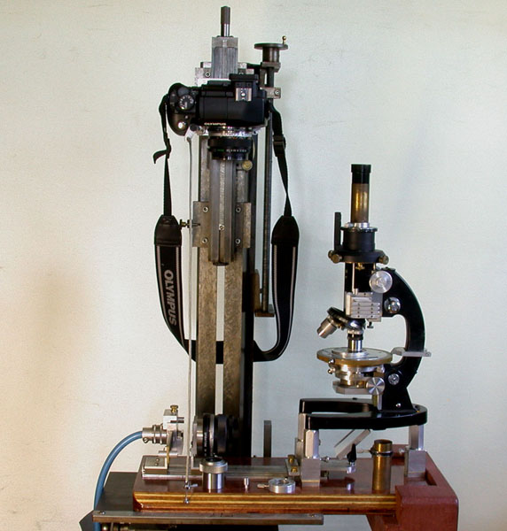
Fig. 1
|
Experiments Using Water Immersion Objectives Ted Clarke, Retired materials Engineer, USA |
I have enjoyed examining lake water organisms using my modified Monolux student microscope using a micro-aquarium slide with water immersion caps on LOMO objectives as described in my earlier article on water immersion caps. My interest in seeing finer details than resolvable with a 40X 0.65 NA objective led me to purchase the 30X 0.70-0.90 LOMO water immersion achromat objective and most recently the LOMO 70X 1.23 NA water immersion apochromat. My previous experiments with high NA COL with the 1.30 NA LOMO apochromat and 1.2-1.3 NA LOMO cardioid condenser convinced me that COL would now become a key illumination method for observing the lake water organisms in addition to darkfield illumination. COL illumination for the 1.23 NA apochromat requires immersion fluid between the condenser and bottom of the slide to acheive the full resolution capability of the objective. Darkfield between 1.25 and 1.33 NA (the index of refraction of the water mountant is 1.33) is not feasible with this objective. I can use my modified LOMO Biolam with the micro-aquarium slide with a ring on top of the condenser to retain glycerin immersion fluid, but loss of fluid coupling to the bottom of the slide is a problem when scanning the 40 mm circular field of the micro-aquarium slide. This has not been a problem with my modified Monolux because of the stage design. I knew that the home-built condenser of the modified Monolux could provide darkfield with a 60X 0.85 NA objective and recently determined that it would provide darkfield for the 0.90 NA 30X objective as well as COL reaching the full 1.23 NA of the 70X objective. Neither the home-built nor LOMO 1.40 condenser is aplanatic when used to provide the high NA COL, but only approximately so because of the narrow illumination annulus. My article on a multimode condenser in Modern Microscopy indicates some loss in image contrast when the COL is not fully aplanatic: . The work by Osamu Oku superbly demonstrates the capability of 1.40 NA COL with a fully corrected objective and condenser in resolving the diatom Amphipleura pellucida to dots with high contrast using blue light.
Fig. 1
My modified Monolux microscope system is shown in Figure1 with the 30X water immersion objective immersed into the micro-aquarium slide without a water immersion cap or cover glass. This cheap student microscope from the 1960’s now has a housing with slider mounted wave retarders and analyzer for polarized light using a rotating stage and focusable condenser with a rotating polarizer and a slot for a darkfield stop. In addition it uses a center-able drawtube. The installed, longer drawtube has the retaining ring set for a 203 mm tube length, adding 43 mm of tube length to correct spherical aberration from using the 30X objective without a cover glass. The added tube length combined with use of a 12.5X Zeiss Kpl eyepiece results in a viewing magnification of 475X. The shorter drawtube with the retaining ring set for 160 mm tube length for use with the 70X objective is shown on the wood base for the external fiber-optic source Koehler illumination system. Also shown on the base is a cap fitting over the eyepiece containing my correction for astigmatism. The Zeiss eyepiece is marked with an eyeglasses symbol for use with eyeglasses, but I find that a higher eyepoint of at least 19 mm is needed for me to avoid contact of my eyeglasses with the eyepiece. To the left of this cap is another cap for the eyepiece. This is a Klein loupe made from a 25X stereomicroscope eyepiece. Being able to view the image of the light source at the rear of the objective with a phase telescope or at the eyepiece eyepoint with the Klein loupe is critical for adjusting the illumination.
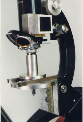
Fig. 2
Fig, 3
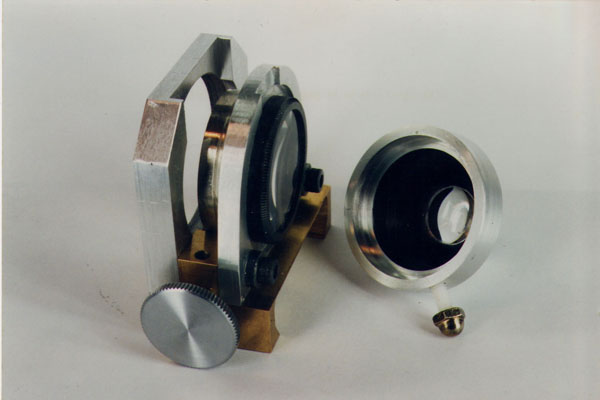
Fig. 4
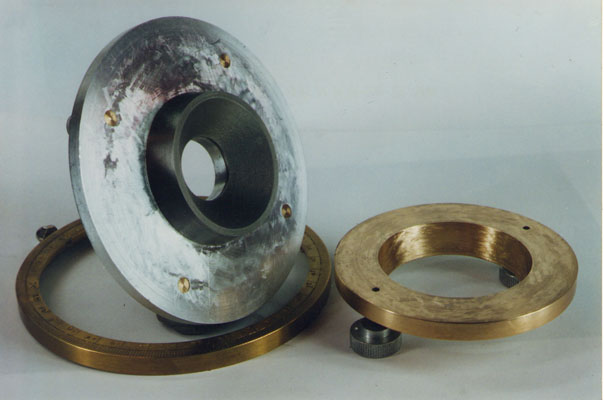
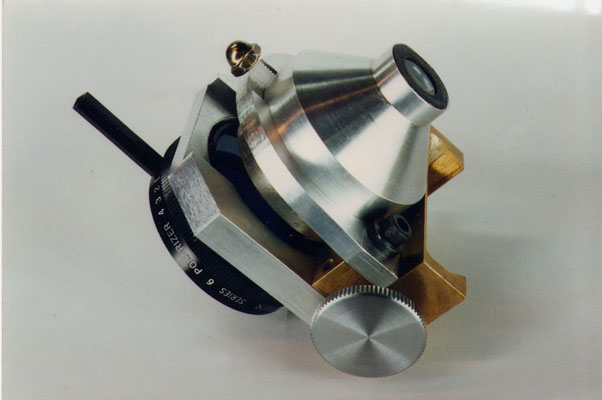
Fig. 5
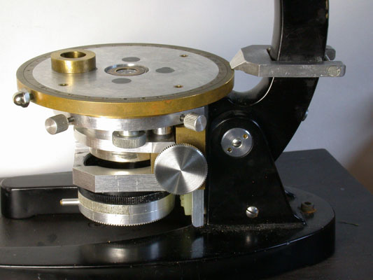
Fig. 6
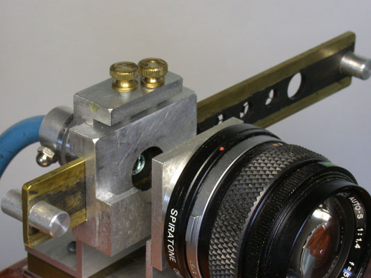
Fig. 7
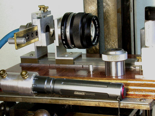
Fig. 8
The original Monolux had only a single element lens mounted in the plastic stage. A bracket was made to mount a homebuilt condenser. This bracket is shown being squared to the objective axis in Figure 2. The vertical leg contains a dovetail and plastic rack for the focusing mount of the home-built condenser shown in Figures 3 & 4. The condenser section contains a doublet with the original Monolux stage lens in the top position. Figure 5 shows the components of the rotating stage. The mating flats and conic sections are hand scraped to precisely mate with a run-out below 5 micrometers and only a thin oil film separating the surfaces. Figure 6 shows the condenser and stage installed on the bracket, two of the four stage centering screws visible. The brass ring in Figure 6 has a very thin internal flange which contacts the brass mount of the top element when the ring is inserted into the gap between the top of the condenser and the opening in the rotating stage. The external illumination system shown in Figure 1 uses a ¼” fiber-optic light-guide as the source with the aperture diaphragm at the end of the light-guide. A slider of stops is located just in front of the aperture diaphragm. The slider of stops is shown close-up in Figures 7 & 8. A 50 mm f/1.4 camera lens is used as the collector lens. The diaphragm of this lens is the field diaphragm when the system is fully aligned for Koehler illumination. Figure 8 shows an LED flashlight coupled to the end of the fiber-optic light-guide. The LED flashlight provided ample illumination intensity for the experiments with the 30X and 70X water immersion objectives. An Olympus E-330 DSLR camera with a 28 mm f/2.8 wide angle lens focused at infinity was separately mounted above the eyepiece to check critical focus and then record the images with a 100 ISO speed setting.
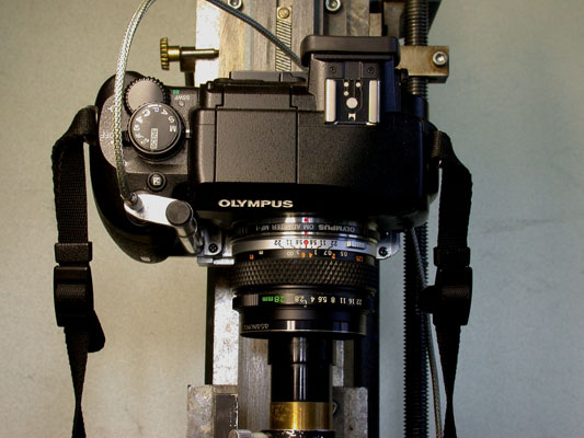
Fig. 9
Fig. 10
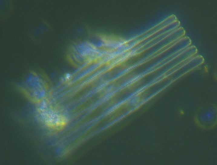
Fig. 11
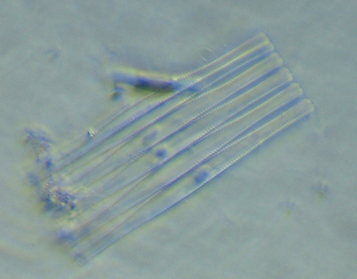
Fig. 12
Figure 10 show the 30X objective immersed in the micro-aquarium slide to record the darkfield image of Fragillaria crotonensis of Figure 11. The slider mounted stop for darkfield with the 40X 0.65 NA objective was inserted in the condenser and a larger NA stop in the slider strip at the end of the light-guide were used for this image. A smaller stop in the slider strip was used to record Figure 12 with COL reaching the full 0.90 NA of the objective. I was pleased to find that the contrast with COL was comparable to that with darkfield. The COL image is sharper as expected based upon it meeting the Abbe resolution criterion with the zero and first order diffracted beams entering the objective. The darkfield image would require an unfeasible condenser NA of 3 times the objective NA for the first and second diffracted orders to enter the objective in order to match the resolution of COL according to the Abbe theory of microscope resolution of 0.5 times the wavelength divided by the NA.1 My earlier work published in The Microscope confirmed the lower resolution with darkfield2. Amphipleura pellucida was resolved with COL and a 1.00 NA apochromat but not resolvable with darkfield from a 1.2-1.3 NA cardioid condenser and the same objective. I was just able to resolve the diatom Frustulia rhomboides with the 1.00 NA objective and darkfield illumination. It seems that the Rayleigh resolution criterion of 0.61 times the wavelength divided by the NA applies to darkfield resolution at high objective NA.
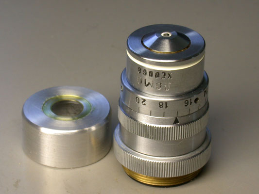
Fig. 13
The 70X 1.23 NA apochromat is shown in Figure 13 along with a home-built water immersion cap. The cap is a push fit over the end of the objective and seats against the shoulder near the end of the objective. A 0.17 mm thick round cover glass is cemented into a counter-bore in the end face of the cap with Canada balsam. The inside of the cap is shown in Figure 14. It has a longitudinal groove to let the air out when the cap is installed on the objective with a drop of water on the inside of the cap to couple the end of the objective to the cover glass window. Figure 15 shows the 70X with its immersion cap in use with the micro-aquarium slide. Figure 16 shows an image of live Fragillaria crotonensis recorded using the 70X objective and COL.
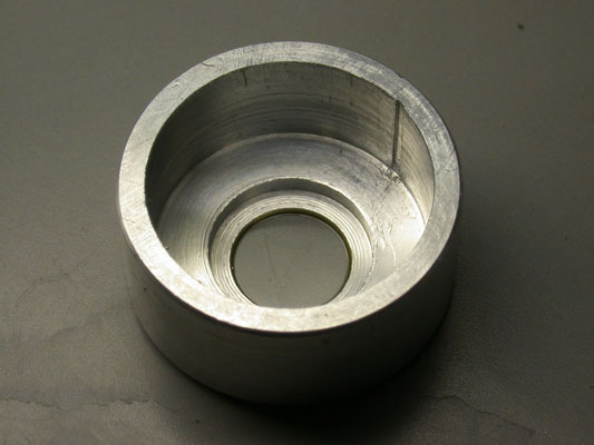
Fig. 14
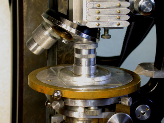
Fig. 15
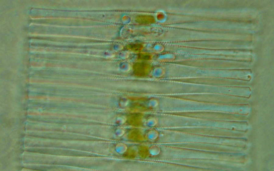
Fig. 16
I am well satisfied with the performance of the LOMO water immersion objectives and look forward to future adventures examining live water organisms using these objectives. Since the live organisms are not fixed in a rigid mountant, I doubt whether image stacking can be done to improve the depth of field at these high NA’s.
Comments to the author are welcomed.
1 Martin, L. C. The Theory of the Microscope; Blackie, 1966, 297-300.
2 Clarke, T. M. “Using the Olympus E-330 DSLR Camera for Photomicrography” The Microscope 2007, Vol 55:4, 163-172.
Microscopy UK Front
Page
Micscape
Magazine
Article
Library
Please report any Web problems or offer general comments to the Micscape Editor .
Micscape is the on-line monthly magazine of the Microscopy UK website at Microscopy-UK .