
AN ARGENTINIAN MICROSCOPE
Manuel del Cerro, Princeton, USA and
Alberto J. Solari, Buenos Aires, Argentina
INTRODUCTION
Argentina, the country enclaved in the southernmost corner of the American continent, is known to the outside world for many reasons. The immense solitude of the “pampas”, the breathtaking beauty of snow-covered high mountains, crystalline lakes and impressive waterfalls are all subject of enthusiastic comment from nature lovers, mountain climbers, and even casual tourists. Argentina is also known to the world for some flamboyant political leaders that gave her fame, if not notoriety, during a good part of the 20th century. Argentina is known to football (soccer in the USA) aficionados around the world for epic victories as well as for catastrophic defeats in World Cup games. But one thing Argentina is not known for is the production of microscopes. Here two Argentinian-born microscope lovers join to describe a microscope signed by the premier Argentinian optical firm of Lutz, Ferrando, and Cia., as well as to discuss personal experiences in dealing with that firm.
Figure 1. The logo of the Lutz & Ferrando Company.
THE MICROSCOPE
[MdC] The following is the catalog description of the instrument in question (del Cerro, 2011).
Microscope #448- Lutz, Ferrando y Cia., Buenos Aires, Argentina. Black and brass amateur monocular microscope. The horseshoe base is 15 cm long and 6.3 cm wide. Two 3.6 cm tall pillars are anchored to a low, circular plate that is attached to the upper surface of the base. The pillars clamp the arm at the junction point; a flat-head, brass screw adjusts the tension of the inclination joint. The inclination of the fork-mounted, plano mirror can be adjusted by means of small side knobs. The mirror is 2.7 cm in diameter. The rectangular stage is 5.8 cm wide and 6 cm deep. A diaphragm wheel is attached to the underside of the stage; it has three openings. The stage retains the two original stage clips. The arm is straight; it emerges at a forward angle from the back of the stage, raises 6.3 cm, and ends in a horizontally placed ring. A brass sleeve screws into this ring; it is signed in cursive with the legend “Lutz Ferrando y Cia 240 Florida Buenos Aires.” This sleeve holds the body tube and the rack and pinion for coarse focus. A side knob located on the right side controls focus. The body tube is 13.8 cm tall; it carries an ocular with no indication of magnification. A cone-shaped nosepiece carries a button-type, divisible, triple objective. The original wooden box is 27.3 cm wide, 9.2 cm tall, and 12.7 cm deep; it contains an ivory-tipped forceps and it has a small drawer for small size-slides. Provenance: Spectra Services, Webster, NY. Date: December 1998. Price: $125.00. OPTICAL PERFORMANCE: Element 1 of the objective operating alone provides a clear image free from curvature of field. Elements 1 and 2 together, provide a sharp image also with minimal field curvature. The field diameters are 2,5 mm for element 1, and 1,030 µm for the combination of 1 and 2. REFERENCES: 1) A nearly identical instrument was auctioned at eBay, item #251492834, on 2000 02 08. The highest bid had reached $177.00. The instrument was called a “Victorian English Microscope”. 2) Dingley (2002) refers to E(duard) Lutz as purveyor of microscopes in Paris during the 1872-1880 period. Could it be that he migrated to Argentina and established there the Lutz, Ferrando, & Cia optical firm? We do not know, and neither do we know if the Company had this instrument manufactured for then locally, or did they buy a generic instrument made for the trade in Europe and had it stamped with their names. Such a practice was not uncommon among the French and English retailers of scientific instruments.
COMMENTS: This is a fine, early 20th century, amateur microscope.
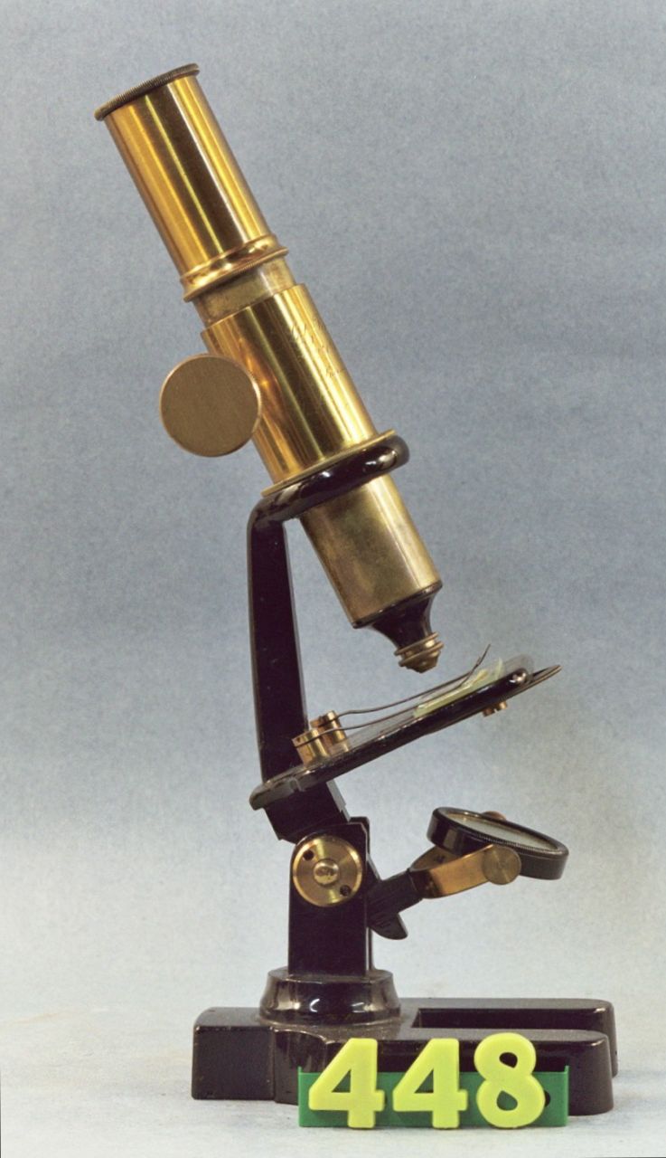
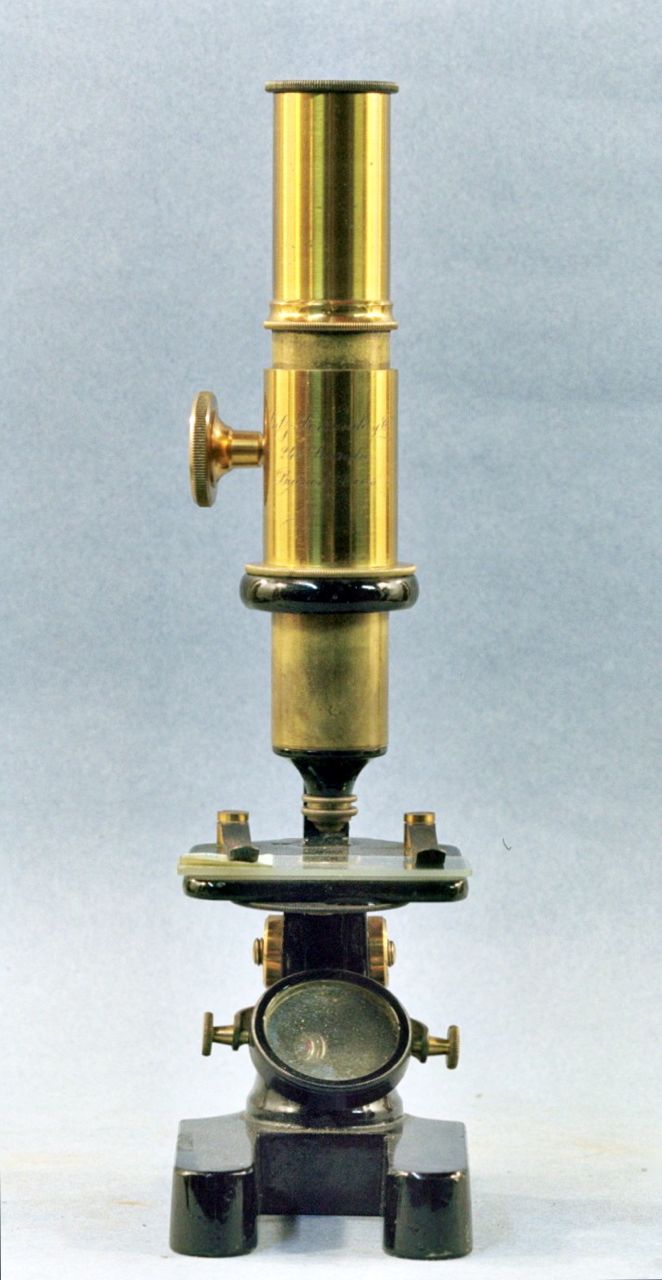
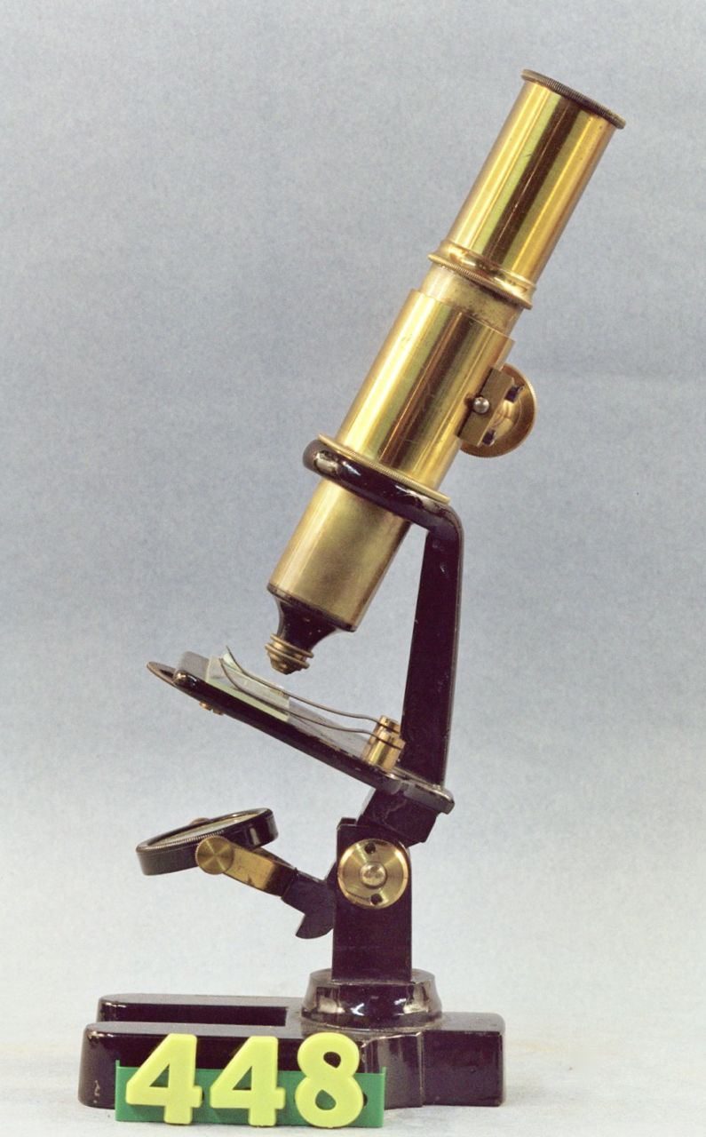
Figures 2, 3, & 4. Front and side views of the Lutz Ferrando microscope.
THE LUTZ, FERRANDO COMPANY
[MdC] As we will comment further, the Lutz Ferrando Company was the highest regarded opticians’ firm in Buenos Aires. Established at the beginnings of the 20th Century and located in the fashionable “Calle Florida” it was still in operation during the first decade of the 21st century. Florida street had at a time the residences of the very rich and powerful in the Argentinian society, and stores to match - a branch of Harrods of London among them. I knew that street as a child in the late 1930s, taken there by my father; incidentally, the street had been by then a pedestrians-only street for many years. In the early 1940s, Father took me to Lutz, Ferrando y Cia. on a less happy occasion, to have my first set of glasses fit there (it was a stigma in those days for a teenager to wear glasses). I still remember the optician trying to convince Father that having glasses for a teenager ground there was a waste of money, but Father insisted that only the best in town should grind the glasses for his only son (some consolation!). Later still, I would, while walking along Calle Florida, stop at the windows of Lutz Ferrando to admire the magnificent display of Leica cameras and attachments (the mythical 135 mm tele!). I do not remember seeing microscopes on display there, although most likely they were, but for sure, I never expected that many years later one would come to me in a very distant foreign country!
[AJS] Lutz Ferrando & Co. was undoubtedly the preferred store for optical and scientific instruments throughout the 20th century in Argentina. The main store located at Florida 240, was the meeting place for scientists, biologists, physicians, surgeons, optometrists and chemists, as this store imported the newest instruments from Europe even without specific orders from customers. For decades, this store was the representative of the German company "E. Leitz, Wetzlar" and in this way provided the famous "Leica", the iconic 35 mm photographic camera. Each camera had also engraved the logo of Lutz Ferrando & Co. A competitor, "Otto Hess" appeared in the mid-1950`s: that store was located at Maipu street, very near the location of Lutz Ferrando & Co. Thus, a possible customer could turn around a street corner and evaluate the quality and prices of both stores. The employees of Lutz Ferrando were technically competent and some of them had engineering or scientific degrees. Furthermore, they were ceremonious and protocolarly collared. To this respect, an author has an anecdote:
"During the late 1950`s I was a student in the medical school. I had begun to make chromosome preparations from onion root cells and the preparations seemed to me very beautiful. However, I wanted desperately to see some further structure in the chromosomes. I thought that if I provided myself with the more powerful microscope objectives I would be able to see the phantasmagoric sub-structures that I imagined in chromosome arms. For that purpose, I had a dream: to get an "apochromat", the finest microscope objective available in the whole world , and to use it in the microscope of the school. With this aim in mind I asked an interview with Carlos Lutz, the son of the owner of Lutz, Ferrando & Co, who told me to get in touch of the manager of optical instruments. I went to the store at Florida 240 replenished with my own enthusiasm. I went through the ground floor filled with common customers and I climbed to the esoteric first floor reserved for scientists and special purchases. There I found Mr. G..., the manager of fine optical instruments. He was a middle-aged man with a shining bald head and a penetrant gaze and it seemed to me that a permanent question mark was seated in his lips. When I asked for an apochromat objective, he stared at me without emitting a word. Then he started to slowly answer, syllable by syllable, while he stared to my face of young student:
- An a-po-chro-mat!
As I was hearing, I felt that I was sinking into the ground five inches by each syllable. He continued with guttural voice:
-An a-po-chro-mat is... a very, very ex-pen-si-ve item!
From my ground level I was able to mumble:
-How much is "expensive"?
He looked at me, bowing towards my low position, with maximum scorn and pronounced his last words:
- Much, very much, my young friend, much more than what you imagine!
I concluded that an apochromat was definitely out of my reach.
After many years, I realized that in fact I would not get much more details of the chromosomal structure with the optical means I had as a young student (see figures 5 and 6).
Even as an electron microscopist during my stay as an NIH Fellow in the University of California at Berkeley with my director Daniel Mazia, I was facing the inextricable paradox that I could study the DNA molecules of chromosomes but the arrangement of the unit fiber of chromatin inside the chromosome remained elusive. On the other hand, the very simple squash — acetic orcein technique for light microscopy, that is still used today, is able to provide valuable data on effects of antimitotic substances and physical variables on the development of the mitotic process. My research career was born using these simple techniques. When I turned to be interested in meiosis, EM and light microscope techniques helped me to discover the "XY body" in mammalian spermatogenesis. But by then the scientific world had become very complex and competitive. The magic of chromosome structure had given place to the fierce competition of molecular biologists who never gasped in front of the beauty of a chromosome picture [or dreamed of the exquisite images given by a Leitz apochromatic objective!].
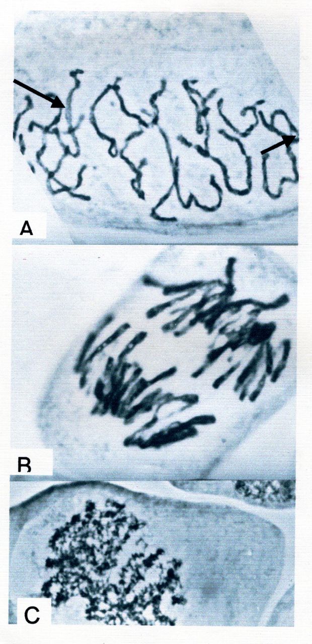
Figure 5. Light microscopic views of chromosomes during different stages of mitosis in meristematic onion root cells in an acetic-acid orcein and squash preparation (see Saez, 1950 for reference). Top: Late prophase chromosomes, showing doubleness of each chromosome (arrows). Middle: Anaphase chromosomes are simple but show an aspect of “bubbles on a string”. Bottom: In early prophase the chromosomal threads show a helical structure. X 3,000.
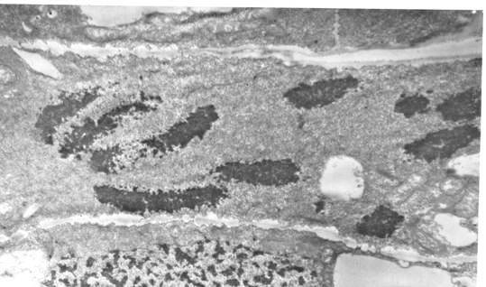
PICTURE 6. Electron microscopic view of chromosomes of an onion root cell in mitosis. This technique does not show further details of chromosome organization. X 8,000. Fixation in Caulfield´s osmium tetroxide fixative, contrasted with uranyl acetate.
[MdC] After going through the previous section the reader may ask whether the chomosomes in mitotic cells just described may be seen with the Lutz Ferrando microscope. I asked that question to myself. The answer is, yes. The four stages of mitosis could be identified both, in a section of the tip of an onion root, and in a section of the thin intestine of a rabbit. In conclusion, the vintage, amateur, Lutz Ferrando microscope is an instrument capable of serving the needs of today’s “supramolecular biologists.” (Selye, 1967).
All comments to the authors are welcomed (via Manuel del Cerro).
REFERENCES
del Cerro, Manuel (2011) The Catalog of the M. and C. del Cerro Microscope Collection. DVD privately released.
Solari, Alberto J.: The molecular organization of the chromatin fiber. In: THE CELL NUCLEUS (Bush H., Ed.) Academic Press, New York, 1974, pp 493-535.
Solari AJ : Structure of the chromatin in sea urchin sperm. Proc Nat. Acad Sci U:S: 53: 503-511 (1965).
Solari AJ: Sex Chromosomes and Sex Determination in Vertebrates. CRC Press, Boca Ratón, 1994 .
Saez FA: Una tecnica sencilla para el estudio de cromosomas vegetales. Ciencia e Investigacion (6)6: 281-282 (1950).
Selye, Hans: In Vivo. The Case for Supramolecular Biology. Liveright Publishing Co., New York (1967) [Selye credits Professor Paul Alfred Weiss (1898-1989) with coining the expression “Supramolecular Biology”]
Microscopy UK Front
Page
Micscape
Magazine
Article
Library
Published in the January 2011 edition of Micscape Magazine.
Please report any Web problems or offer general comments to the Micscape Editor .
Micscape is the on-line monthly magazine of the Microscopy UK website at Microscopy-UK .
© Onview.net Ltd, Microscopy-UK, and all contributors 1995 onwards. All rights reserved. Main site is at www.microscopy-uk.org.uk .