|
Mysteries Surrounding Dried Stuff by Richard L. Howey, Wyoming, USA |
Several years ago, I came across a couple of dealers in the Philippines who were offering a fairly wide variety of dried marine life and I obtained some wonderful specimens of sea urchins, starfish, sponges, corals, bryozoans, bristle worms, basket stars, brittle stars, small crabs, including some porcelain crabs, small strange fish, hydroids, tube worm shells, bits and pieces of detritus, and a number of unidentified thingies sufficiently distorted and undistinguished in terms of gross morphology to make them highly interesting puzzles.
Today, I was sorting through some of that latter material and began to wonder how to deal with it. My first thought was to give some samples to friends who are also natural history enthusiasts and tell them that if they could identify the general group of the organism, then they would win a prize from me. (I didn’t tell them that the prize would be a sweat shirt with my picture on it.) My second thought was to donate them to a university or museum but, knowing first-hand the disposition of such institutions, I figured they would probably just discard them. So, finally, I decided to keep them and see if I could find enough clues so that, at least in a few instances, I could determine what these things were.
To me, rehydration was the obvious first step. One might think that making up a batch of artificial sea water (or if you live near the ocean, going down and getting a bucketful of real sea water) would be the right approach. However, it occurred to me that a fair amount of sea salt and minerals had already adhered to the specimens during drying, so I simply used artesian or distilled water for rehydration. Sometimes, I would add a bit of glycerine in the hope (probably in vain) of lubricating the tissue a bit. However, before any of these baptismal rites, I would, using a sharp scalpel, take some scrapings and, in some cases, small, thin surface sections for microscopic examination. In one instance, this paid off immediately since the scrapings clearly contained holothuroid (sea cucumber) spicules. Few dried items are, however, that forthcoming!
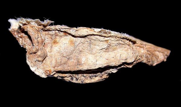
In the particular batch I was looking at today, I concentrated on 7 of the items, all of which are between 1 and 2 inches long. Five of them look like pieces of old, brownish or blackish, shriveled leather with sand and other debris imbedded on them. The sixth which we look at in a minute, however, is white with some pinkish blotches. These are all descriptions before any microscopic examination. Here is a typical “leathery” one.
The first one which I examined, as I mentioned above turned out to be a sea cucumber. How did I find that out? In this instance, it turned out not to be very difficult, in part because, over the years, I have spent a fair amount of time studying echinoderms. This little critter gave itself away in two significant respects. First, there were distinct, round disks arranged in such a manner that they were clearly retracted tube feet. Secondly, by examining the surface carefully at 80x, I could see the dense packing of calcareous spicules which is a typical feature of some sea cucumbers, such as Eupentacta.
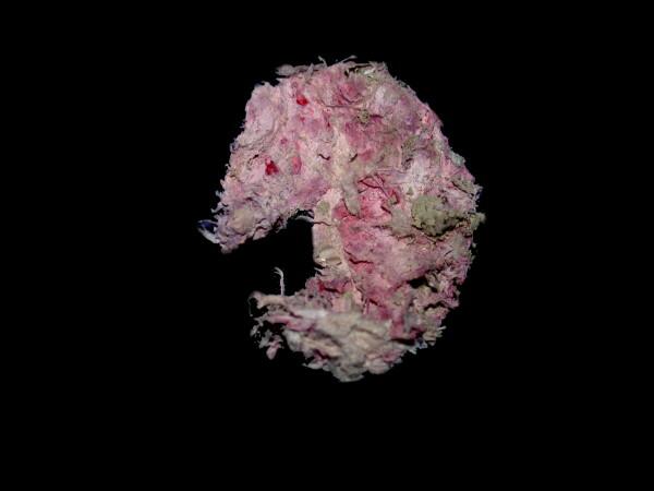
However, I was even more intrigued by another specimen, an odd, flattened white thing that from its outer appearance suggested that it could have been an old, discarded wad of chewing gum–the pink and white thing.
However, before I proceed to it, I wish to digress into a minor rant. (I don’t want to disappoint my fan base of fellow curmudgeons.) A couple of groups of specimens arrived with each item moist, wrapped in toilet tissue, and then placed in individual plastic bags. By the time the parcel arrived several weeks later, the specimens were either dry or smelled like Lazarus and required immediate preservation in strong alcohol. These specimens were being provided by a family with several children, modest means, but a splendid determination. I corresponded with them briefly and they asked me if I had any particular types of specimens which I especially wanted to study. I gave them a fairly short list, but a number of the items really required preservation in fluid. However, as we now all live in High Paranoia (see Mel Brooks), shipping anything in fluid internationally is not only bureaucratically complex, but expensive, especially when it involves a toxic substance (formalin) or a volatile one (alcohol). All of this makes life more difficult, expensive, and exasperating for us peaceful microscopists and natural historians and their suppliers. With all of the technology and database capabilities which are now available, surely some semblance of sanity could prevail; but alas that is the way of bureaucracies. Finis.
So, I took my flattened, white-pink thing, put it in a small dish and began to examine it with my Olympus stereo microscope. The first think that struck me was that the pink stuff was more a deep orange-red than pink and was distributed in powdery lumps. This was entirely unhelpful. As I continued scanning the surface, I was beginning to despair of getting any sort of clue to this thing’s identity and then I saw, down in one of the dried wrinkles, a little bit of something glistening. I took a micro-needle and began gently probing around the area and then suddenly, I was flooded with pure delight. After I carefully isolated it , cleaned it, and put it on a slide, here is what I had uncovered.
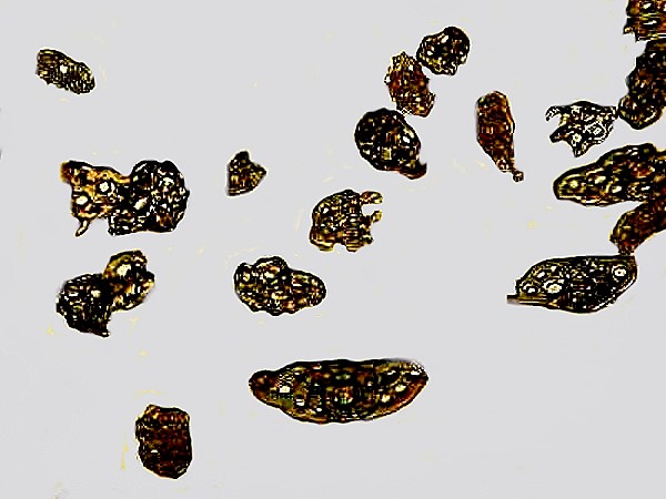
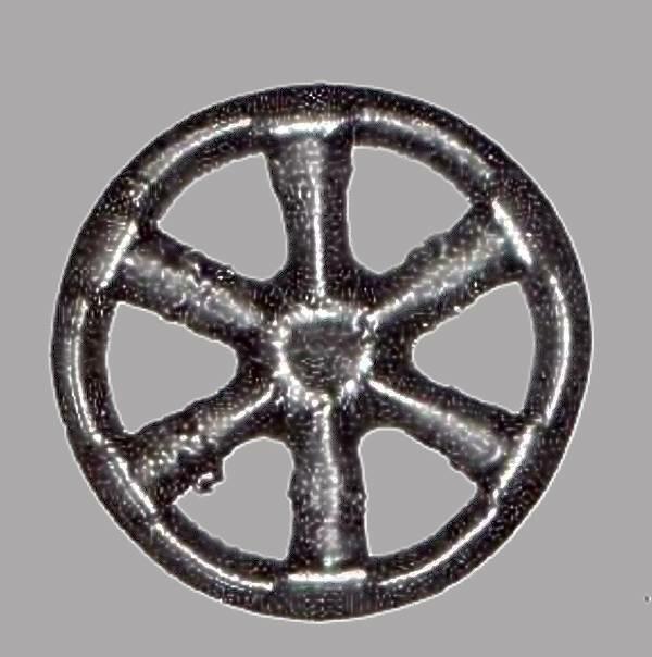
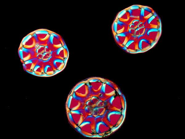
Now, I knew what this dried blob had been–a small sea cucumber which I strongly suspect is of the genus Chirodota or, if not, a closely related one. [Here I should note that there is a difference in spelling on this species. Some of the older accounts use the spelling I have here but, many more recent researchers spell the genus “Chiridota”. I have primarily used the old spelling since this was the first one I encountered.] I say this on the basis of having dealt a number of times with 2 other species which I definitely know belong to the genus Chirodota and which have little clumps of spicules embedded just under the skin and which are easily mistaken for aggregations of sand grains at first sight, but here’s what the spicules of one of the other species look like.
These marvelous wheels are indeed distinctive indicators that what we’re dealing with is a special group of sea cucumbers. To find just one such wheel spicule on this flattened and badly damaged object was, of course, not significant. One or two or even several could have floated down from a genuine Chirodota that was injured or dead and these spicules could have just happened to fall on this white lump, especially since I had also found a small mollusk shell and a tiny jointed appendage of a crab embedded in this blob and then there was the additional problem of picking off the dried toilet tissue which had undoubtedly helped hold in some detritus that was on and around this organism when it was collected. What I needed to find was several wheels in close proximity, actually embedded in the tissue. After about 20 minutes of careful prodding, poking, cutting, and cursing, I came across exactly what I was looking for–a group of spicules in a piece of the dermal tissue. Success! Well, not quite yet. There was still a possibility that this enigmatic pink-white thingy might be an ascidian tunicate onto which the spicules had fallen while it was still alive and it began to engulf them with tissue growth. Some kinds of tunicates are notorious repositories. So, at this point, we needed to take some tissue samples. “Igor, bring me a scalpel.” “Yes, master.” Oh, I would have been a superb 19th Century mad scientist.
What is enormously helpful to us here is the radical difference in the tissue composition in the echinoderms as compared with the ascidian tunicates. If I were to treat this tissue sample with sodium hypochlorite (or household bleach) and after a few hours or even days still had lots of undissolved tissue, then it’s a good bet that this is from an ascidian. Their outer tissue or tunic is largely composed of cellulose compounds which require specialized enzymes, in this case, cellulase, to dissolve them.
If, on the other hand, most of the tissue dissolved, then in this instance, we can be relatively sure that it’s from a sea cucumber. Well, the test results are in and the winner is —— Chirodota, the sea cucumber.
Sometimes, such experiences make me think I should have been a forensic scientist and then I remember that I generally don’t like working with vertebrates at all and that I can’t stand the sight of blood.
Ah, yes, earlier I mentioned 7 specimens which were puzzles. The seventh, as it turned out, was not much of a challenge and turned out to be a chiton, a very nice and interesting one, I might add.
I have continued to poke and probe at my pink and white Chirodota and this odd little lump continues to pose puzzles. I’ll confess that I haven’t yet tried to rehydrate any of these specimens yet. As I said previously, when I first got them, I popped them into 70% isopropyl alcohol, but in small plastic jars whose lids unfortunately don’t seal tightly and allow for complete evaporation over a period of months, but I have already vented on this subject in a previous essay.
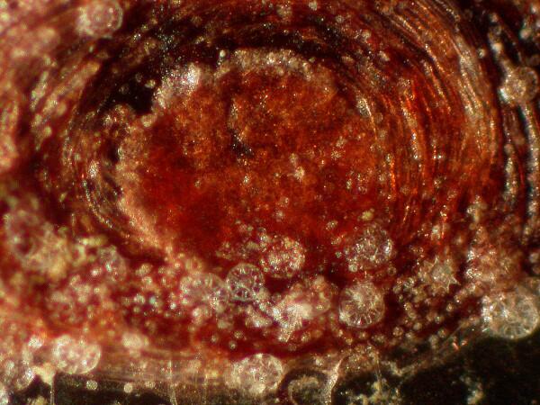
So, what I have been doing is continuing to take scrapings and, as a consequence of the extreme degree of distortion, it is in most cases, very difficult to determine whether particular bits are intrinsic to this organism or whether they are something extraneous that attached themselves to the Chirodota. Among the scrapings, I found an interesting, dirty reddish-brown clump with some glassy, rod-like structures projecting from around its edge.
This afternoon, I was browsing the Internet for information on Chirodota and came across an article by a Mr. F.M. Hamlin published in 1882 on the spicules of Chirodota in a species found in Bermuda. Elatedly, I pulled up the article and was presented with a provocatively interesting page 1 and the information that if I wanted access to the rest of the article, I had to either 1) subscribe and pay a fee, 2) be a certified member of a subscribing institution such as a university or research institution or 3) belong to a dysfunctional, recognized groups such as the Senate, House of Representatives, or Supreme Court. I called the Outreach Service at the University Library and talked to a wonderfully helpful woman, who helped me access my old, dormant account and–brilliantly–I now have access to all the electronic journals to which the university subscribes and in the comfort and convenience of my own home without having to stagger up to the library. I give my sincerest thanks to the library people at the university in the reference, outreach, and document divisions; over the years, they have been exceptionally helpful and patient and they helped maintain my faith in a kind of civility that seems to have disappeared from so many aspects of our current social lives.
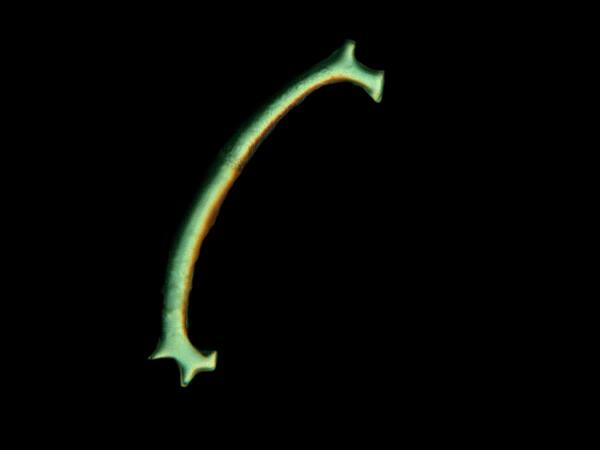
Hamlin’s account is of interest for several reasons. First of all, it reminded me that there are at least 2 other kinds of spicules in some species including his Bermuda one which have elongated
“C”-shaped spicules scattered throughout the tissue and a set of calcareous plates forming a ring which surrounds the base of the tentacles. The species he is studying almost always has wheels with six spokes in the adult stage, although he reports that specimens in the very early stages of development may have 5,6, 9, or 13 spokes, so something interesting happens to narrow this down to 6 spokes in the adult. At one point, I had a couple of specimens of the 6 spoke variety and here is an image of one of the “C”-shaped spicules .
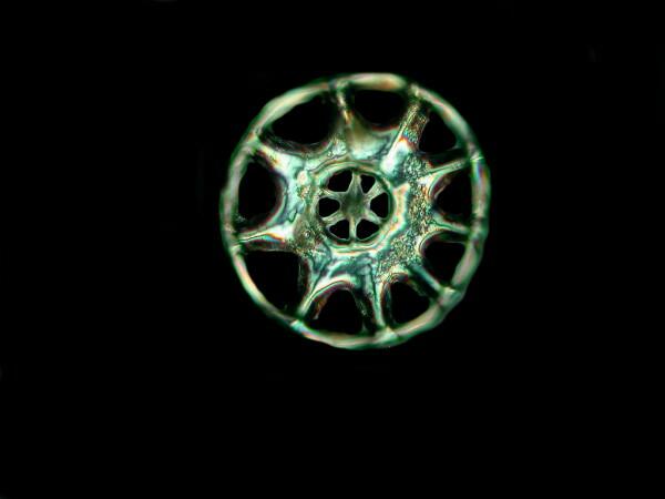
The Philippine specimen which I have been examining poses some other puzzles. My wheels have 9 spokes and then a central disk which has 6 spokes. The one thing that is clear is that this species is different from Hamlin’s. Our usual expectation regarding echinoderms is to find pentagonal symmetries, though there are many exceptions, and here Mother Nature tosses in another one (actually, several). Furthermore, I have been finding some short, calcareous, curved rod-like structures which seem to be in proximity with the wheels but, as yet, I have been unable to discover any clear connection. “Who cares?” you might ask. Clearly, I do, but then I’m ancient and eccentric. Nonetheless, I have faith that the young naturalists also have an obsessive curiosity and a love for trying to solve odd puzzles.
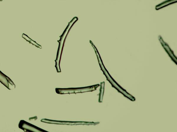
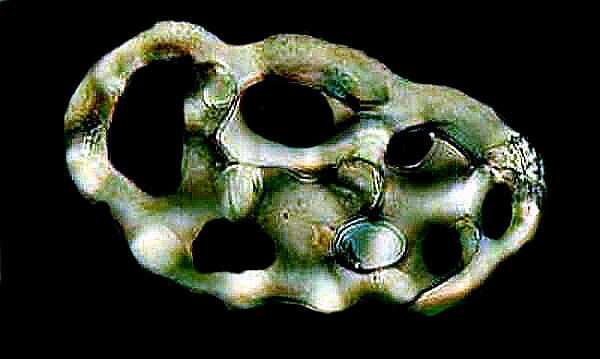
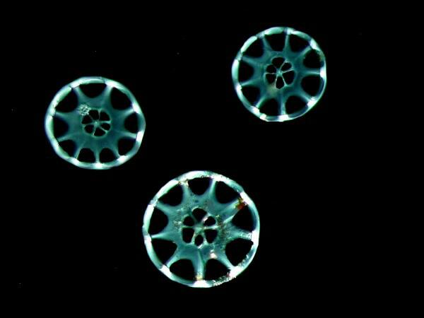
These are the “branching” spicules which may be just fragments or might, in fact, not even be an aspect of the Chirodota. Here again, Hamlin’s article is provocative. He describes how in the Bermuda species, there are small pockets of wheel spicules enclosed by a very thin membrane which is difficult to see. He thinks, nonetheless, that he can see tiny tubules connecting some of the wheels to form this roughly spherical mass. This may very well be and could explain some of these branching structures; however, in my Philippine specimens, the spicules are widely dispersed and while several may occur in proximity to one another, there is no suggestion of a cluster, but this may simply be a consequence of the fact the specimen is so badly mangled and distorted. At this point, I am already delighted with the result of being able to identify this as a species of Chirodota and so I want to show you 3 more images of the spicules. Here is one that is a composite of 25 images stacked using Helicon Focus software.
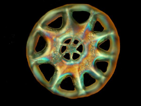
After a considerable amount of maneuvering accompanied by threats, curses, and a dose of good luck, I was able to isolate and group 3 of the spicules for your (and my) viewing pleasure.
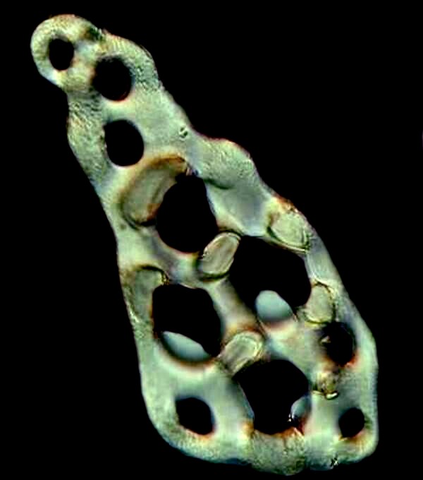
This was taken with Nomarski DIC; I then adjusted the prism to introduce a bit of optical staining and here is the result.
How these funny, fantastical creatures manage to make these elegant structures still remains largely a mystery.
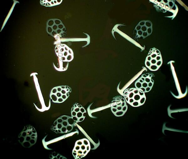
While rummaging around on my shelves, I came across a plastic 8 oz. jar from Maine, one of those awful things that virtually guarantees extreme evaporation—hey, we live in a culture where everything else is labeled “extreme”, so why not evaporation?–and there at the bottom were 2 shriveled, dried specimens of Chirodota that would easily have fit in a half-ounce jar with room to spare. However, once I put them under my stereo-zoom microscope, I felt that wonderful tingling sensation that said “the game’s afoot, Watson!” These 2 small brown things had several rows of what look like little clumps of sand and I knew right away that we had some interesting spicular structures to investigate. Immediately, using a micro-scalpel, I isolated one of these aggregates and transferred it to a drop of immersion oil on a clean slide. I was able to remove it and transfer it readily and when I examined it, I discovered a nice, roughly spheroid clump of wheels which strongly suggested that there was a thin, dried membrane holding the spicules together in the neat, compact package. I have not yet tried to observe the membrane but, if the oil doesn’t make it visible, I’ll try distilled water and then possibly some stains. I’ve mentioned this before but, it’s worth repeating: Alizarin Red S has an affinity for calcium and so for calcareous spicules, forams, bone, etc., it is usually worth trying. Make up a stock solution and then dilute it down to a small quantity for regular use. It does tend to form sediments, so it is a good idea to filter a small quantity (1 or 2 cc.) just before use. Holothuroids produce a variety of types of spicules and certainly those of the genus Chirodota are among the most distinctive. Perhaps, its major rival are those plate and anchor spicules of some of the synaptids. Here are a couple of images from a 19th Century slide of these forms. Here you see a cluster of both the anchors and the plates.
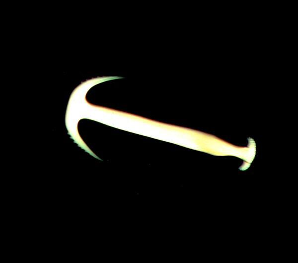
And now here is an individual anchor.
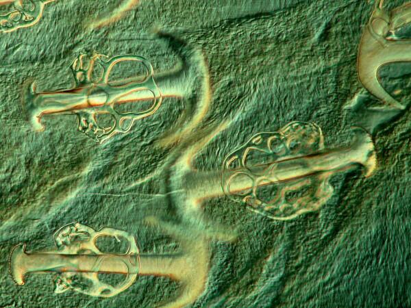
And finally anchors and plates still embedded in the tissue of a section of skin from a synaptic sea cucumber. These last 3 images were taken from an amazingly fine 19th Century slide.
When we take the time and effort to look carefully and probe and dig and ask unorthodox questions, nature can sometimes be induced to reveal wonderfully surprising and aesthetically pleasing answers and mysteries to us.
Addendum
If this gets too lengthy, you have my permission to take a nap. Actually, if it gets too long, I’ll make this into Part 2 and leave you on tenterhooks for a month. The problem is that my smashed, distorted, white and pink wad-of- chewing-gum sea cucumber (the Philippine Chirodota) keeps posing new problems and questions, even in its sorry state. Two days ago, I decided to rehydrate it, put it in a small Petri dish, added distilled water and today, when I looked at it I thought–Oh, Yuck! At first glance, it had turned into a slimy, semi-gooey mess.
O.K., I guess we’d probably better continue this as Part 2, so I’ll see you next month.
All comments to the author Richard Howey are welcomed.
Editor's note: Visit Richard Howey's new website at http://rhowey.googlepages.com/home where he plans to share aspects of his wide interests.
Microscopy UK Front
Page
Micscape
Magazine
Article
Library
Please report any Web problems or offer general comments to the Micscape Editor .
Micscape is the on-line monthly magazine of the Microscopy UK website at Microscopy-UK .