|
Introducing Children to the Micro-Life of Fish Lake Theodore M. Clarke, Retired Materials Engineer JoAnn M. Burke, Ph.D.. Director, Gerontology Program, Saint Mary’s College |
My friend’s family has enjoyed exploring the live micro-life of Fish Lake in rural Indiana. We collected our water samples near the shore. The specimens were examined in a micro-aquarium slide shown in my earlier article in The Microscope (1). We used a Meiji stereomicroscope with my multimode trans-illuminator and the 2X and 4X paired objectives. The same specimens were then examined with my modified Biolam equipped with a multi-mode condenser and water immersion caps on the objectives. The girls were fascinated with watching a live copepod and a worm with the stereomicroscope using transmitted darkfield illumination. The Meiji stereomicroscope with my multimode trans-illuminator was demonstrated at the Inter/Micro 2010 conference.
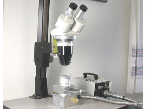
During the summer of 2009 we conducted a brief experiment to determine the level of interest of my friend JoAnn’s granddaughters in using my Meiji stereomicroscope and novel, multi-mode transmitted light illuminator to examine living organisms in a lake water sample. They were fascinated and preferred darkfield illumination. Their father was more interested in the interference color patterns exhibited by calcite particles in the hard water lake when viewed between crossed polars. I subsequently realized that a more detailed and documented experience of JoAnn’s granddaughters exploring the water organisms early the next summer could be a good topic for a presentation at Inter/Micro 2010. The multi-mode condenser is shown in Figure 1. Figure 2 is a schematic ray diagram for this illuminator used with a ½” diameter fiber optic light-guide. The illuminator has a slider, shown in Figure 3, with a clear opening for brightfield and inserts in the other openings for polarized light, first order red retardation, and darkfield. This time the study would include using my modified Biolam with its multimode condenser (2). The same water specimen in a micro-aquarium slide could then be viewed with both microscopes. Water immersion caps on the Biolam objectives allow direct immersion into the micro-aquarium slide (3).
JoAnn’s granddaughters watched as we gathered the water sample near the lakeshore, see Figure 4. The exploration of the organisms in the lake water began with the girls viewing some a lake water specimen in a micro-aquarium slide with the stereomicroscope, as shown in Figure 5. The micro-aquarium slide, instead of a conventional specimen with a cover glass, allows a much larger specimen volume to be examined. Darkfield was once again the preferred illumination. The girls found copepods, see Figure 6, and worms, see Figure 7, most interesting to observe as they scanned the slide. (Brian Ford noted that the copepod shown in Figure 6 has been parasitized.) Upon closer examination of Figure 7, diatoms and desmids are evident. The sheets of the diatom Fragillaria were the subject of a higher magnification study using the 20X objective of the Biolam and darkfield, see Figure 8. Darkfield illumination was used with the water immersion cap equipped 20X objective. The ¼” diameter light guide was coupled to an LED flashlight rather than the 150 watt quartz halogen illuminator used with the stereomicroscope. The girls viewed the organism through the secondary eyepiece while I controlled the microscope and used the eyepiece pointer to indicate key details such as the fragillaria shown in Figure 9. This image was recorded with an Olympus E-330 DSLR equipped with a 28 mm f/2.8 wide angle lens over the secondary eyepiece.
My daughter and granddaughter visited the lake house just before Inter/Micro 2010. My granddaughter was very interested in looking at insects through the stereomicroscope with top lighting. She observed that a housefly caught, by my daughter, was covered with hairs. I subsequently purchased a LOMO SF-10 stereomicroscope for my daughter to use with her daycare kids and my grandchildren, see Figure 10. JoAnn subsequently purchased a beginners microscope for her granddaughters. We both hope the grandchildren will continue to use home microscopy to further their interest in science.
Intergenerational learning encounters such as these have been shown to enrich children’s knowledge base in the sciences (Ruby, Jessel, Gregory, & Arju, 2007). This experiential learning complements the science curriculum used in formal educational settings and encourages children to seek further scientific exploration. Intergenerational learning experiences such as these are not limited to family interactions. With the growth of the older population, there are many opportunities for older adults in the science community to share their knowledge with children (Karaski and Wallingford, 2007). Perhaps schools at all levels can act as catalysts to bring adults with expertise in the sciences together with children in the community so we can encourage our children to pursue scientific exploration.
All comments to the author Ted Clarke are welcomed.
(First published in 'The Microscope', 59:1, pp. 29-31, 2011.)
References
1
Clarke,T. M.”Multimode Trans-Illuminator for the
Stereomicroscope”; Microscopy Today; July 2007, 40-43.
2 Clarke, T. M. Using the Olympus E-330 DSLR Camera for Photomicrography; The Microscope 2007, 55, 163-172.
3 Clarke, T. M. “Water Immersion Caps for Pond Water Microscopy”; The Microscope 2000, 48:2, 87-91.
4 Karasik, R.J.; and allingford, M.S.”Finding Community: Developing and Maintaining Effective Intergenerational Service-Learning Partnerships”: Educational Gerontology 2007, 33: 775-793.
5 Ruby, M.;Kenner, C., Jessel, J.; Gregory, E.; Arju, T. “Gardening with Grandparents: an Early Engagement with the Science Curriuculum”; Early Years 2007, 131-144.
Figure 1: Stereomicroscope with the home-built multi-mode transmitted light illuminator.
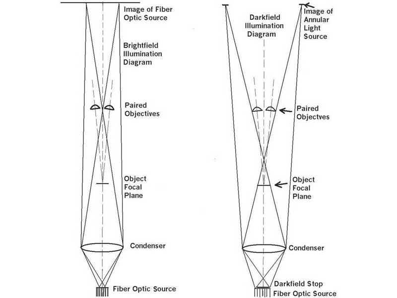
Figure 2: Schematic ray diagram for the multi-mode illuminator shown in Figure 1
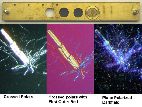
Figure 3:
Top of view shows the slider from the illuminator shown in Figure 1
with a clear opening for brightfield and inserts for polarized light,
first order red, and darkfield in the other openings.
Lower
photographs illustrate use of the slider with Chrysotile asbestos in
1.550 mountant.
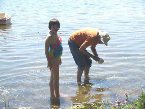
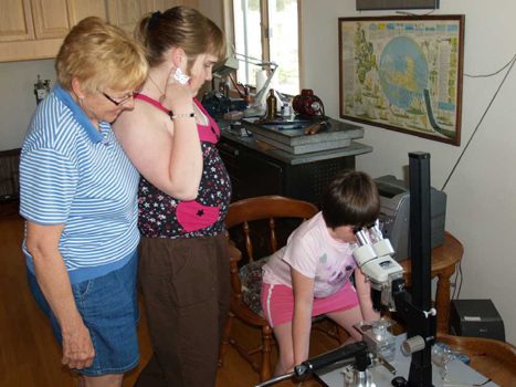
Figure 4 (L): Collecting the lake water sample.
Figure 5 (R): JoAnn with her granddaughters examining the lake water organisms using the stereomicroscope.
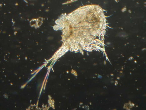
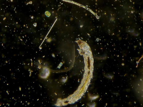
Figure 6 (L): Photograph of a parasitized copepod taken through one of the eyepieces of the stereomicroscope using darkfield illumination.
Figure 7 (R): Photograph of a worm taken using darkfield illumination with the stereomicroscope.
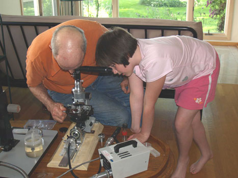
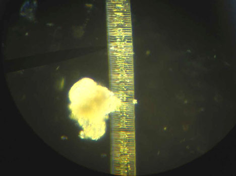
Figure 8 (L): One of JoAnns’s granddaughters viewing water organisms through the secondary eyepiece of the modified Biolam.
Figure 9 (R): Photomicrograph of Fragillaria taken through the secondary eyepiece using the 20X objective of the Biolam and darkfield illumination.
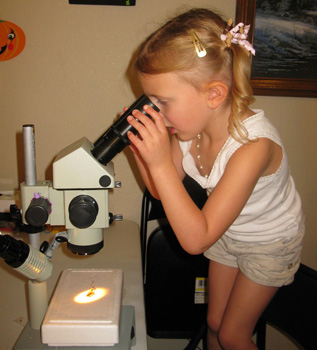
Figure 10: Photograph of my granddaughter using her stereomicroscope to examine an insect.
Microscopy UK Front
Page
Micscape
Magazine
Article
Library
Please report any Web problems or offer general comments to the Micscape Editor .
Micscape is the on-line monthly magazine of the Microscopy UK website at Microscopy-UK .