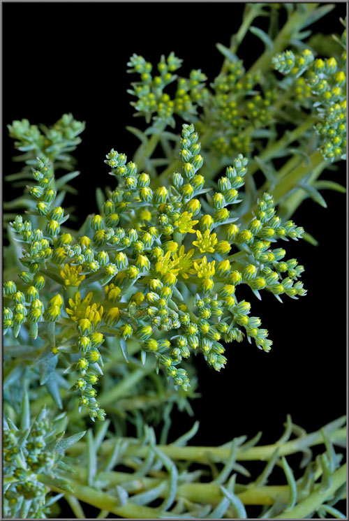
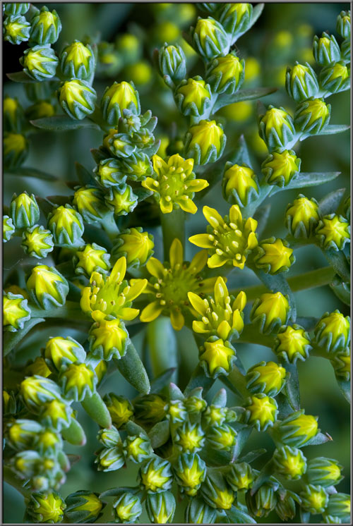
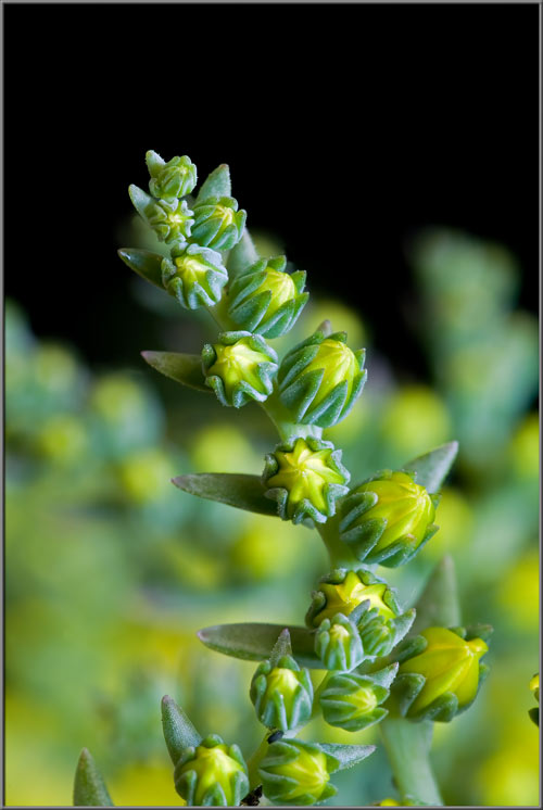
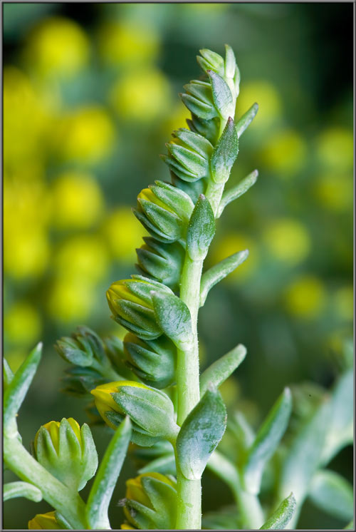
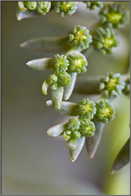
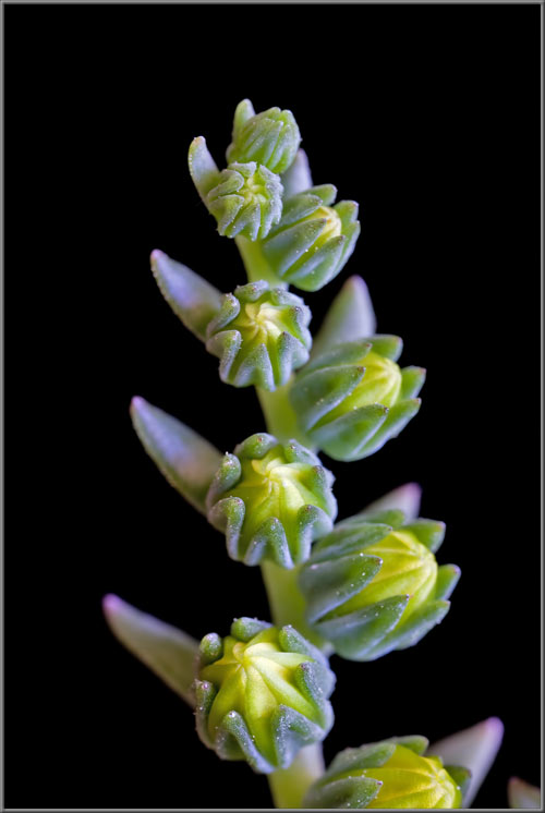
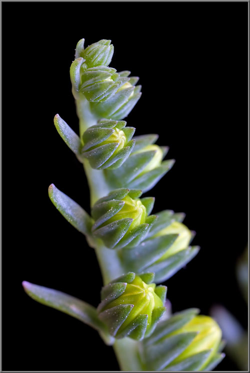
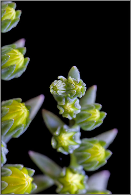
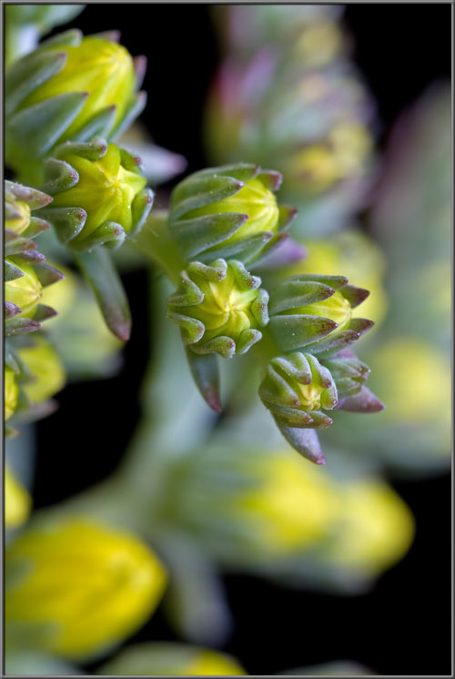
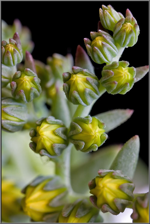
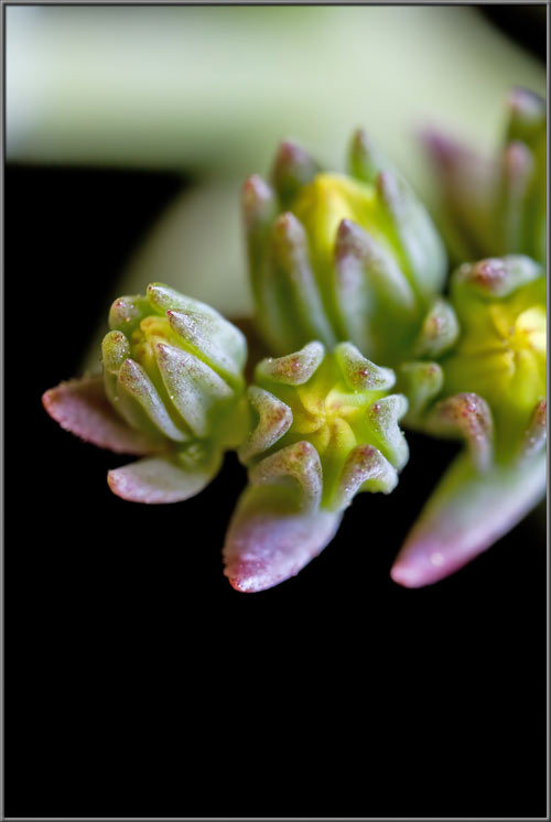
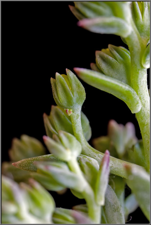
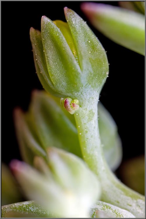
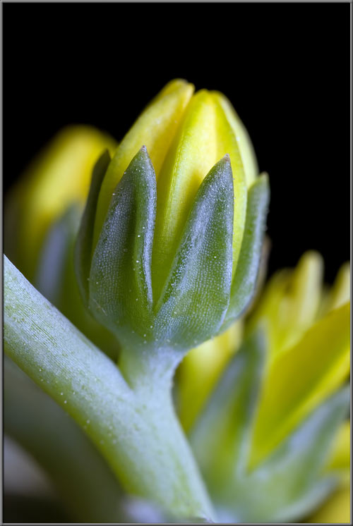
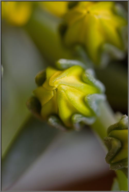
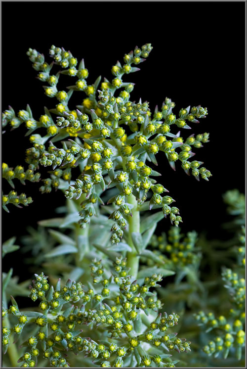
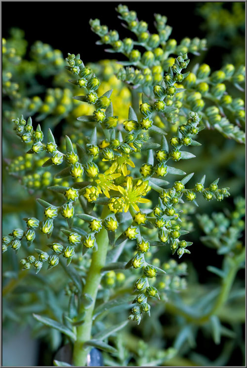
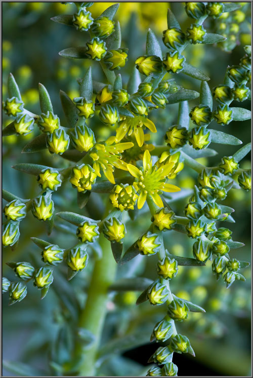
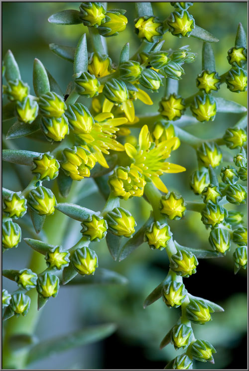
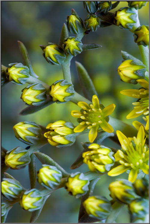
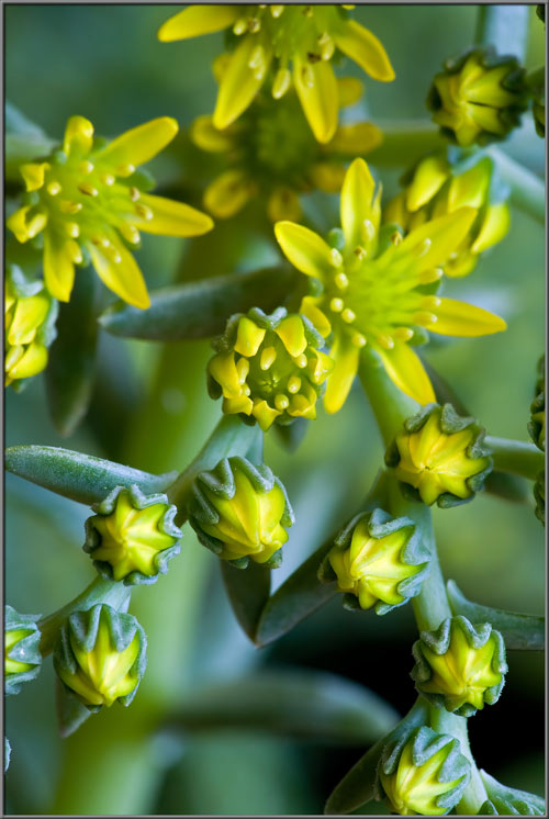
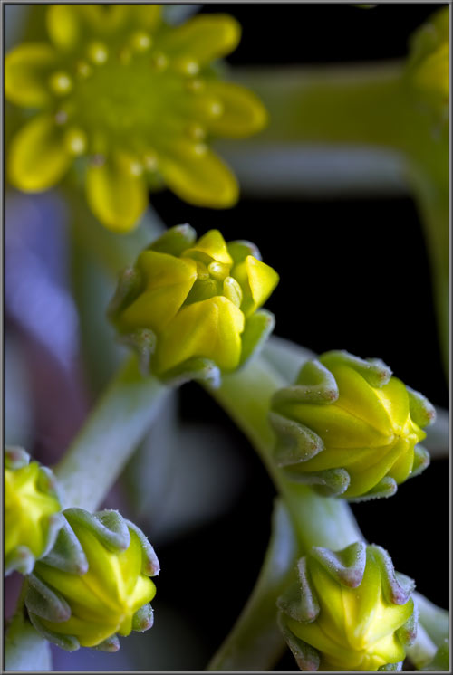
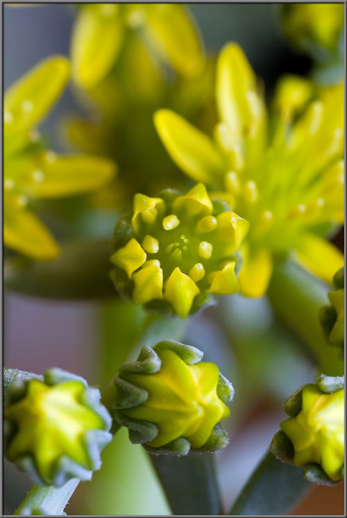
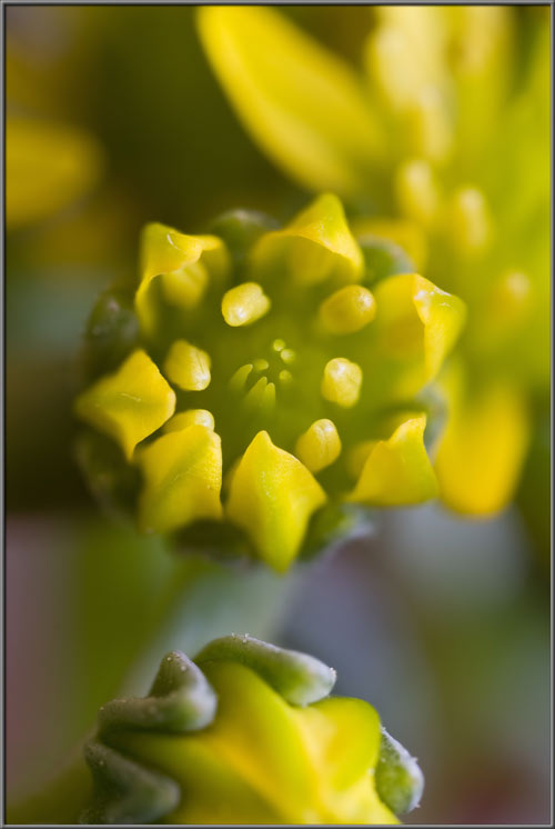
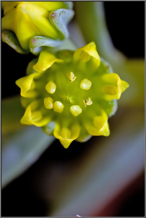
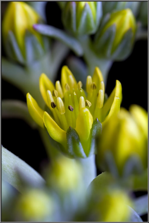
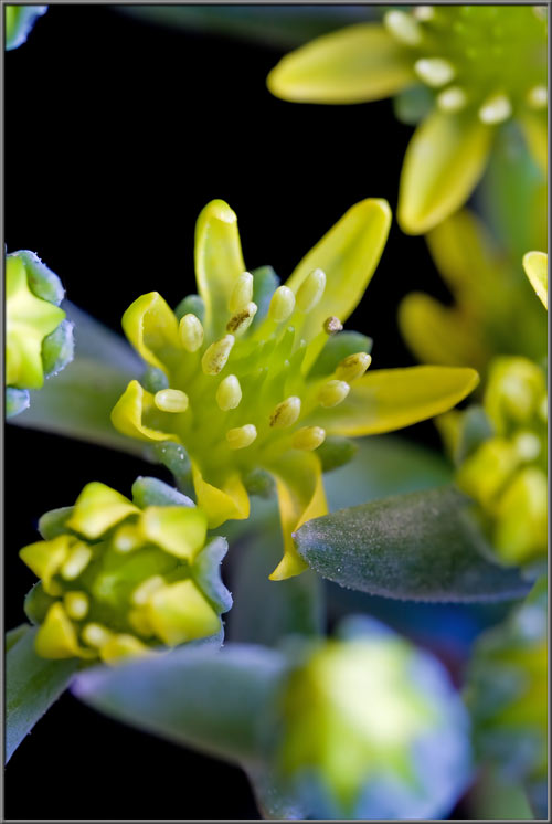
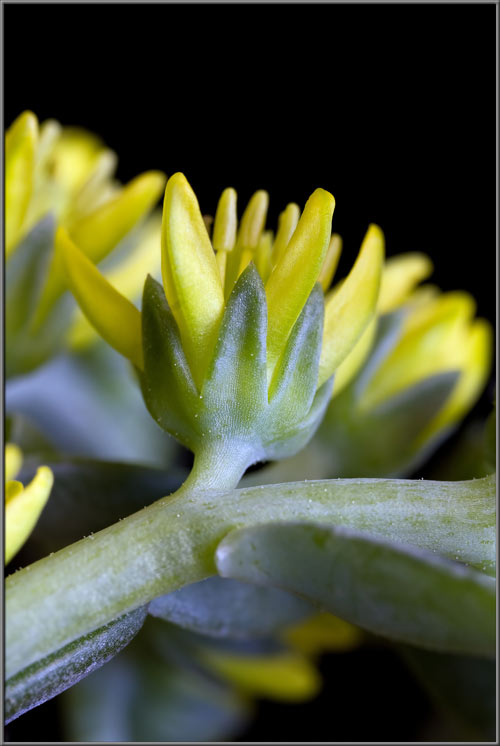
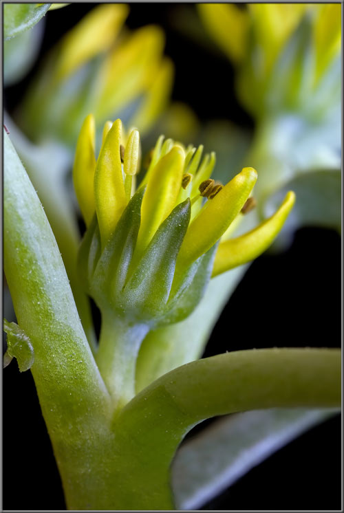
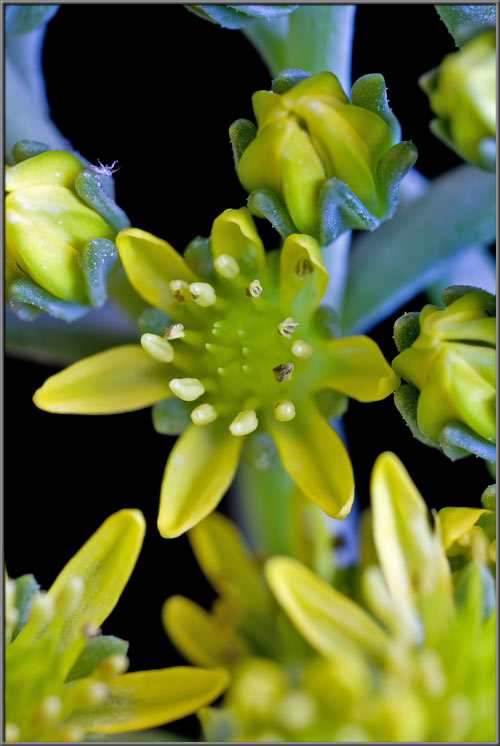
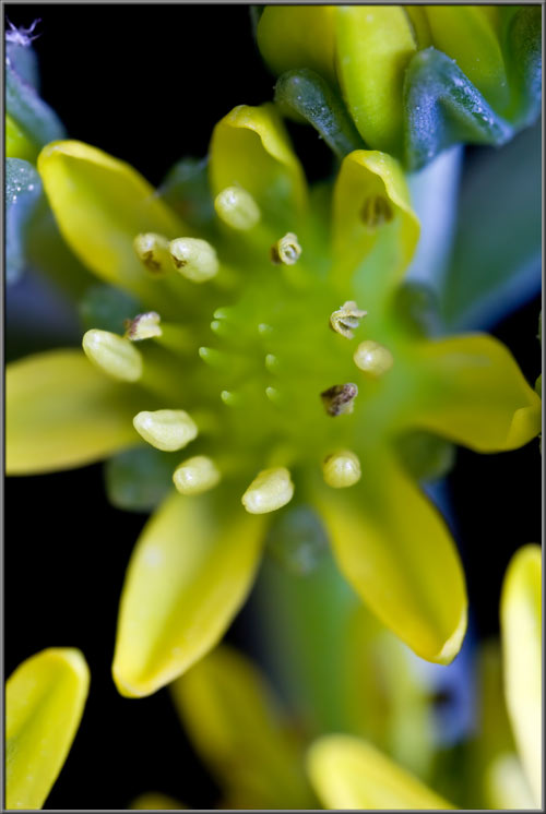
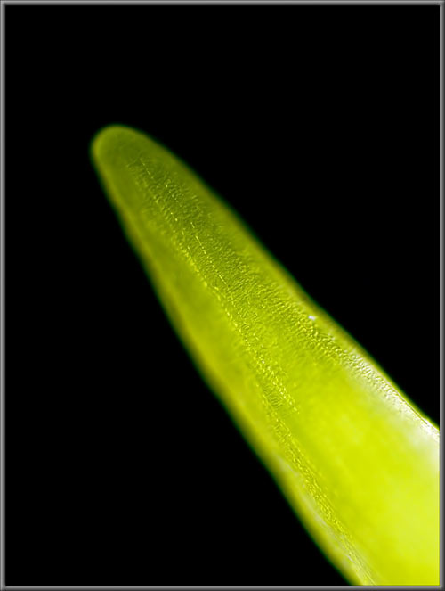
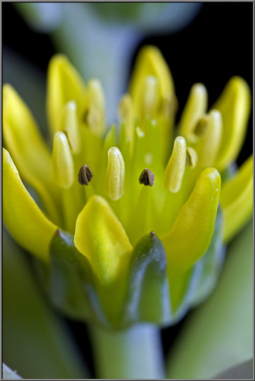
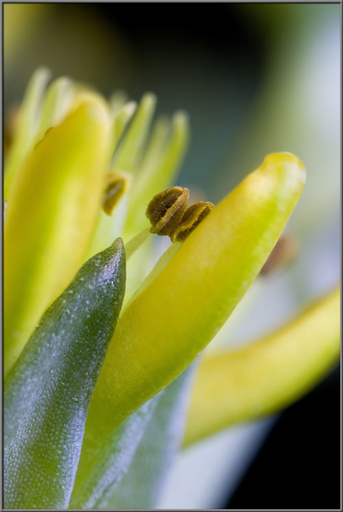
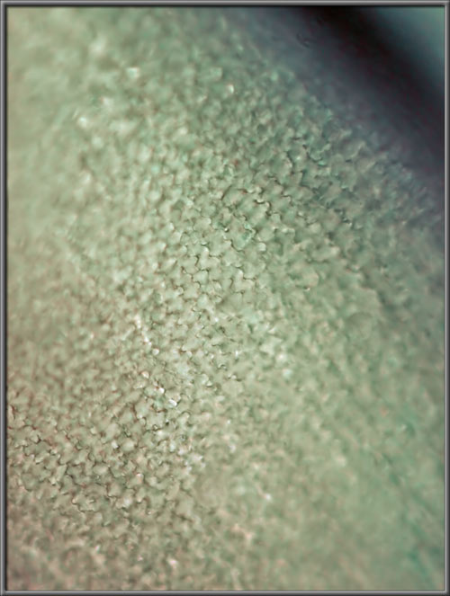
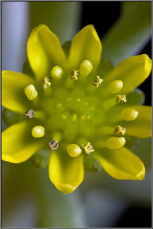
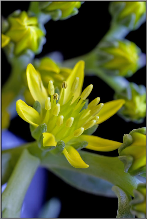
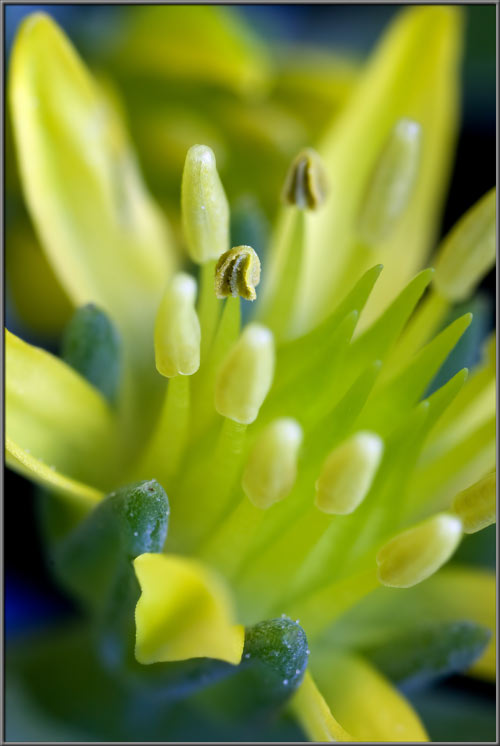
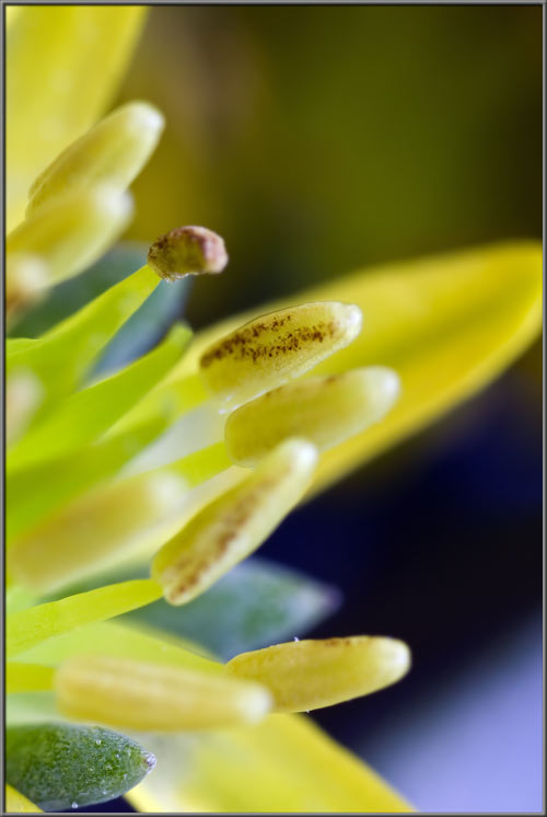
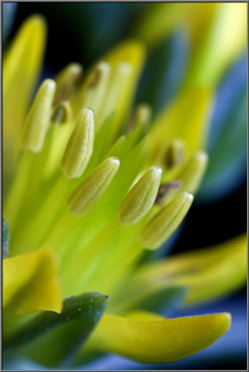
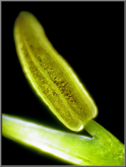
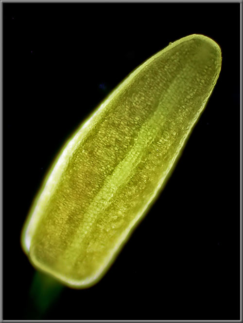
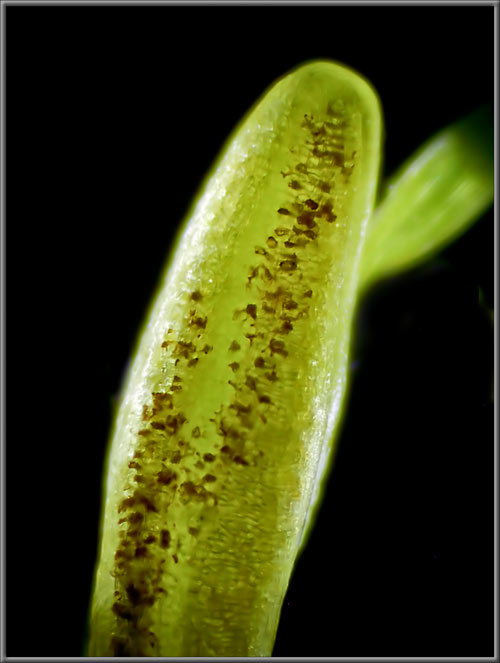
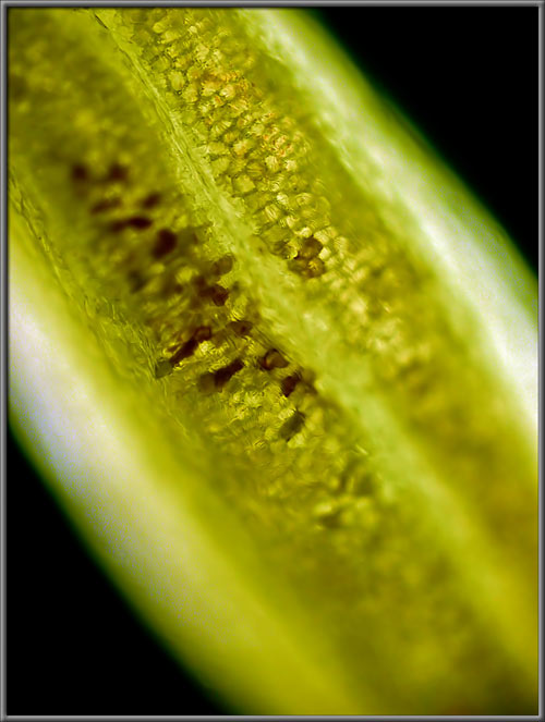
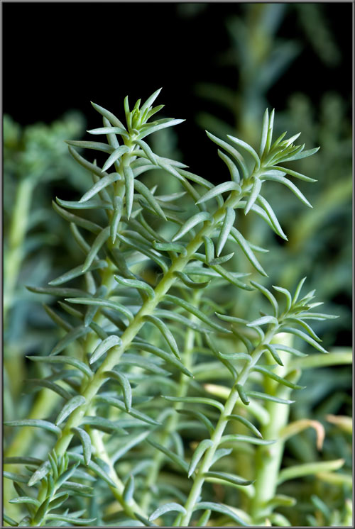
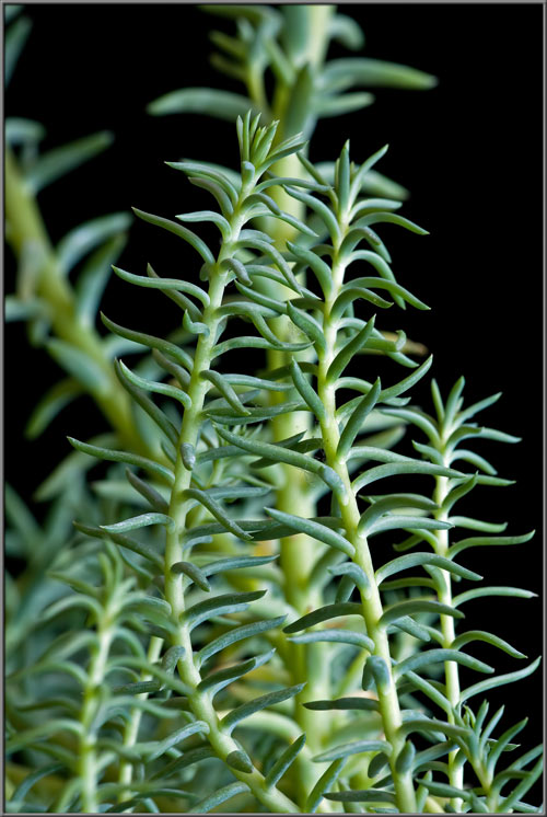
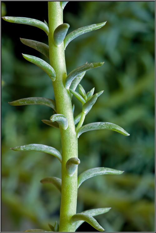
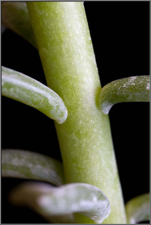
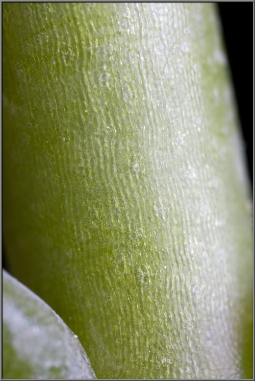
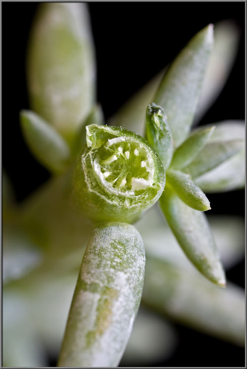
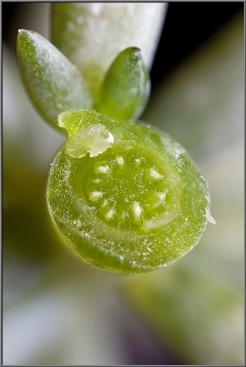
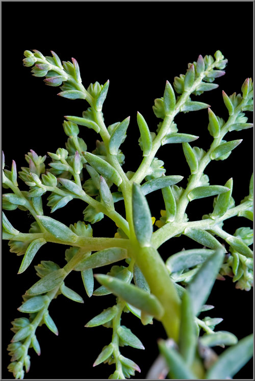
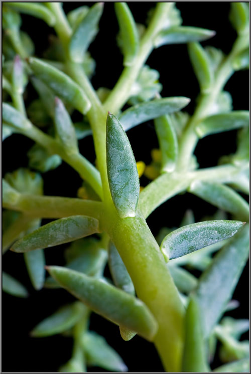
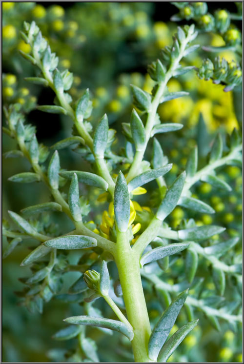
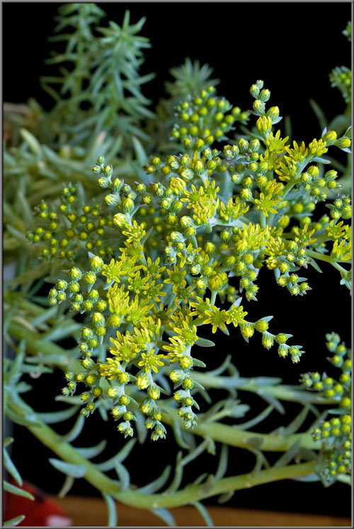
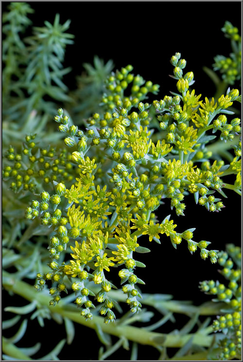
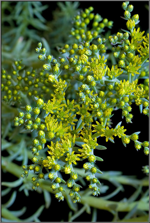
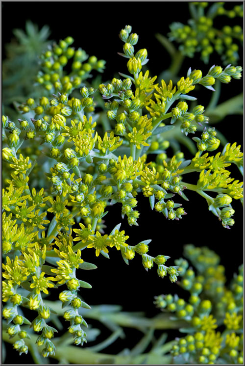
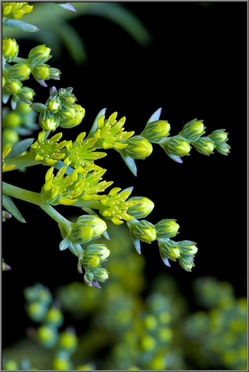
A Close-up View of the Stonecrop Hybrid

'Iceberg'

Sedum reflexum 'Iceberg'


by Brian Johnston   (Canada)

Sedum is the large stonecrop genus that belongs to the Crassulaceae family.  Members of this genus, native to the northern hemisphere, store water in their leaves, and are thus referred to as succulents.  The species name reflexum is from the Latin, and refers to the tendency of the plant’s leaves to curve, or bend backwards.

The particular cultivar studied here, called ‘Iceberg’, grows to about 30 centimetres in height, and has a very appealing colouration, with its lemon yellow flowers, and pale green leaves and stems.  As can be seen in the first image in the article, flower-heads have a ‘star-burst’ appearance.  The two images below show closer views of the flower-head.


The number of buds and flowers on this particular plant is simply amazing!  Although small in size, they make up for this deficiency with their elegant, muted colouration, and striking shape.  Buds and leaflets grow profusely along the radial stems of a flower-head.


The tightly packed yellow petals of buds peek out from between the seven, pointed, green sepals that form the flower’s protective calyx.


Buds near the ends of flower stalks often have a reddish tint to their sepals’ tips.


I thought at first that the tiny greenish-yellow object attached to a bud at the base of one of its sepals might be an insect, but it turned out to be an aberrant, grotesquely distorted  bud.


It is not only this stonecrop’s flowers that are beautiful; the mature buds are also!


The next stage to be looked at is the blooming process.  As can be seen below, buds usually bloom from the centre of the flower-head out to its circumference, but there are exceptions to this rule.


As we move closer, the images show the flowers’ petals beginning to open to their final, almost horizontal positions.


The sequence of images below shows this process in more detail.  Notice that the anthers appear completely yellow at first, but soon begin to turn a light beige colour.


Images taken from a different angle show this difference more clearly.


Side views of a partially open buds show that the sepals are fused at their bases.


The flower’s seven pistils are difficult to distinguish because of their similarity in colour to the base of the inside of the flower.  Look for seven very light green points in an almost circular array at the flower’s centre.


A photomicrograph of one of these pistils reveals that the stigma tip is just a continuation of the cone-shaped supporting style.  Unlike in most other plants, the surface of the stigma appears to have no tiny lobes or hair-like projections to catch passing pollen grains.


If the surface of a petal is examined under the microscope, its cellular structure becomes visible (third image).  Note that Photoshop’s ‘Levels’ function was used to increase contrast and this results in the image being false-colour.


The process of pollen release by an anther (dehiscing) may begin before a bud is fully open.  In the image below, every other anther is dehiscing – a very, very unusual example, since the normal situation is completely random.  It appears as though the immature anthers are covered by an egg-shaped yellow green membrane, which dries and shrivels up to reveal the light brown dehiscing anther beneath.


An immature anther is much larger in size than a mature, pollen releasing one.  (Most of the flowers on the plant have 7 petals like this one.  If you look at the previous image again, you will count 8 petals.  Obviously there is some variability!  Usually members of this genus have twice as many anthers as they have petals.)


The first sign of imminent membrane disintegration is the appearance of brown areas on the membrane’s surface.


Below are photomicrographs showing pristine membranes, and later stage ones.


The appearance of an anther changes dramatically from the original bloated form, to the leaner pollen covered form.


This stonecrop’s leaves grow out from lighter coloured cylindrical stems.  Notice how most bend back at their tips (reflexum).


Few stems that I have photographed have been as perfectly circular in cross-section as this one.  Notice that the leaves are without stalks.


Two images showing cross-sections through stems, reveal their internal structures.


If the entire plant is tilted away from the observer, one gets a view as though looking up from beneath the plant.  The leaves are arranged alternately on the stem, and as mentioned before, are stalkless.


While examining bits and pieces of a flower under the microscope, I noticed a couple of interesting things.  Along the edges of petals, there are a number of randomly spaced glandular hairs with swollen tips.


One normally associates stomata and guard cells with the underside of leaves, but those seen in the image below are from the green base of a petal.  The very fact that the base is green indicates that chlorophyll is present, and that photosynthesis is occurring.


In the area where I live, lush carpets of green grass cover the lawns of the majority of houses.  Most homeowners take great pride in their ‘perfect’ lawns.  (I have seen my next door neighbour on his knees with nail scissors, clipping errant blades of grass to the right height!)  The municipality has recently outlawed chemical weed-killers, and has suggested that some homeowners might do well to dig up their grass lawn and replace it with a front yard flower garden.  I have noticed that the (very) few that have done so have many stonecrop cultivars to provide contrast with the colourful flowers.


Photographic Equipment


The low magnification, (to 1:1), macro-photographs were taken using a 13 megapixel Canon 5D full frame DSLR, using a Canon EF 180 mm 1:3.5 L Macro lens.

A 10 megapixel Canon 40D DSLR, equipped with a specialized high magnification (1x to 5x) Canon macro lens, the MP-E 65 mm 1:2.8, was used to take the remainder of the images.

The photomicrographs were taken using a Leitz SM-Pol microscope (using a dark ground condenser), and the Coolpix 4500.


A Flower Garden of Macroscopic Delights

A complete graphical index of all of my flower articles can be found here.


The Colourful World of Chemical Crystals

A complete graphical index of all of my crystal articles can be found here.


 All comments to the author Brian Johnston are welcomed.

Microscopy UK Front Page
Micscape Magazine
Article Library


© Microscopy UK or their contributors.

Published in the January 2014 edition of Micscape.
Please report any Web problems or offer general comments to the Micscape Editor.
Micscape is the on-line monthly magazine of the Microscopy UK web
site at Microscopy-UK  


© Onview.net Ltd, Microscopy-UK, and all contributors 1995 onwards. All rights reserved. Main site is at www.microscopy-uk.org.uk with full mirror at www.microscopy-uk.net .