|
A Festive Gallery Dedicated to Dessication Part 1: Echinoderms by Richard L. Howey, Wyoming, USA |
Years ago when I was still teaching and had an adjunct appointment in Zoology in addition to my regular appointment in Philosophy, I discovered a couple of large cabinets which house a very nice reference collection of specimens in the Invertebrate Teaching Laboratory. They ranged from sponges to tunicates and about 95% of them were preserved in fluid (or were supposed to be); the other 5% were dried specimens. Unfortunately, time and neglect were wreaking havoc on this collection. In over 50% of the jars, significant evaporation had taken place to a degree which had or would very soon affect the usefulness of the specimens as teaching tools. I found this disturbing and went to the department chairman and volunteered to replenish the preservative and seal the jars if the department would provide the materials. Well, budgets are always tight unless you’re a billionaire CEO, so I got the preserving fluid and then bought, out of my own pocket, a couple of large roles of vinyl tape to put around the lids of the jars–by no means a good solution, but better than nothing. After several weekends, I felt that the collection had been restored to a reasonable point and could prove useful for some time to come. A few months later, I had occasion to go over to the Invertebrate lab to see some new equipment for marine aquaria and afterwards, I stopped to look at the cabinets. To put it mildly, I was very annoyed. The cabinets were in complete disarray. Everything was out of order, the vinyl tape was gone from many of the jars, and once again, significant amounts of the preservative had evaporated. I give you this account only to explain why, at that moment, I decided that my collection of specimens would be reversed–95% dry, 5% preserved in fluid. Of course, it did not work out precisely to those percentages, but I did make sure that the large majority of my specimens preserved in fluid would still be valuable for my research by deciding to focus on “hard structures”–spicules, spines, plates, shells, etc. So, in a significant sense, dried things became my specialty. Here, I want to share with you some of the delights and puzzles that I have encountered over the last few decades.
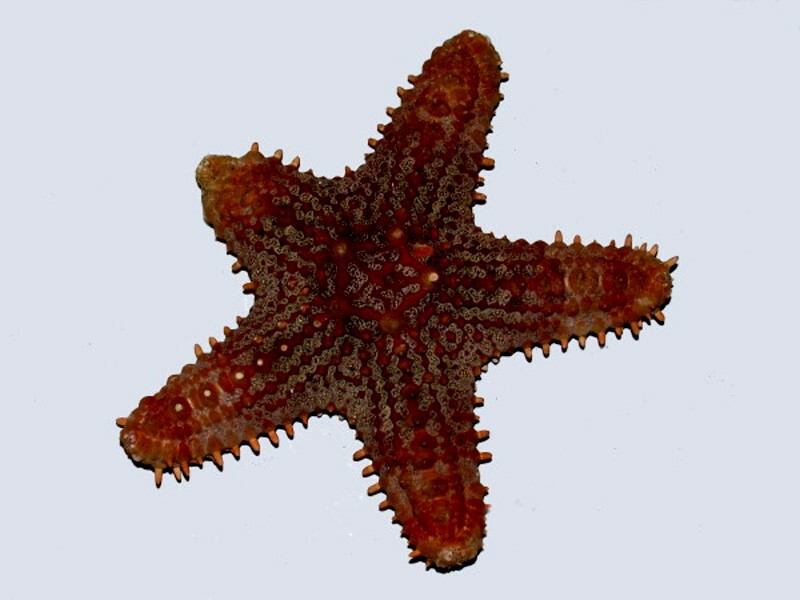
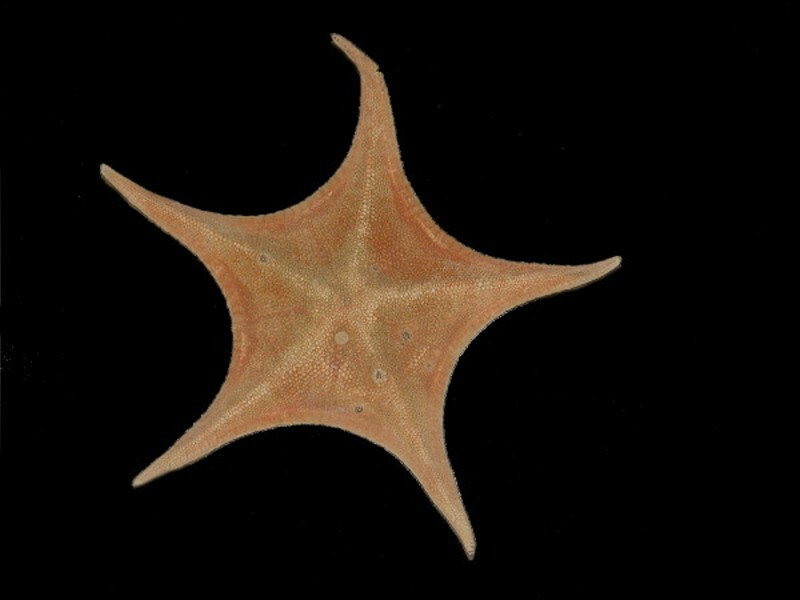
Let’s begin with some familiar beasties that most of us encounter only in a dried form, namely, starfish. We’ll start with some small ones, less than 2 inches across, and work up. This first one still retains a rich, deep red color and one can see an edging of small spines around the arms–altogether, a handsome little asteroid that fits our usual notion of a starfish.
The next one is a bit less typical in that the arms are shorter creating a very pleasing geometric shape. Here we find a nice variegation of subtle colors.
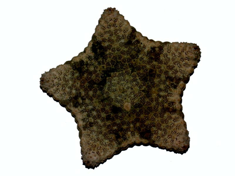
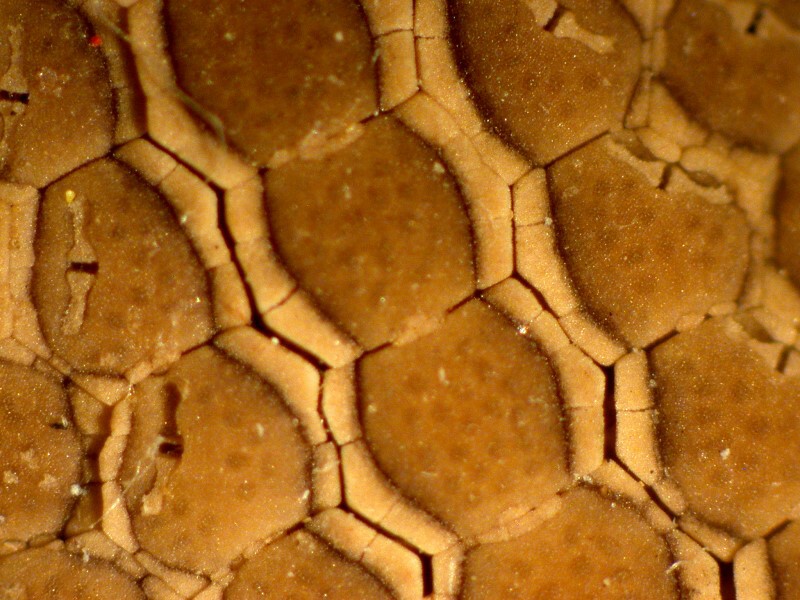
The third example is especially intriguing aesthetically in that the dorsal surface looks like it was carefully sculpted and tiled in remarkably intricate patterns and I’ll show you a complete image and then 4 closeups. In this first image, notice the mosaic-like character around the edges of the arms.
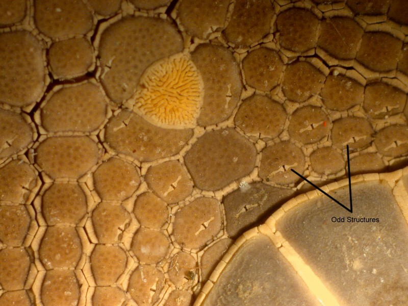
In the next image, we see a bit of the square tiling around an arm edge and a triangular wedge extending up from the central part of the body.
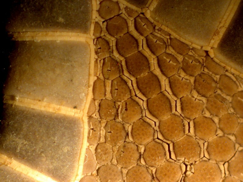
A closeup of the triangular area reveals yet more interesting detail including some odd little slit-like structures, the function of which remain, at this point a total mystery to me.
The next image is labeled to point out these structures and also shows the madreporite (the filter for the water vascular system).
And here is a closeup of that amazing little filter which looks like a bit of embedded coral.
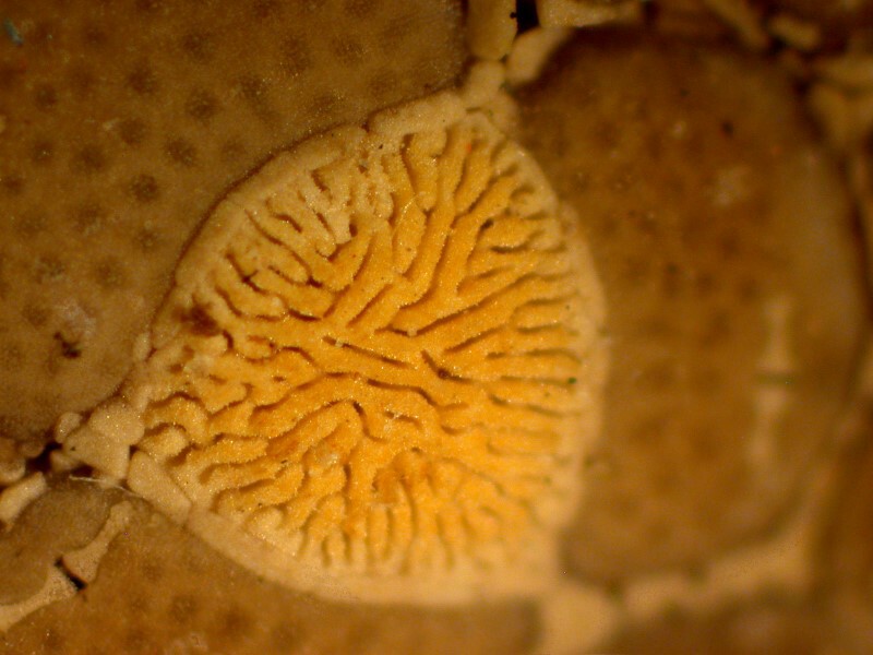
This marvelous structure is the sieve and gateway to an elaborate network called the water vascular system which controls the extension and retraction of the tube feet. I’ll save elaboration of that for another essay, especially since it doesn’t survive drying well.
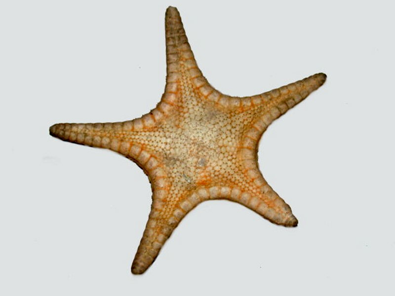
I want to show you three more examples of asteroids, two medium-sized specimens and one quite large one. The first one looks fairly standard except that the arms come to sharp points at the tips. However, this particular specimen shows something highly unusual on the dorsal surface where there are small pits which have been excavated by some aggressive little snails.
Here is a closeup to show you a clearer view.
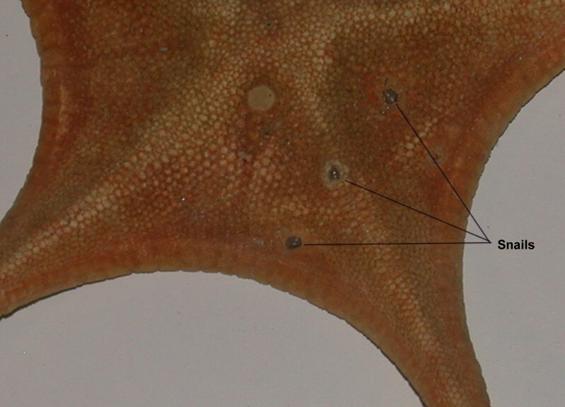
Many mollusks have bands of calcareous teeth controlled by powerful muscles. So, it’s quite possible that these critters scooped little nests for themselves and moved in.
The next starfish, a somewhat larger one, is a beautiful blue–oh ho, you think, some unscrupulous craft shop dyed these–but no, these particular asteroids are actually this splendidly vibrant color.
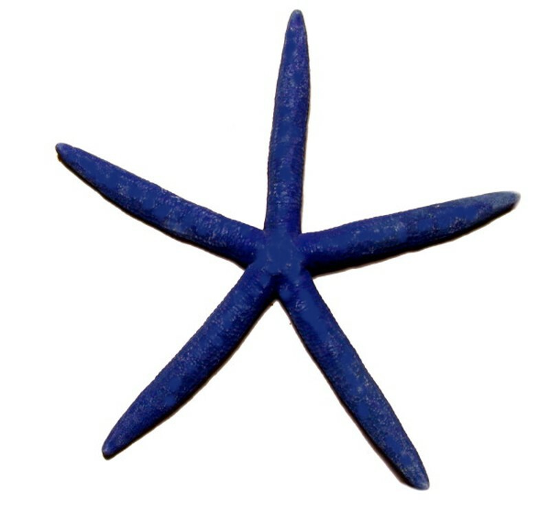
This is a species of Linckia, namely L. laevigata and this genus, even for starfish, has extraordinary regenerative capabilities. The 19th Century oyster fishermen used to drag large mops over the oyster beds and pull up hundreds of starfish which they would chop in half and toss back into the ocean. Unfortunately for the fishermen, both halves often survived and regenerated lost parts to once again prey on the oyster beds. Usually, to regenerate, a starfish requires a substantial portion of the central disk and at least a significant portion of one arm. However, there are some species of Linckia that can regenerate from a single arm! They are often misshapen and certainly wouldn’t meet the high standards of the starfish Quality Control God, but they can survive and prey.
Personally, I would like to see much more extensive, well-funded research on a wide variety of aspects of regeneration rather than pouring billions of dollars into high-tech, Neo-medieval devices such as artificial organs, implants, transplants, and drugs with paragraph after paragraph of possible dangerous or even lethal side affects.
Some starfish get quite large and one large one has a particularly nasty reputation, Acathaster plancii, also known as the Crown-of-Thorns starfish. When you see the image you’ll know why.
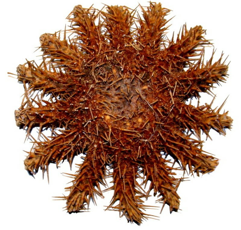
As is immediately evident, these are prickly characters. To make things even worse, those spines secrete a toxin dangerous to divers. Well, you say, why don’t the divers quit bothering them. Unfortunately, Acanthaster attacks coral and eats the polyps and an unexplained population explosion of these beasts has led to enormous damage to South Pacific reefs including the Australian Great Barrier Reef over the past couple of decades. This is not a problem which could be ignored and one can’t poison them without harming the reefs and obviously chopping them up isn’t a viable option. So, divers have been employed to painstakingly collect them in the attempt to save specific areas. So, as striking and interesting as this organism is, it is also, from our point of view, a destructive monster. It dries beautifully and because they are so plentiful, one can obtain specimens quite inexpensively and if, in the course of studying them, you happen to come up with a means of controlling them, you can be almost certain of getting substantial grant funding and, if your method is practical on a large scale, assurance of earning a fortune. But, of course, we pursue such knowledge not for crass commercial gain, but for the love of science. Yeah, right.
Ah yes, a word or two of warning about odors and dried specimens. If you’re buying them in a shop, then you have a chance to check that they have been properly prepared and don’t reek unless, of course, they’re sealed in a packet. If you are ordering by mail, I have found that most shops that advertise on the Internet are quite reliable. If, however, you are doing the collecting and drying yourself, be sure to carry out the procedures either outdoors or in an exceptionally well-ventilated area where you spend relatively brief amounts of time. I speak from profound olfactory experience. When I was a young teenager, our family traveled from Lincoln, Nebraska to Los Angeles to visit relatives and the ones we were staying with lived relatively close to the ocean. For me, with my interests in Natural History, this was a golden opportunity. I collected about a dozen nice, juicy starfish and I brought them back to my aunt and uncle’s house and spread them out on the concrete driveway to let the sun bake them. After 4 or 5 days, we packed up to leave, including a box of starfish. We drove off and my, father, my mother, and my sister, after only a measly single mile, agreed to stop the car and force me to throw out my precious starfish which were admittedly a tad odiferous. Only 2 years ago, I ordered from a dealer in the Philippines, 2 large sponges; each is about a foot across. They have been sitting on top of some metal shelving in my basement storage lab for the whole time and, even now, when I get within 3 feet of them, I can smell them. It’s not overwhelming, but it’s not pleasant either. Another strategy is to take specimens which have been well-preserved in formalin, transfer them to strong alcohol, and then let them dry. In most cases, this will solve virtually all of the odor problems.
Echinoderms, which I find especially fascinating, can generally be dried quite successfully allowing for a careful investigation of all those mind-boggling skeletal and incidental calcareous hard structures.
Visually similar and closely related to starfish are the ophiuroids or “brittle stars”. The name comes from the ancient Greek for serpent and the movement of their arms when they are living is indeed snake-like. I’ll show you 2 whole specimens from the dorsal aspect and then a closeup of the oral side of the second specimen.
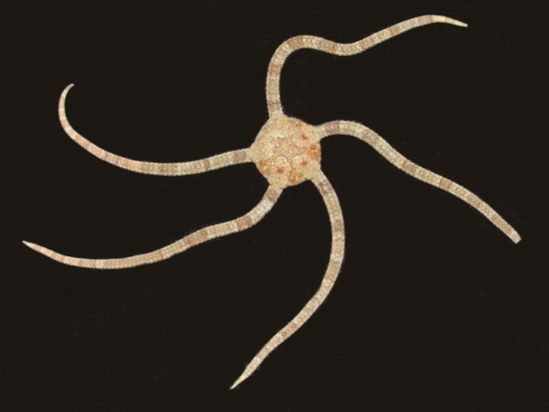
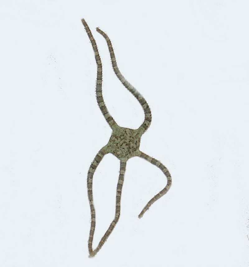
As you can see, these have subtle, but complex surface patterns.
In the closeup below, there is a good view of the central disk.
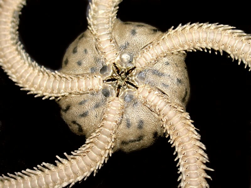
Also clearly visible are the 5 slits around the mouth aperture and, as you can see, lots of lovely spines along both sides of each arm. The number of plates and spines in asteroids and ophiuroids is phenomenal as you can readily discover by taking a specimen and immersing it for a few days in a full strength solution of household bleach.
However, in terms of massive numbers of calcareous bits and pieces per specimen, I think probably the crinoids (sea lilies) and comatulids (feather stars) are the champions.
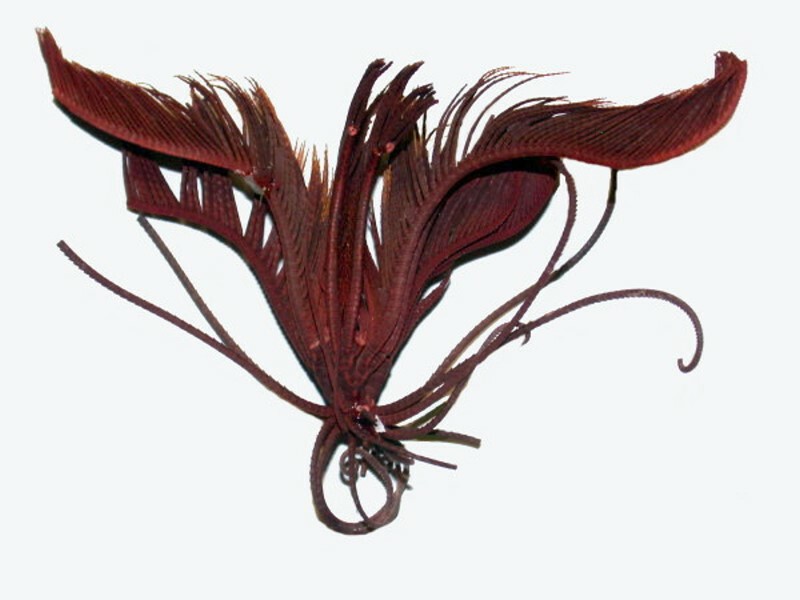
Each feathery “arm” has thousands of tiny calcareous plates to allow for great flexibility. These are marvelous and somewhat obscure creatures and at some point, yes, they need a full essay or 2 or 3.
However, now we need to move on to a splendidly diverse group: the echinoids or sea urchins. Below are a few examples spread out on a styrofoam tray and they are from the Philippines.
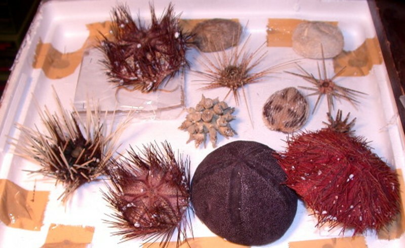
A wonderful collection of toothpicks for a Philistine!
Echinoids are bundles of marvels and mysteries and fortunately properly dried specimens can provide endless hours of enchantment and puzzlement.
Ideally, for each species, one would have 6 specimens with spines and 6 tests (no spines) but, only philosophers and fools live in an ideal world. However, being both, I’ll tell you 12 is a magical number in spite of the fact that I think only once have I ever had that ideal number of a given species.
1) A reference/display specimen with spines.
2) A reference /display specimen test
3) & 4) A specimen to carefully examine using a variety of methods that do minimal damage to the specimen, thus allowing repeated investigations and verifications of earlier observations for both an intact specimen and a test.
5) & 6) A spined specimen and a test for slow and careful disarticulation to study their composition and construction.
7) & 8) A spined specimen and a test for quick and complete disarticulation by immersion in Potassium hydroxide or a strong household bleach. This will provide you with a daunting number of bits and pieces to examine. However, in this mass of material, unless you are obsessively systematic, you will miss certain quite small structures which can be of great interest. So, here’s where specimens 9) & 10) as reserves can become important if you are looking for disks in the tips of tube feet, rods in the elastic part of the tube feet, jaws and rods in pedicellaria or if you are attempting a detailed closeup examination of the crystal arrangement in the suturing of the plates in the test.
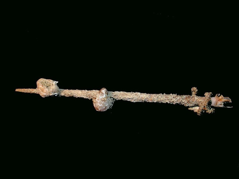
Finally 11) & 12) where you may find and wish to investigate anomalies in the spines or on the surface of the test itself. The test in a given species may show slight to moderate variation from specimen to specimen. And, when it comes to spines, one can find an impressive variety of “hitchhikers”. Here on a single spine, you can see a barnacle, a small bivalve, a tiny brittle star all firmly ensconced on the spine.
As with asteroids, let’s begin look at echinoids by starting with some rather small specimens and work up in terms of size.
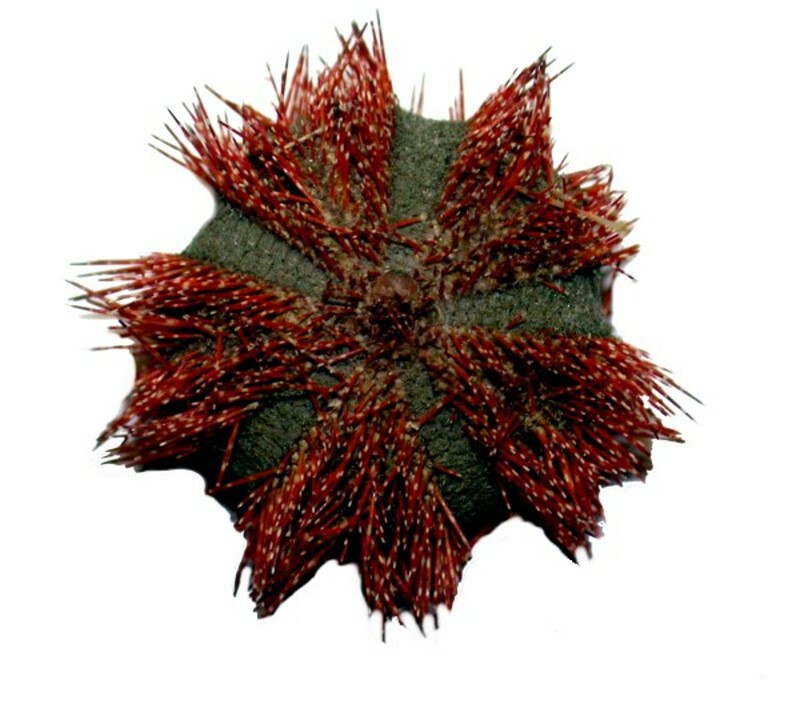
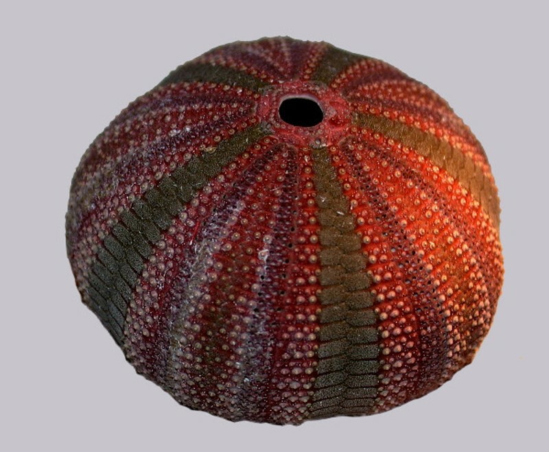
This first little marvel is Christmas-colored, red and green and belongs to the genus Mespilia. It is less than 2 inches across even with its spines. First, a view with the spines and then a denuded test (for adults over18 only!)
A common notion of a sea urchin is that it is more or less spherical and has lots of small sharp spines and certainly this next specimen fits that description.
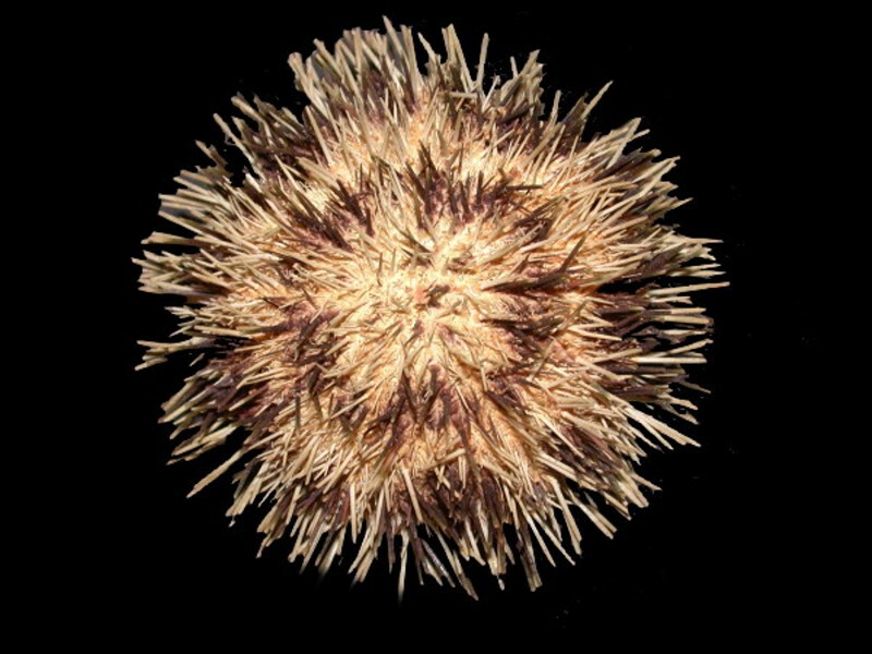
This particular image intrigues me because of its pattern and shape which rather resembles certain instances of crystallization of Ascorbic acid
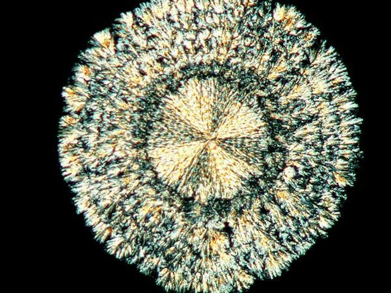
Shield crystal of ASC
As extraordinarily diverse as nature is, there are certain types of patterns that occur over and over. D’Arcy Wentworth Thompson, the splendidly eccentric Scottish biologist produced a provocative study of a series of such patternings in his huge work On Growth and Form which was published in the early part of the 20th Century. Get a copy! Dover publications has reprinted the entire work; however, if you want a somewhat modernized, selected version, there is a paperback edition edited by the development biologist from Princeton, John Tyler Bonner.
However, getting back to echinoids, I’m quite convinced that it is a mistake to argue that there are “typical” organisms that represent a type or group. So, let’s look at some other examples which will, I think, illustrate my point. First of all, take a look at this little beauty.
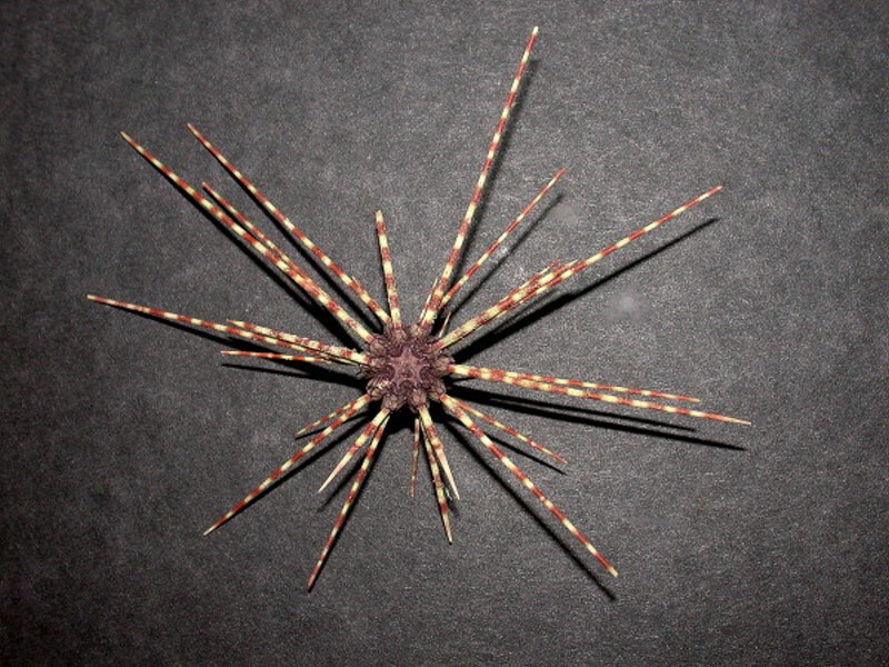
Here we have a very small test, long, and relatively sparse peppermint candy-striped spines. I was going to strip this of the spines as it was the only specimen I could find at the moment, but then I decided I couldn’t do it, so instead I’ll show you the test of a very closely related species. The one with the spines is Coleopleurus maillardi and the test I will show you is from C. exquisitus. However, just look at the lovely surprise underneath when the spines have been removed.
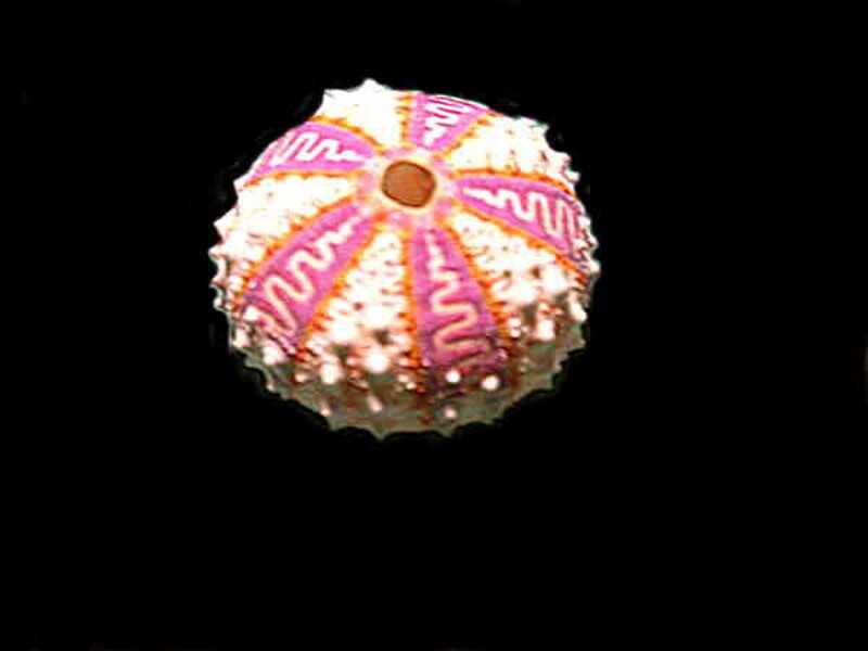
Test only
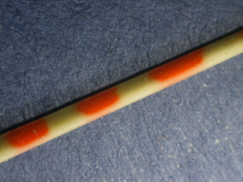
When we look at a closeup of one of these spines, we see that they’re not really striped and they’re not really red and white. Instead, there are circular spots spaced along the spine and the colors of the spots are orange rather than red and the spine is a light yellowish-green rather than white.
Tropical species often have very prickly spines as you can see in the next 2 images.
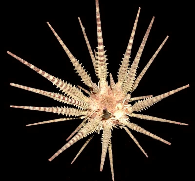
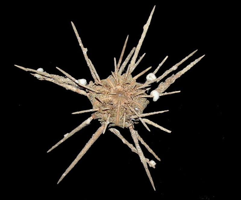
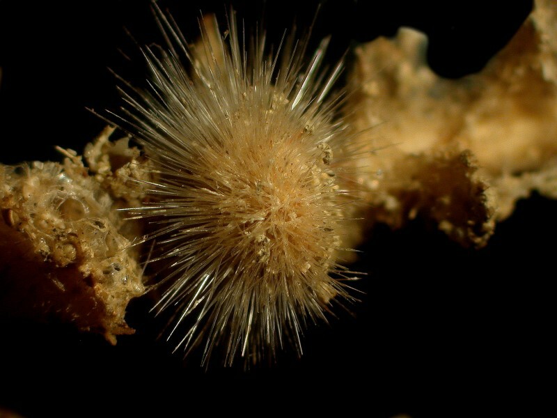
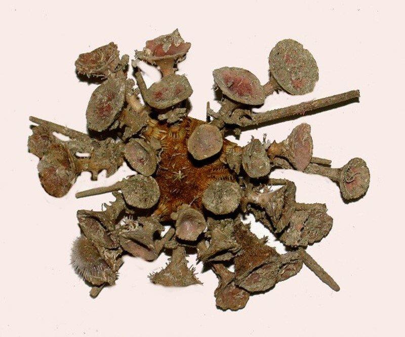
All of these spines provide foothold for all kinds of opportunistic small creatures looking for a home–especially tropical and semi-tropical cidaroids with long spines. We saw one example earlier and now I’ll show you 5 more. The first two I’ll show you are complete urchins with their opportunistic commensal hitchhikers.
A closeup look at spines can reveal fascinating examples of colonization. First up, a spine with a sponge blatantly astride it.
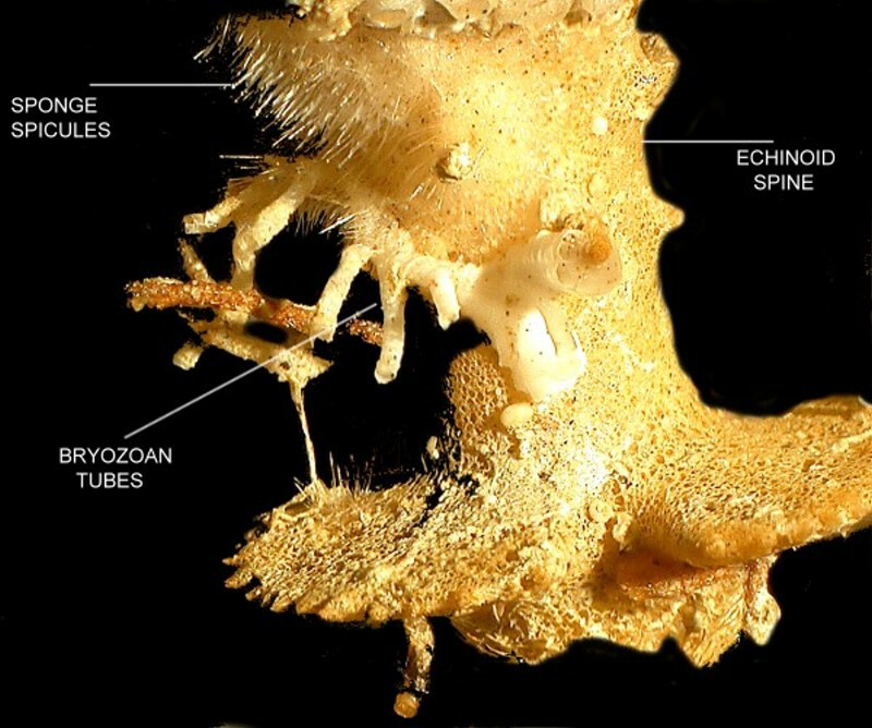
Here is the base of another spine and I have in this image labeled it the “invaders”.
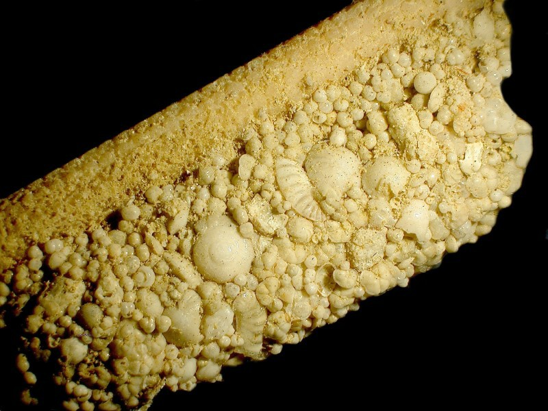
One of the most remarkable finds was a spine with an encrustment along one edge which is twice as thick as the spine itself. A close look reveals that we are seeing a mass of foram shells that have been cemented together. Even from a quick glance, one can tell that there is a considerable variety of species. However, the critical question is how did this come about? I had the advantage of being able to examine this structure from several perspectives including taking it apart. Amazingly, this is the outer surface of a hollow tube–the one-time house of a tube worm! Unfortunately, I know little of the wondrous habits of tube worms, but this is certainly an intriguing bit of architecture.
Did the tube worm arrive at the spine already possessing a prefab foram housing unit? Highly unlikely. I suspect that what transpired was that the larval form of the worm took up residence on the spine and proceeded from there. Tube worms are incredible architects and like human ones, they run the gamut from “Let’s throw this thing together and use all these scrap pieces lying around here” to very fussy, meticulous constructors that like everything to fit neatly and nicely. They secrete an adhesive mucous to which all kinds of things can adhere and different worms utilize different materials much as freshwater caddis fly larvae do. Some species will cut bits of plants, other use smooth pebbles all of about the same size and, yet others will glue together random sand grains of just about any manageable shape and size. The same sort of thing can be found with tube worms. The tube we’re looking at is composed almost exclusively of foram shells of various species and sizes.
Next come the hard questions. How in the devil does the worm get all those forams up there onto its sticky matrix, particularly if the inhabited spine is projecting upwards. Can the worm secrete a foram-specific chemical signal? Again, that seems highly unlikely. Is the worm wriggling down along the bottom gluing forams to construct the tube and then afterwards crawling up onto the urchin spine? That also seems unlikely. So, mystery # 12,829,321,222,876,581,192 (I keep track of all those things I can’t figure out.)
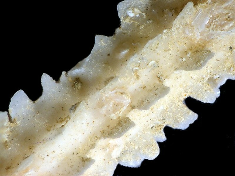
Spines are an endless source of fascination and I want to show you one more closeup of a cidaroid spine; it almost looks as though it was sculpted out of ice.
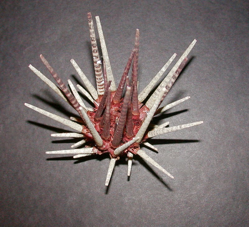
But magnificently stranger spines are yet to come. Consider Goniocidaris.
Cup spines! Inverted umbrellas! Imagine, an urchin that takes strolls in the rain. Clearly, these neat little declivities are an open invitation to small visitors. Note that it also has a few traditional toothpick-like spines.
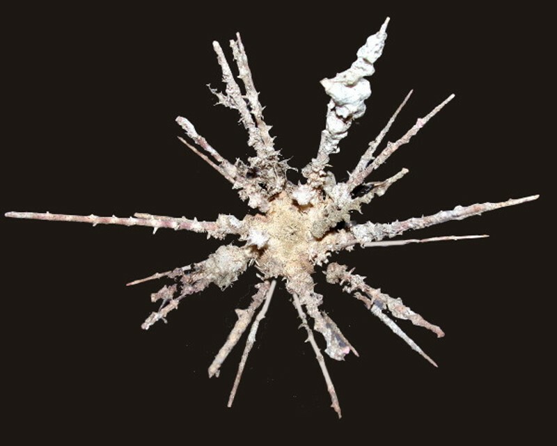
But something even stranger lies in wait–Psychocidaris and after getting a glimpse of its crazy spines, you may well regard it as aptly named.
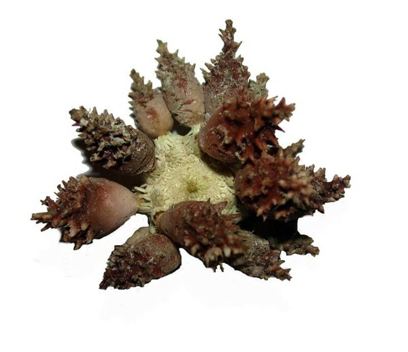
One look at these lumpy, prickly projections and one is tempted to say: “You’ve got to be kidding me! These can’t be spines!” Well, they are and I suspect that their internal composition may be rather strange as well. One of these days, I’ll check that out.
There are so many wonderful, strange, intriguing, and puzzling structures in the echinoids and we have barely scratched the surface, so in Part2 we’ll take a look at a number of cross sections of spines, look more closely at some tests, examine some pedicellaria and look at a large slate pencil urchin.
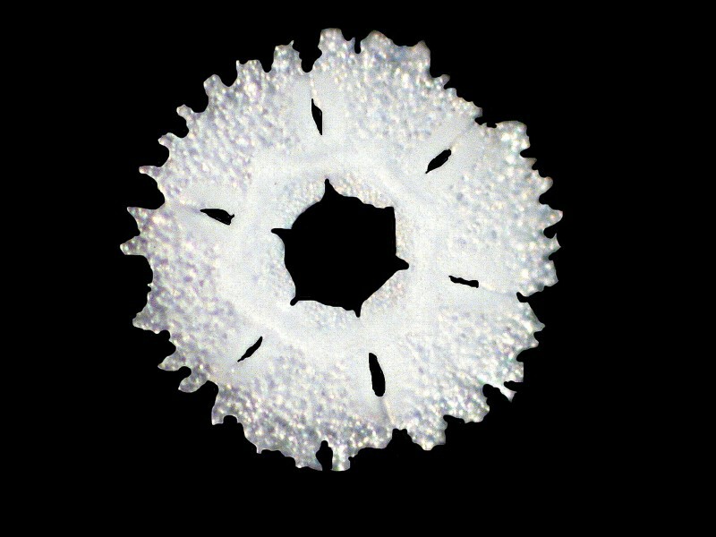
However, just to convince you that there are striking secrets yet hidden among these marvelous creatures, I want to show you one more image–a disk in a tube foot of Stronglyocentrotus fransicanus, a red urchin which is fairly common along the coast of California. This is a beautiful structure and makes me think of a snowflake.
All comments to the author Richard Howey are welcomed.
Editor's note: Visit Richard Howey's new website at http://rhowey.googlepages.com/home where he plans to share aspects of his wide interests.
Microscopy UK Front
Page
Micscape
Magazine
Article
Library
Published in the January 2014 edition of Micscape Magazine.
Please report any Web problems or offer general comments to the Micscape Editor .
Micscape is the on-line monthly magazine of the Microscopy UK website at Microscopy-UK .
©
Onview.net Ltd, Microscopy-UK, and all contributors 1995
onwards. All rights reserved.
Main site is at
www.microscopy-uk.org.uk .