A Brief Survey of the
Handbook of Protoctista
by Richard L. Howey, USA
This book is
an enormous undertaking with 914 pages, a large format, and is filled
with both optical and electron photomicrographs, and has 60
contributors who are experts on specific groups of organisms. If you
can find a copy of it for $200, jump at it. In fact I just checked
Amazon and there is a used copy available for $182.50 on up to a new
copy for $2,432.64 + $3.99 shipping. However, a second edition is to
be published in early January of 2015 for $474.95 but Amazon has a
pre-published guaranteed price of $452.20.
I
bought a copy when it first came out and felt distressed to have to
pay $80. Now it is a classic reference work and even though it was
published almost 25 years ago (1990). In a way, I am surprised that
a second edition is forthcoming given the cost of production and
competition from projects like the Internet
Encyclopedia of Life inspired by E.O. Wilson. Nonetheless, this is
an exceptionally informative work and I won’t ever sell my
copy; they’ll have to pry it from my cold, dead hands. (Out
here in Wild Wyoming, we like to talk that way.)
The volume
has 4 distinguished editors:
Lynn
Margulis from the University of
Massachusetts,
John O. Corliss from the University of Maryland at College Park,
Michael Melkonian from the Botanical Institute of Cologne University
in Germany, and David J. Chapman from the University of California at
Los Angeles. In addition, there was an editorial coordinator,
Heather I. McKhann also from UCLA. The editors had the difficult
task of bringing together contributions from 60 specialists, imposing
a consistent format, and trying to establish terminological
uniformity. This last issue became one of the most contentious and
given the strong personalities involved, it is not surprising that
there were some strong disagreements. Two major examples need to be
mentioned.
1)
Margulis insisted upon using the awkward name “protoctista”
for this kingdom whereas Corliss and others much preferred the term
“protista” which has a tradition dating back at least as
far as Ernst Haeckel. This issue was never resolved and Margulis
wrote about protoctists and Corliss about protists. (Not that it
makes any difference, but I’m afraid that I’m on
Corliss’s side on this one.)
2)
Margulis also insisted on employing the term”undulipodia”
instead of “flagella” when applied to eukaryotes. She
rightly points out that this structure is different from that
possessed by the bacteria which belong to another kingdom or two.
Again, Corliss retains the more traditional term and speaks of
flagella when discussing protists. (Once again, I’m afraid I’m
on his side here.) Perhaps it is important to reinforce this
distinction by way of terminology, but surely something a bit more
felicitous than “undulipodia” could be concocted. I
think one of the barriers to attracting more students into the
serious pursuit of the biological sciences is the massive maze of
terminological thickets and brambles. All scientific disciplines
require a certain amount of technical jargon. At least, in
contemporary physics, one finds a certain amount of creative whimsy
with quarks (borrowed from Lewis Carroll) which have properties such
as upness, downness, and strangeness. One physicist has suggested
that if 2 more are discovered, they should be named Truth and Beauty.
There is also String Theory and the T.O.E. (Theory of Everything),
not to mention black holes, antimatter, and worm holes.
By way of
contrast, consider this passage from the handbook:
“The
antigenic nature of this glycoprotein can be changed repeatedly; each
variant surface glycoprotein (VSG)
is
associated with a serologically identifiable variable antigen type
(VAT). As trypanosomes multiply by fission in the ascending
parasitemia, as particular VAT, the hemotype, forms the major part of
the population. When the host mounts an antibody (IgM) response to
the homotype, the parasitemia goes into remission as trypanosomes of
the homotype VAT are killed off.” (P. 229)
Or a passage
by a different contributor:
“Within
the cytoplasm of developing thraustochytrid thalli is a normal
complement of eukaryotic organelles: mitochondria with tubular
cristae, lipid granules, multivesicular bodies, and Golgi bodies with
the forming face usually closely associated with the nuclear
envelope. The centrioles, present throughout interphase, are located
in a shallow pocket of the nuclear envelope. Rough endoplasmic
reticulum, which may form arrays of parallel cisternae (Moss, 1980),
is usually associated with the cytoplasmic side of the dense plug of
sagenogen.” (Pp. 393-4)
So, I’m
glad we got these things cleared up. Clearly, this is not a book for
beginners and I don’t mean to minimize the difficulties of
writing about the morphological, behavioral, and ecological
intricacies of these organisms, but some of this jargon is
unnecessarily technical and obscure. It becomes parallel to
Bureaucratese and Pentagonese. In my own discipline of philosophy, I
used to tell students that they can be clear without being
superficial and that abstruse and obscure modes of expression are
rarely profound or insightful. One can use tables, graphs, charts,
drawing, photographs, equations, and technical jargon to transmit
information, but unless there is also some passion, some excitement
and clarity, then the communication is deficient. When science
becomes entrenched in obscurantist modes of writing and talking, the
scientists should not be surprised that few students are attracted to
their disciplines. This is especially aggravating in this kind of
case where the kingdom Protista contains come of the strangest, most
intriguing, and downright alien organisms imaginable.
The Handbook
of Protoctista has
two subtitles, the first of which is:
“The
Structure, Cultivation, Habitats and Life Histories of the Eukaryotic
Microorganisms and Their Descendants Exclusive of Animals, Plants,
and Fungi.” This is fairly straightforward and helpful once
you understand that ‘eukaryotic” simply means organisms
which have nuclei that are surrounded by a membrane. One might
quibble about whether or not all of them are truly micro-organisms,
since some are macroscopic (i.e., big enough to see with the naked
eye) and others are, what we might call “meso-scopic”,
that is, big enough be examined with a low power magnifying glass.
The real point, however, is that the only way you’ll really
learn much about these organisms is through microscopic examination.
So, sometimes we have to be a bit flexible and generous in our
interpretations of terminology.
Naturally,
there are complicated exceptions and editorial disagreements arise as
is evident in the Introduction written by Lynn Margulis. In section
12 of the Introductions, she addresses the issue of “Size and
multicellularity: Protoctist vs. protist”. She asserts:
“Thus
we restrict the term ‘protist’ to an informal usage.
Protist, in this handbook, refers to the protoctista members of the
microcosm that require the use of microscopes for their
visualization. Whereas the term ‘protoctist’ includes
all members of the kingdom ; ‘protist’ refers to only the
small organisms, generally composed of a single or only a few cells.”
A minor, but
not insignificant tissue, is reference to this tome as a
“handbook”–it is anything but, requiring two arms
just to lift it. Earlier, she points out that Protoctista range in
size from organisms like the chlorella which are only about 1 micron
in size to the giant kelps which range up to 150 feet.
The second
subtitle is:
“A
guide to the algae, ciliates, foraminifera, sporozoa, water molds,
slime molds and the other protoctists.”
This is
modestly helpful, because it informs us that many of our old
friends–Amoeba
proteus,
Paramecium,
Euglena,
forams, and slime molds are to be found here. This kingdom, as
presented in this volume, has 36 phyla and 19 classes! Too many! As
a consequence I suspect that in the next few decades we’ll see
the creation of a number of new kingdoms in order to tidy things up a
bit.
Now,
however, we need to go back to section 3 of the Introduction: “There
are no single-celled animals or plants.” Being a bit
slow-witted, it took me a while to understand what she was getting at,
since she does talk about unicellular and multicellular protoctists.
In brief, Margulis views the terms “unicellular” and
“multicellular” as acceptable when talking about
protoctists, but when talking about plants and animals, it is a
mistake to talk about “unicellularity” because they are
by
definition multicellular.
Here I strongly disagree with the first part of her claim. In my
view, as I have briefly argued elsewhere
The Unicellular Fallacy protists, such as Paramecium,
Amoeba,
Navicula,
etc., are by
definition NOT
unicellular, but are rather complete, self-contained organisms
radically different from neurons or epithelial cells. This is a
difficult issue, because the Margulis kingdom of Protoctista includes
organisms such as the giant kelps which are clearly multicellular.
This is one major reason why I prefer the notion of the kingdom
Protista which as she says “refers to only the smaller
organism, generally composed of a single or only a few cells.”
It is appealing to me (with a major qualification which I’ll
discuss in a minute.) I suspect that the giant kelps and most of the
other clearly multicellular protoctists require new kingdoms. As
Margulis rightly argues size alone is hardly a sufficient criterion
for making fine discriminations, but neither is it irrelevant. In
the Mammalia, one has everything from the pygmy shrew to the elephant
and the blue whale, but they are clearly multicellular and share a
significant number of characteristics that make them mammals.
However, in
this kingdom of Protoctista, it is important to ask: what is the
relationship between slime molds and foraminifera, between
dinoflagellates and coccolithophores, between ciliates and giant
kelp? With this conception of Protoctista as defined by Margulis,
these questions become extremely difficult, if not impossible, to
answer. If, though, we accept the restricted sense of Protista as
Margulis describes it, namely limited to smaller organisms and
consisting of “a single or only a few cells,” then, I
think, we end up with a more comprehensible kingdom. Here is where I
would introduce my major qualification. We must
not think
of them or describe them as cells; they are individual organisms or
aggregates or colonies, but not
cells.
Giant kelp do consists of cells, so what do we do with them and other
such anomalies? Well, a couple more kingdoms won’t hurt and
might even help keep things better organized. In any case, though I
have been a lone voice crying out in the wilderness of Wyoming, I
think that we should be well-served by abandoning the concept of
unicellularity.
There are
reasons for resisting this suggestion and they depend in large part
on the fact that there are some colonial organisms with “cells”
that are very much like independent non-colonial organisms. For
example, sponges (and many other creatures including humans) have
amoebocytes which can serve a variety of functions within the larger
organism. However, admittedly, the matter with sponges is not easy
and straightforward. Experiments have been done with 2 different
species of sponges which have been disassociated and vitally stained
so that one group is perhaps red and the other blue. These
components are then mixed together and reaggregation does seem to be
species specific at least in certain species. There have been other
experiments using 2 specimens (of the same species) to study whether
or not, at this level, there is a “self/not-self”
biochemical recognition response. The results are largely ambiguous.
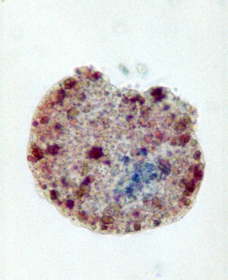
A student of mine (Mr. Hanly) and I tried this experiment using the
primitive Placozoan, Trichoplax
adhaerens and
using the vital staining technique, found the there was no
“self/not-self” recognition response in the organism.
After we finished the project, he took a photograph of one of the
Trichoplax showing the mixed colors of the cells, had it framed and
gave it to me. You can see it below.
Another sort
of difficulty arises with organisms such as Volvox,
Anthophysis,
Synura,
and
Conochilus
The voluptuous Volvox
is
a delight to almost every microscopist. It consists of hundreds to
thousands of “units’ or “cells” depending on
the species. Here, for me, the question arises: Can a colony be
composed of cells or is it an aggregate of organisms? Let’s
start with Conochilus
which
is an elegant colonial rotifer. I first found these in a reservoir
at an altitude of about 6,000 feet and they are wonderful to observe.
Google Images provide a selection of images of this intriguing
rotifer.
During the
process of collecting, individuals occasionally break off from the
colony and single rotifers separate from their original colony. Can
the individuals survive alone? Can they start a new colony? I’m
not sure anyone knows. Rotifers are multicellular
so, there
is a fair chance that they may be able to establish new colonies once
separated from their original one. Clearly, there are colonial
organisms, such as, siphonophores where the individual members that
constitute the colony are interdependent for their survival
suggesting that
we need to find clearer language for talking about these different
sets of configurations. Off the top of my head, I’m inclined
to talk about Kellicottia
as
an aggregation of rotifers rather than a colony. While there is a
relatively small number of individuals in a Kellicottia
aggregate, I would estimate that the upper limit is around 30.The
important thing to note is that it is not a fixed number. There are
certain algae where a given species will have a specific number of
components or “cells”, but there are many protists where
the number is variable even within a given species.
Anthophysis
vegetans poses
some intriguing problems. It is, in one sense, an Über-colony.
It is in fact a remarkably strange organism. If you encounter a
fully-formed mature colony, it looks like a series of brown branching
stalks which at the very tips of the branches have wheel-like
colonies of flagellates. When the larger colony is disturbed–when,
for example, they are transferred from a culture dish, put on a slide
and subjected to the pressure of a coverglass–some of the
colonies at the tips of the branches may separate from the tips and
go spinning off through the water.
So, here are
my puzzlements: How do these brown branches get formed by this
aggregate of flagellates? When the aggregates break off from the
tips of the branches and go swimming away, do they form new
Über-colonies
with branches? What triggers the creation of a new branch and the
development of a new spherical colony? These are not trivial
questions, because each such puzzle we can solve tells us something
more about the incredible subtlety and complexity of life. This
complexity, however, has nothing at all to do with the fabrications
of intelligent design “theory”; nature frequently
displays complexity in the form of Cruelly, Random, Ephemeral,
Whimsical Design (CREWD). As far as my questions regarding
Anthophysis
are
concerned, I suspect that nobody yet knows the answers to them, in
part, because governments prefer to support the creation and
production of new weapons of destruction over and above the study of
protists, perhaps with the exception of a few pathogens that might be
modified for use in biological warfare.
Synura
uvella is
a somewhat larger aggregate of flagellates than Anthophysis,
but Synura
doesn’t
have a stalk. It can reproduce at an incredible rate and produce
“blooms” which give the water a distinct smell of
cucumbers. Nature is always pushing at the edges, the limits, and I
find this consoling since, in my eccentric (and often random and
unintelligent fashion), I keep trying out experiments which, on a
strictly rational basis, seem either wildly eccentric, absurd, or
just silly.
Volvox is a world-class enigma. The largest species Volvox globator consists of thousands of cells and has a wild model for reproduction. Here again Wim van Egmond provides us with splendid images.
A large
specimen can be 2000 to 3000 microns in diameter and so is just
visible to the naked eye. It appears to be a hollow sphere, but
clearly there is liquid within and some special elements or “cells’
floating within. However, let’s first discuss the outer
surface. The “cells” are small and each possesses 2
flagella and a red “eye spot” or light sensor. These are
crucial in keeping the organisms properly oriented with regard to
sunlight since the “cells” also have chloroplasts for
generating food. The “cells” are connected by minute
fibrils and these undoubtedly have something to do with coordinating
movement for the sphere. It wouldn’t do to have half of the
cells striving to go north and the other half striving to go south.
How does all of this work? Again, no one knows for sure. Wichterman
wrote a book on Paramecium,
Tartar wrote a book on Stentor,
Giese wrote a book on ‘Blepharisma–why
hasn’t
anyone done a book devoted to Volvox?
There’s a project for some bright, young researcher who wants
to age gracefully into becoming an expert on the mysteries of this
marvelous organism.
Frequently,
in fact almost always, when looking at a good collection of Volvox,
one will observe specimens which appear to have a miniature colony or
two or several within the meta-colony and that is exactly what they
are. A Volvox
with
these sub-colonies or “daughter”
colonies as they are known, is reproducing. Volvox #1 may have all
female sub-colonies, Volvox
#2
might have all male sub-colonies, and Volvox
#3
might have a mixture of male and female sub-colonies. Now here’s
challenge for a sexologist! Is there some way of determining in
advance which meta-colonies will produce which set of sub-colonies?
I suspect there is by means of some highly sophisticated molecular
biological procedures, but I also suspect that no one has ever yet
tried to make such determinations in Volvox.
What
this Handbook most certainly does do is raise a series of fascinating
conceptual issues, including, but by no means, limited to taxonomic
considerations and, in addition, presents intriguing information
about some of the weirdest and most bizarre, as well as beautiful,
creatures to be found on planet Earth. For some of the larger
groups, one may find the discussions too abbreviated and rather
disappointing. This is especially true for organisms such as
amoebae, ciliates, diatoms, and desmids to mention a few. However,
one has to remember that this volume is a survey and covers a very
wide range which is simultaneously a strength and a weakness. It
does, however, provide extensive bibliographic information for
further reading and research. It also has the virtue of providing a
glossary of terminology.
One of its
greatest strengths is that it introduces us to a large number of
quite small groups, many of which are very likely completely
unfamiliar and gives us further information and splendid optical and
electron photomicrographs regarding organisms with which we have only
a nodding acquaintance, such as, coccolithophores. These are very
small creatures which can occur in such phenomenally large numbers in
the North Atlantic as to produce aquatic “clouds” that
can be detected by satellites. Coccolithophores are relatively
well-known among microscopists because of their elegant structure as
revealed by scanning electron microscopy as you can see here.
However,
how many of you are familiar with the members of the phylum
Xenophyophora? (Pp. 135-38) These odd lumps have been known since the
latter part of the 19th
Century during the period of extensive deep sea dredging by a series
of major expeditions. These organisms have posed great challenges in
terms of classification, because all of the material examined has
been preserved or studied briefly, usually in fragments when dredged
up and brought on shipboard. These oddball creatures illustrate
beautifully what happens when human beings start trying to get
everything in order. Dr. Øle Secher Tendal of the University of
Copenhagen, who is the contributor on this phylum comments:
“As
members of an unrecognized group they were often mistaken for poorly
preserved fragments of sponges, coelenterates, bryozoans, or
ascidians, or they were regarded as inorganic concrements consisting
of foraminifera tests, radiolarian skeletons, sponge spicules, or
mineral grains.
Like many
small groups of organisms, their eccentricities pose major problems
in terms of classification even at the phylum level. This is readily
seen with the exclusively fossil groups like graptolites which have
been bounced around taxonomically for over a century. It’s not
surprising that the Xenophyophores have been a challenge. Most of
them range in size from a few millimeters to about 7 centimeters,
although one specimen of a “Stannophylum”, was of a flat
form and measured about 25 centimeters in diameter, but was only
about 1 millimeter thick.
So, what are
these weird thingies–animal, vegetable, or mineral?
Interestingly, they have long been regarded as some bizarre variant
of a Rhizopod, in other words, something related to the amoebae. If
you look at the photographs in Tendal’s article, you would
never think that these had any relationship to anything amoeboid and,
in fact, Haeckel thought they were sponges. Tendal describes
Xenophyophores as:
“...a
plasmodium enclosed by a branched tube system made of a transparent,
cementlike organic substance. The complex is called ‘granellare.’
Besides numerous nuclei the cytoplasm contains huge numbers of
barite crystals (‘granellae’). Pseudopodia are supposed
to be extended through the free ends of the granellare branches.”
A plasmodium
is a mass of protoplasm with many nuclei, but no internal boundaries,
it is without any walls or “cell” membranes. In this
respect, it bears some resemblance to the true slime molds.
Seemingly
a distinctive feature is the occurrence of large quantities of barite
crystals. Their appearance is highly variable in large part due to
the sorts of foreign objects (xenophae) which they glue to
themselves. Since these are deep sea organisms, most of the usual
sampling techniques cause extensive damage to the specimens. As
intriguing as these creatures are, much of what has been reported
about them is conjectural and much more research remains to be done.
But, alas, they have no economic significance, so studying them is a
very, very low priority for most researchers.
As it happens, this is true for a large number of the other strange and splendid creatures which appear in this handbook. As a consequence, this volume is in many respects more a guidebook than a handbook, a book of hints, suggestions, and conjectures which one can hope will lead both professionals and amateurs to try to discover more about these remarkable creatures.
All comments to the author Richard Howey are welcomed.
Microscopy UK Front
Page
Micscape
Magazine
Article
Library
Published in the January 2015 edition of Micscape Magazine.
Please report any Web problems or offer general comments to the Micscape Editor .
Micscape is the on-line monthly magazine of the Microscopy UK website at Microscopy-UK .
©
Onview.net Ltd, Microscopy-UK, and all contributors 1995
onwards. All rights reserved.
Main site is at
www.microscopy-uk.org.uk .