A New Method of Illumination for the Microscope
Michael Pyshnov, Canada
The method described below gives excellent images with high magnification objectives. The illuminator is using an LED light source and a relatively simple system of collector lenses. It is mounted under the microscope condenser as shown on Fig. 1.
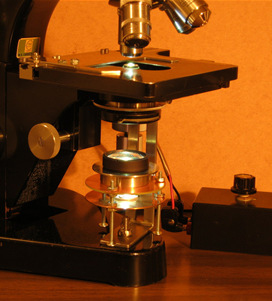
Fig. 1. LED is on the lower round plate, collector is on the upper.
The assembly.
- The LED light is 3 Watt with a regulator of voltage.
- The collector can be made of 2 or 3 convex lenses, each having one side strongly convex. It is most essential that this strongly convex side of the upper lens must look towards the condenser, and the strongly convex side of the lower lens looks towards the LED. The lower lens is only about 3 mm above the LED; this distance needs to be finely adjusted. The collector has a very short focus. All lenses should have a diameter not smaller than 3 cm. The lower lens of the collector used here is the lower element of an Abbe condenser.
- Condenser is simple, apparently having 1.25 aperture, old Leitz.
- The objective used in the photographs here is a Leitz Fluorite 95x, aperture 1.32.
The results of this method are as follows.
- The back focal plane of the objective is fully and almost evenly illuminated. There are almost no colour fringes. The image of the LED is visible in the center, but it is only slightly brighter.
- There is no image of the light source in the field of view which is absolutely evenly illuminated, and no structures of any kind are visible.
- The beam of light coming from the collector is essentially parallel (can have some deviations one way or the opposite). However, if the diaphragm is inserted, it functions not as a field diaphragm, but as an aperture diaphragm The field diaphragm is absent in this method.
- The distance between condenser and the slide is a fraction of a millimeter, so, there is no problem with retaining oil between them.
- The most striking feature is that the objects appear very sharp in focus. There are no haloes on the edges. The depth of focus is very small, which allows to see the object sufficiently free from the images of the other objects above and below it.
- Full aperture of the objective is used. That, however, does not produce parasite light, and the contrast (especially in stained objects) is indeed unusually high.
Below, are the photographs of two diatoms made with a Lebeca web camera (320X240 pixels). No Photoshopping was used, although, of course, the parameters on the camera properties were adjusted.
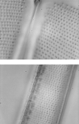
Fig. 2. Diatoms, S. phoenicenteron and N. sigma, from the eight form diatom test plate preparation of Klaus Kemp (with great thanks to him).
This method was found empirically. I have no explanation of how it works in terms of geometric optics. The experiments show that the chosen here outward orientation of strongly convex sides of the collector lenses (together, of course, with short focal distance of the collector) creates all these features.
All comments to the author Michael Pyshnov are welcomed.
Microscopy UK Front
Page
Micscape
Magazine
Article
Library
© Microscopy UK or their contributors.
Published in the January 2016 edition of Micscape Magazine.
Please report any Web problems or offer general comments to the Micscape Editor .
Micscape is the on-line monthly magazine of the Microscopy UK website at Microscopy-UK .
©
Onview.net Ltd, Microscopy-UK, and all contributors 1995
onwards. All rights reserved.
Main site is at
www.microscopy-uk.org.uk .