|
A MICROSCOPIC LIFE (or) THE MEMORIES OF A VERY OLD MICROSCOPIST
MANUEL del CERRO, MD PRINCETON, NJ, USA |
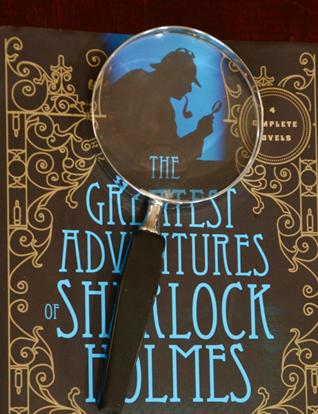
I was introduced to the marvels of the Small World at the age of 9, when my father gave me a hand-held magnifier. It may have been a 4X or so, a small version of the Sherlock Holmes iconic magnifier, with a wooden handle and a metal rim. With it I soon made a monumental discovery: that the spiders (or at least the one I found dead in a plant pot) have multiple pairs of eyes!
With my scientist career starting in such an auspicious manner, I continued searching the Small World. I remember discovering that the color figures in the Sunday cartoons were in fact collections of colored dots, the colors mixed in various proportions to achieve the desired effect (it was about fifty years later that I learned that Roy Lichtenstein had achieved deserved fame and fortune by using similar dots, conveniently magnified, in some of his distinctive paintings).
Figure 1, A low power, handheld magnifier like the one that introduced me to the Small World.
Once started, I was not going to stop. Next step: a microscope! Anybody who has read “Greg’s Microscope” (and everybody, young or old still should), knows how intense the wish for a microscope can be. It soon became clear to me that to have my own microscope, I would need government support. In my household, “government” was, officially at least, my father (my mother was the power behind the throne). I approached Father with the request, and he came with a counter offer. He would provide one half of the cost; the other half would have to come from my allowance. I wanted the microscope bad enough so I accepted. Years later I realized that: a) my allowance also came from my father, and b) that he only wanted to be sure I REALLY wanted that microscope, that it was not a passing whim for another toy. Then the microscope came! Oh, how beautiful it was! In reality, it was a post-WWII Japanese contraption, with only coarse focus, flat mirror, and a button-type, triple component objective. To change magnification one had to screw in, or remove, one or two of the components. Few more inconvenient forms of magnification change have ever been devised. But since it was the only one I knew, I thought it was a marvelous achievement of the human mind. However, one thing could be said in favor of the optics of my first microscope. The magnification levels were about 50X, 100X, and 150X. The insanity of offering children microscopes claiming a top magnification of 1,200X had not yet been inflicted upon the uninformed.
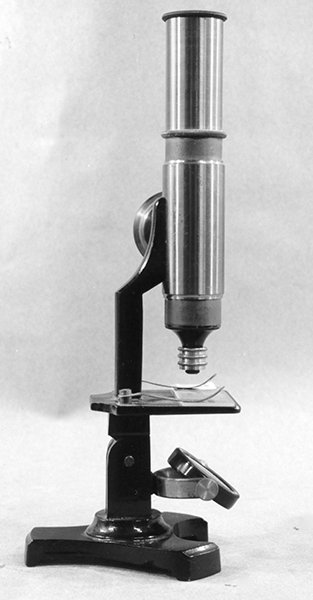
Figure 2. A “Student”, or “Amateur” microscope with a design similar that of my first microscope.
Years went by, and I found myself in Medical School. There we used (and much abused) microscopes in Human Histology, Physiology, Microbiology, and naturally, Pathology. Our approach was brutally utilitarian. We did not know, were not told, or much cared, for the fine details such as the proper use of the iris diaphragm, the proper positioning of the condenser, or the meaning of the that esoteric inscription “N.A.” that was stamped on the barrel of each objective. The microscope was a tool (they were for the most part Leitz monoculars), what mattered was that we could identify the structures under consideration well enough to recognize them when examination time came. At that time teaching microscopes were fitted with that infernal devise called the teaching ocular, or demonstration eyepiece. For the benefit of those lucky enough not to have confronted one, let me describe the teaching ocular. It consisted of an ocular inserted into the microscope tube in the regular manner, that was the part used by the professor or instructor. A lateral side-tube supported a second eyepiece, the student ocular. An indicator simultaneously visible to the examiner and to the student could be moved to indicate any structure visible in the field. I can still hear a voice asking, “Well, and what is this?” Incidentally, the demonstration ocular was not inexpensive. The 1931 Arthur A. Thomas Catalog of Laboratory Apparatus lists the Bausch & Lomb version at $41.00. Forty-one dollars was a very significant amount of money in the days of the Great Depression.
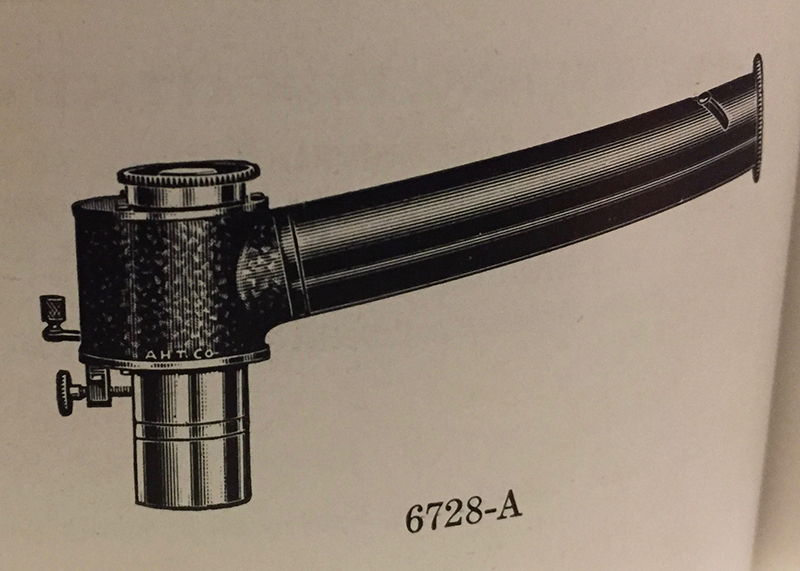
Figure 3. The Bausch & Lomb teaching ocular sold by the Arthur H. Thomas Company.
A residency in Intensive Care Medicine didn’t allow much time for the microscope, only an occasional look at the Neubauer haemocytometer or to urine sediments. “The Lab” made most of the observations. Once the residence requirements were fulfilled, the question was, should I continue in this specialty, or move to other area of medicine?
Intensive Care practice is high-adrenaline medicine. Time to act is of the essence; time to think, necessarily limited. That was not what I wanted to do for the rest of my life. Instead, I moved to “The Cloisters” of Academic Medicine, and back to microscopes! And microscopes we had! To begin with, the Cell Biology Institute of the University of Buenos Aires, Medical School received at that time (1956) an RCA electron microscope, one of the first in Latin America. This was soon followed by two TEMs, a Siemens 1 and then Siemens 1a. The RCA was a good instrument by the standards of the times, but the Siemens were marvelous machines, decades ahead of their times.
As many (too many!) Argentine physicians and scientist of my generation, at some point I found it prudent to leave my country and take my family to calmer lands. That was in 1964.
A long journey, in distance and in time, ended in 1998, with me retiring as an Emeritus Professor of Neurobiology and Ophthalmology, of the University of Rochester, NY, Medical School. Microscopes of all kinds have been part of my academic existence. Another Siemens 1a, this time “my” microscope, received me on arrival to Rochester in 1965 (figure 4).
The Rochester Medical School and I liked each other; my initial two-year contract resulted in a thirty-two year tenure.
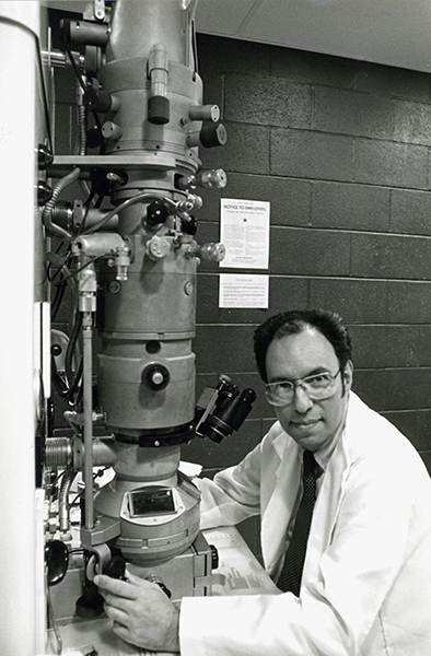
Figure 4. “My”microscope at the Center for Brain Research, U. of Rochester Medical School. The first time I visualized individual molecules (ferritin) inside brain tissue, I felt the thrill that a pilot may feel the first time he/she breaks the sound barrier.
As my work evolved, I worked with Zeiss TEMs, and with SEMs of various makers. The Zeiss, I particularly liked, but as other investigators, I found more and more that the light microscope, far from being made obsolete by the EM, was having a major come back as an instrument able to give a view of the micro world different, but totally integrated with that provided by the EM.
I became aware of the power and the beauty of fluorescence microscopy. Using a Leitz fitted with the Ploem fluorescence system was pure joy!
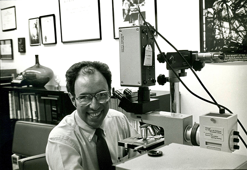
Figure 5. Using the Leitz fitted with an incident UV source made me smile.
A turning point in learning to take full advantage of the capabilities of light microscopes was my attending in 1982, the Light Microscopy Course annually offered at Woods Hole by Professor Robert D. Allen (igure 6). Bob Allen was a world leader in the use of advanced light microscopy (Figure 7), having visualized in 1981, the action of microtubules within living cells, a feat considered impossible by many at the time. Bob, a gentleman and concert level cello player, remained a friend and advisor until his untimely death.
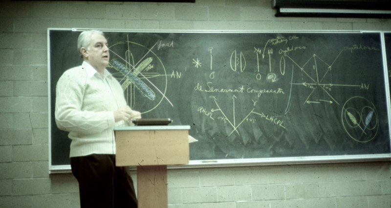
Figure 6. Professor Robert D. Allen lecturing on polarization microscopy at Woods Hole, Mass.
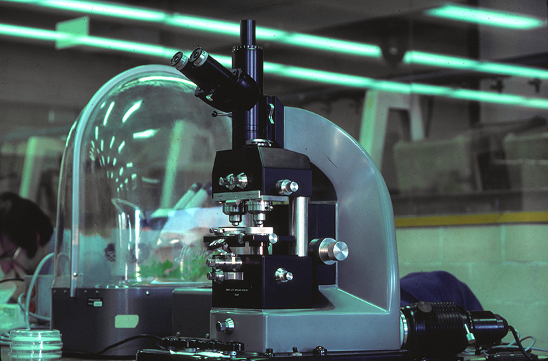
Figure 7. Bob Allen’s Leitz Mach – Zehnder quantitative interference microscope used for demonstrations at the Woods Hole course. This marvel of opto-mechanical technology allows the determination of the dry mass of living cells and organelles – it is having a revival these days.
A few words of recognition should be devoted to two forms of low power stereomicroscopes that are the unsung heroes of the microscope world. I refer to the slit-lamp and the surgical microscope.
The slit lamp is essentially a horizontally placed stereomicroscope attached to a very ingenious illumination system, from which the “lamp” misnomer arises. All of us who have been at an ophthalmologist or optometrist’s consult know how it does feel to be at the receiving end of the apparatus. Those of us who have look through one of them know the beautiful, highly detailed, three-dimensional view of the anterior segment of the eye that is seen through it.
The surgical microscope too is a stereomicroscope, used most of the times at low magnification, with side ports for the assistant surgeon, co-axial illumination, and video capabilities. They come into a variety of sizes, prizes, and configurations, the choice dictated by the type of patient, laboratory animal or human (Figures 8 and 9), or by the complexity of the mechanical controls that allow operation by hand, or preferably, foot controlled pedals.
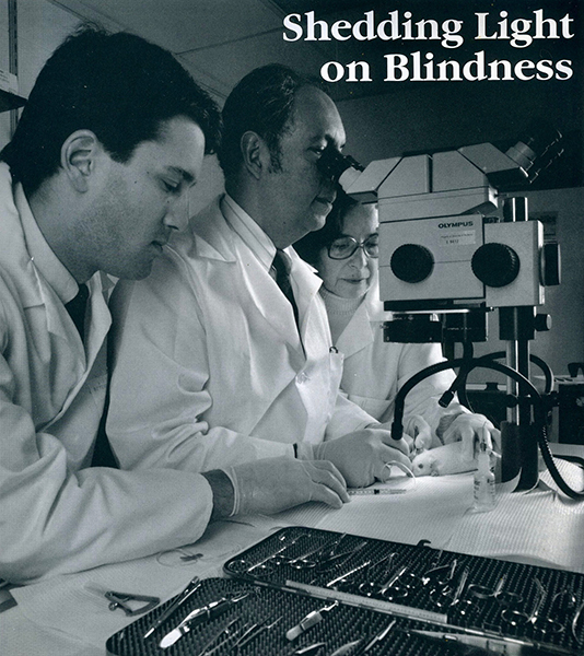
Figure 8. A table-top surgical microscope facilitating cell transplantation into the retina of a genetically blind rodent. A medical student (David A. DiLoreto, now an MD, Ph.D) and Research Associate and Laboratory Supervisor, Coca del Cerro, complete the surgical team.
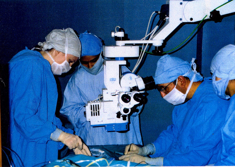
Figure 9. A floor-mounted surgical microscope facilitating cell transplantation into the retina of a blind human patient.
Having lived with microscopes for so many years, I could not fail to notice the progressive integration of electronic components into microscope stands. The trend culminated, as far as my experience went, in the Olympus Vanox 2. The electronic components of that microscope provided push-button change of objectives, automatic focus, and automatic Köhler illumination for each selected magnification. It was excessive to my taste but the fact that it also featured extensive fluorescence capabilities was the deciding factor in purchasing one for the lab.
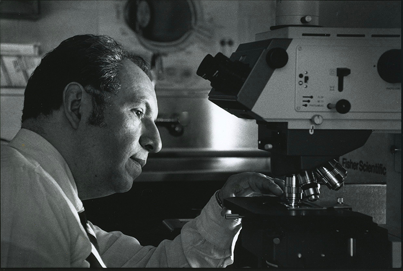
Figure 10. The ponderous Olympus Vanox 2. It was customary to turn off the ceiling lights while observing fluorescence-stained preparations.
An interest in the history of the microscope developed slowly but became intense (Figure 11). It was nourished by visits to important collections such as those at Washington, Leyden, and Utrecht, where the curator show us the van Leeuwenhoek iconic microscope, the little instrument that changed the world of science .
Figure 11. Using microscopes, collecting microscopes, and talking about microscopes. Is that the definition of an obsession?
We visited the collections at London, Cambridge, and the impressive RMS Collection at Oxford. A less well-known collection, but a highly a significant one, is that at the Cajal Institute in Madrid. It shows many of the microscopes used by the Father of Neurobiology (Nobel Prize in Medicine, 1906), and by his associates. Without ignoring other contributions, it is fair to say that at this place, and from those instruments, modern Neuroscience was born (Figure 12).
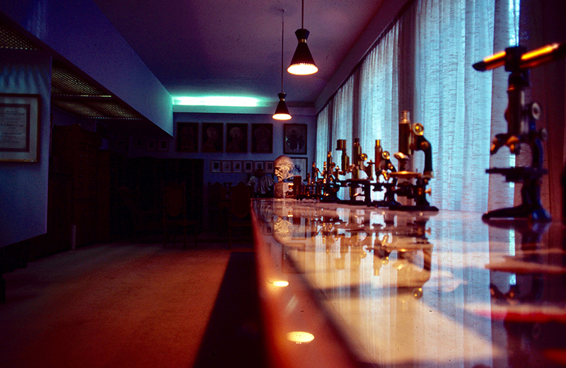
Figure 12. A partial view of the microscopes in the collection of the Cajal Institute. A bust of “El Maestro” is in the background.
The Chemistry Nobel prize in 2014 recognized the triumphal return of the light microscope as a premier search instrument. But by now my days of professional research are over. I’m still using the microscope, just for fun (Figure 13), and describing my new adventures in articles published in Micscape. Of my microscope collecting efforts I have already spoken in this publication (Micscape, January 2008). I only like to add that I still carry in my pocket a 4x magnifier in case I can make some other “discovery” as significant to me, as it was seventy-some years ago finding that spiders have multiple pairs of eyes. Who knows, the micro-world is full of wonders!
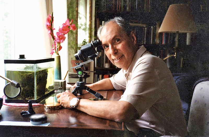
Figure 13. Just as the text says, this is using the microscope just for fun. This resulted in hours of pleasure and in an article in Micscape (April 2007). The stereo microscope was the one mounted on “my” Siemens (Figure 4). It was rescued when the EM was discarded in the early 1990s.
PS: Dear reader, I will not vex your patient with more verbiage but I thank for your indulgence in reading this. I wish you a long and enjoyable life in microscopy!
Acknowledgements: "No man is an island." Many persons helped me to become a microscope lover, and later a professional using the microscope on daily basis. My father, Señor Manuel Perez del Cerro, opened the doors of the Micro World for me. He was the first and best of my teachers. I owe a great debt to my mentors, professors, colleagues, and students. I hope my students learned from me at least a fraction of what I learned in teaching them. My wife, Coca del Cerro, was my devoted research associate for many years. She tolerated (even encouraged!), my collecting habits. These days, as the sun is setting in my life (Figure 14), my microscopic adventures have continued thanks to the enthusiasm and encouragement of Mr. Dietmar R. Krause, a dear friend and fellow perpetrator of “Just for Fun” articles published in Micscape. I am grateful to my daughter, Marilu del Cerro DeCoste, for fine editorial assistance. As always, the help of our Editor was extraordinary.
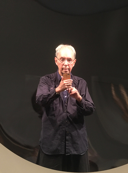
Figure 14. The Old Microscopist in front of a large reflecting lens exhibited at the Art Museum, Princeton University. He holds an Apple iPhone that doubles as a low power, hand held, photographic microscope.
All comments to the author Manuel del Cerro are welcomed.
Microscopy UK Front
Page
Micscape
Magazine
Article
Library
Published in the January 2017 edition of Micscape Magazine.
Please report any Web problems or offer general comments to the Micscape Editor .
Micscape is the on-line monthly magazine of the Microscopy UK website at Microscopy-UK .
©
Onview.net Ltd, Microscopy-UK, and all contributors 1995
onwards. All rights reserved.
Main site is at
www.microscopy-uk.org.uk .