|
||
|
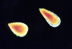
Haematococcus.
A common inhabitant of bird baths
by Steve Durr
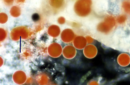
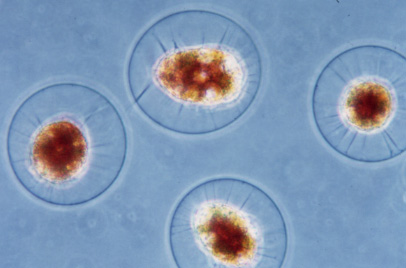
This member of the Volvocales is usually encountered by people who have the good sense to install birdbaths in their gardens. Very often the first indication that something strange is happening is that the water begins to change to an orange-red colour. If you're lucky enough to have a microscope at hand a strange sight awaits you. The culprit for this unusual coloration is a micro-organism called Haematococcus pluvialis. The red colour is due to the pigment called astaxanthin, which possibly protects the organism from the harsh sunlight, especially the ultraviolet rays from the Sun.
A pigment is a molecule that transmits or reflects light and the colour is due to the way that the pigment reflects or absorbs different wavelengths of light. E.g. Haemoglobin & chlorophyll are two other well-known pigments.
|
||
|
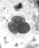
It has been my experience that whenever these micro-organisms are found they nearly always appear to be in a state of encystment at the bottom of the bird bath. Only when the cell is placed under the cover slip does it appear to come to life. It is remarkable that this organism can withstand extremes of climate, many bird baths completely dry up for long periods of time. The spread of this alga is probably due to the habit of birds coming down to preen their feathers and carrying away with it some of the cells attached to the legs or body.
|
||
|
||
|
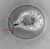
Haematococcus is a very easy micro-organism to keep and requires no special conditions except the birdbath where you found it. The life cycle can be followed with ease and photographs can be taken to supplement your observation through the microscope. Haematococcus does not always appear a bright reddish colour and therefore can be mistaken for Chlamydomonas by the inexperienced. Watch out for the cytoplasmic strands and the clear gelatinous margins at the cell wall. Certain euglenids can also turn the water red but can easily be identified.
An excellent book to introduce the Algae, which has some excellent photographs and text, is Freshwater Algae, by Hilda Canter-Lund & John W G Lund. ISBN 0-948737-25-5 and published by Biopress ltd.
Looking back at some of the old Quekett newsletters I found a few people who have commented on this micro-organism. My good friend Eric Hollowday wrote a small piece stating that there are recorded instances of these animals being kept dry for up to 7 years in the laboratory. He also made the observation that they are usually found in the company of the rotifer Philodena roseola.
Dr Thain also made some observations back in the 1980's that his birdbath, which is stored over winter in the garden shed, has produced Haematococcus over the past ten years. It appears that this alga thrives well in such inhospitable conditions without too much trouble.
| Ultraviolet Radiation, electromagnetic radiation that has wavelengths in the range between 400 nm, the wavelength of violet light, and 15 nm, the length of X rays. (The nanometre, nm, equals a millionth of a millimetre, or 40 billionths of an inch.) These figures are reproduced from the Encarta encyclopedia. (back to text) |
Comments to the author Steve Durr are welcomed
Prepared for the web by Anne Bruce
Published in January 1999 Micscape Magazine.
Please report any
Web problems or offer general comments to the Micscape Editor,
via the contact on current Micscape Index.
Micscape is the
on-line monthly magazine of the Microscopy UK web
site at Microscopy-UK