Hydras
by
Jan Parmentier, Netherlands
You will not find many Coelenterata in fresh water. To this
phylum belong jellyfishes, corals, sea anemones and polyps,
characterized by stinging organelles, called cnidae or
nematocysts. Only a few genera, all belonging to the Class
Hydrozoa, occur in freshwater, due to the fact that the phylum of
the Coelenterata evolved in the sea and not many species
succeeded in conquering fresh water.
Hydras managed to overcome this barrier and are found almost
everywhere in clean water where they can attach themselves to a
substrate (plants, stones). Hydras had already been studied by
Van Leeuwenhoek in 1703 and by Trembley in 1744, who did a lot of
ingenious experiments and wrote a fine monograph about hydra. I
was very happy to buy a modern reprint of this monograph. They
were followed by many other investigators.
The best way to find them is to take home small waterplants
(duckweed, pondweed) and to let these stay in a small vessel.
After a while you often see hydras attached to the glass. They
are coloured green, grey and brown. It is difficult to determine
the grey and brown ones; you have to look at the nematocysts to
decide the name of the species. However, there is in Europe only
one green species, Hydra (or Chlorohydra) viridissima.
Symbiotic green, unicellular algae (Chlorella vulgaris)
are responsible for its green colour. This hydra is typical of
small ponds and ditches and is less tolerant for water pollution
than other species. I did not have to go far to find them, they
are abundant in my own small garden pond (1.5 square meters).
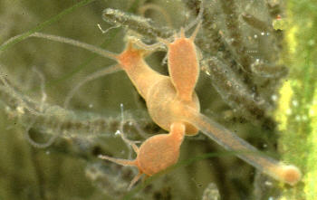
Reproduction in hydras occurs most of
the time by budding; in the photograph shown right, you
will see a brown hydra with two other budding hydras
already completely formed but still attached. Hydras can
also reproduce sexually. The green hydra is
hermaphroditic, meaning that male and female organs
(gonads) are located on the same animal.
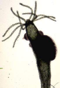
|
 |
| In the image shown right you will see the male organs
just behind the arms; the female organ, much larger, is
situated a bit lower on the animal. |
 |
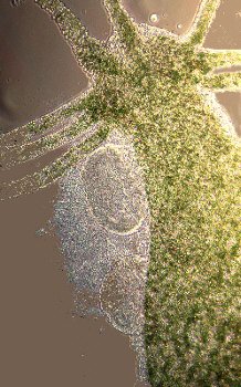
In the image shown right the sperm is
oozing out of the male organ, in the image shown below a
ripe egg is visible. The egg is fertilized by the free
swimming sperm (I could detect only one flagella in each
sperm cell), becomes surrounded by a tough covering and
is then released from the hydra. It sinks to the bottom.
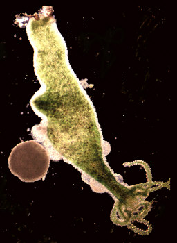
|
|
| The green symbiotic algae are already present in the
egg, coming from the parent. The fertilized eggs develop
into ciliated planula larvae, which can develop very
quickly into new polyps. Due to the presence of these
symbiotic algae, this hydra is attracted by light,
contrary to other species and can survive long periods
without food. A lot of other things can be studied on a
hydra. The stinging cells (you need an oil immersion
objective for that), the catching and digestion of prey,
movement, specialization of skin cells and many other
interesting features. |
 |
Comments to the author Jan
Parmentier welcomed.
Photographs by the author.
Texta and images © Jan Parmentier 1998
Web page prepared by Dave Walker
Please report any Web problems to the Micscape Editor.
Published in Micscape Magazine, June 1998 (
ISSN 1365 - 070x )
Micscape is the on-line monthly magazine of the
Microscopy UK Web site at
http://www.microscopy-uk.net/mag/indexmag.html
WIDTH=1
© Onview.net Ltd, Microscopy-UK, and all contributors 1995 onwards. All rights
reserved. Main site is at www.microscopy-uk.org.uk with full mirror at www.microscopy-uk.net.