Image Gallery:
The Desmid Cosmarium reniforme. 
Three photomicrographs from my archives.

by William Ells, UK

 
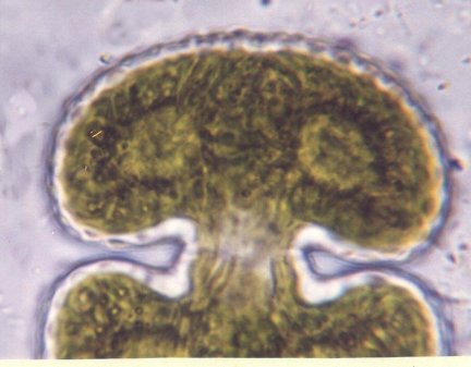
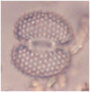
The author's enclosed images show the following:

The desmid genus Cosmarium, with the apt species name reniforme, the semi-cells are obviously kidney shaped in face view as they are usually seen. In apical view elliptical. The cell walls are granulate. The species are widely distributed throughout Britain.


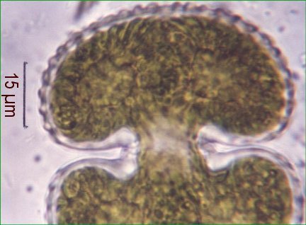
Fig. a) shows the prominent solid looking granules
around the margins of a semi-cell.


Fig. b) shows the green chloroplast, the lighter
green spots are the pyrenoids said to secrete starch.

 
Figs. a & b were photographed using a 100:1 oil immersion objective and a 4:1 projection eyepiece.
Fig. c) shows an entire but empty cell of another smaller specimen. Cells without their chloroplast content are often found, sometimes this has been depleted by fungi. I have also found empty cells of many species of desmid in the faeces of microscopic and macro-fauna. Such cells are very useful in enabling any punctae or scrobiculations (depressions) to be seen, or as here the granulations showing up as white spots.

Fig. c) was taken with a 40:1 plan fluorite objective. All on a Nikon Skt. microscope with trinocular head .

All comments to the author W H Ells are welcomed.


Microscopy UK Front Page
Micscape Magazine
Article Library


© Microscopy UK or their contributors.

Published in the July 2002 edition of Micscape Magazine.

Please report any Web problems or offer general comments to the Micscape Editor,
via the contact on current Micscape Index.

Micscape is the on-line monthly magazine of the Microscopy UK web
site at Microscopy-UK

WIDTH=1


© Onview.net Ltd, Microscopy-UK, and all contributors 1995 onwards. All rights reserved. Main site is at www.microscopy-uk.org.uk with full mirror at www.microscopy-uk.net.