Diatoms have been popular with microscopists since Victorian times and even if they do not devote themselves to diatoms in particular, microscope users will be familiar with such mellifluous names as Pleurosigma angulatum or Frustulia rhomboides because these have been standard test objects to check the quality of their lenses - and, especially, the quality of their microscope expertise
From a more seriously scientific point of view, diatoms are even more fascinating. They abound in almost any body of water worldwide. That makes them major primary producers, meaning that they drive the Earths food web and are a rich source of the oxygen we breathe. Secondly, diatoms are incredibly diverse, with estimates of the number of species running from 30,000 to 100,000 and more. Thirdly, diatom studies make it possible to demonstrate environmental changes spanning tens of thousands of years. And finally, diatoms build their external skeleton (the valve) by catching silicate from the water and depositing it with amazing precision in specific patterns. In fact, they run a super-secret ceramics process, which - if we could duplicate it - would open up fantastic vistas of nano-engineering: manufacturing complicated nanostructures out of silicate, an ultrapure, inert and refractory material.
So lets take a second look at two common test diatoms to illustrate two aspects of diatom research.
Taxonomy
Taxonomy addresses the single most important issue in any form of biological study: the identity of the critter youre talking about. If you publish a DNA sequence of Mr A showing that he is the culprit in a criminal case and it turns out you actually examined the DNA of Mr B, justice will not be done. In science, the equivalent would be the single most horrible mistake imaginable, because the research would be unverifiable and irreproducible. Unfortunately, this situation has not been at all rare in diatom studies, because the vast species diversity makes misidentification easy.To avoid this, zoologists and botanists have worked out a rigidly codified approach. Each organism is assigned to a genus (a group with common characters) and given a first, generic, name - like a human family name. To place it within the genus, its also given a species name. And to honour authorship, the discoverers name follows. If a revision is done at a later moment, the revisers name is added. This nomenclatural procedure led to the name Frustulia rhomboides (Ehrenberg) de Toni.
The second measure to ensure reproducibility is that the author describing a new species must deposit a type in a recognized collection, which serves as a reference from then on. For a new diatom, one would make a slide, ring several specimens with a diamond marker and deposit the slide in a museum of natural history. Such collections are a treasure trove for research and we should cherish them and the kind people curating them.
Detective story
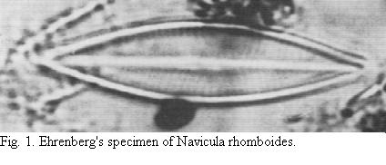
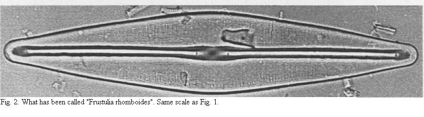
If you suspect that something went wrong in the identification of a species, taxonomy can become a thrilling detective story. It starts with a lot of pondering and careful literature research and has to end up with searching for and examining the type. I can well remember the first time I lay my hands on one, an irreplaceable slide from 1853. It scared the hell out of me and I put a pillow on the bench to prevent breaking itIn a lovely bit of sleuthing, two redoubtable taxonomists, Lange-Bertalot and Jahn examined Frustulia rhomboides. Ehrenberg described Navicula rhomboides, which de Toni transferred to the genus Frustulia, so Lange-Bertalot and Jahn dug up Ehrenbergs original stuff of that diatom and for the first time in about 150 years (!) actually looked at it. The result was shattering: although Ehrenbergs preparation (its not a slide but a sort of cell) does not permit good imaging, you need not be a specialist to see that his diatom (Fig. 1) is not in any way similar to what we now call Frustulia rhomboides (Fig. 2). Lange-Bertalot and Jahn showed that everybody had uncritically copied previous writers. I have found similar such cases and if you have a slightly iconoclastic streak, I can assure you its really cool if you can prove that authorities have goofed. The everyday equivalent would be that seagrasses have been claimed to grow on Scottish crags (spurious ecology) and heffalumps are said to roam Flanders Fields (spurious biogeography). The correct name is now Frustulia saxonica Rabenhorst.
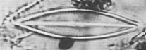
Fig. 1. Ehrenberg's specimen of Navicula rhomboides.
Length 40 microns. Click on image for larger view.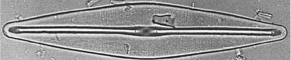
Fig.2. What has been called "Frustulia rhomboides". Length 100 microns.
Same scale as Fig.1. Click on image for larger view.
True images
The structure of diatoms has been another bone of contention. The dots of Pleurosigma angulatum, for instance, have been interpreted as holes but also as thickenings of the valve and in Edwardian times there was quite some feuding about this. Even now, you see descriptions as pits or as hexagons.
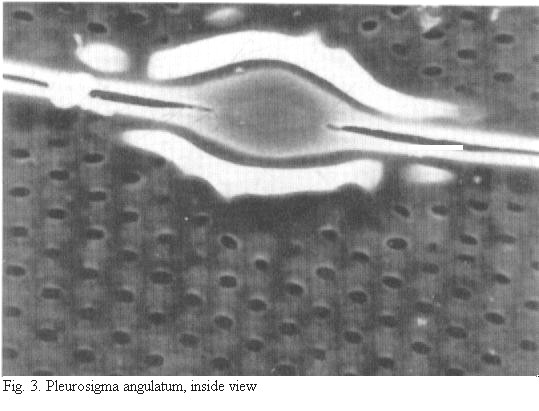
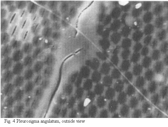
Really good images of diatoms only became possible with the scanning electron microscope (SEM). In SEM, the valve of Pleurosigma species turns out to be a sandwich. There is an internal layer with roundish pores (Fig. 3) that are closed off by a thin sieve-membrane in the living plant, and an external layer with tiny fissures (Fig. 4). The layers are joined by an internal framework.
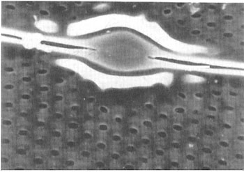
Fig.3. Pleurosigma angulatum, inside view.
Click on image for larger view.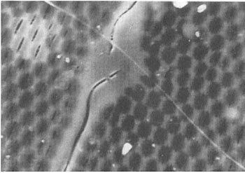
Fig.4. Pleurosigma angulatum, outside view.
Click on image for larger view.That this actual structure does not correspond to previous interpretations of the light-microscope (LM) image is because the size of these structures is simply beyond the LM. The LM can separate (resolve) two tiny details if they are about 0.2 microns apart, but it cannot show the shape of these details. All you get is a rough equivalent of reality. When the light passes through the internal layer of a Pleurosigma valve, it is diffracted and when it passes through the outer layer its again diffracted. The end result is a scramble and its surprising that this results in a crisp LM image at all!
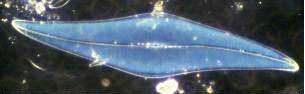
Fig.5. Pleurosigma angulatum, darkfield 20/0.46 objective.
Length 280 microns.Because of their regular structure, Pleurosigma valves are indeed impressive diffraction gratings, as is evident from their colours with darkfield illumination (Fig. 5). But also because of this regularity, you should be aware that the picture in the LM is a physical-optical construct rather than the true image!
All comments to the author Frithjof Sterrenburg are welcomed.
Acknowledgement:
Horst Lange-Bertalot and Regine Jahn gave permission to use photomicrographs from: Lange-Bertalot , H.and R. Jahn (2000): On the identity of Navicula (Frustulia) rhomboides and Frustulia saxonica (Bacillariophyceae). Syst. Geogr. Pl. 70, 255-261.
The quality of the originals is better than youll see on screen.