|
Science as Art
by Howard Webb (St. Louis,
MO, USA)
|
Background
One of the joys of photomicroscopy is the beauty
of what we see within the small structures of the creation. At times the most
accurate (or content-full) image is less than artistic, and sometimes we relax
the scientific discipline for the sake of a good image. Occasionally the science
and art come together in something that we find truly amazing. Several things
came together recently to give me such a moment.
I had put half a dozen Daphnia pulex in a watch
glass, intending to sort some out for observation; but got busy and did not
return to them till the next day. While checking them under a dissecting microscope
I noticed several flecks of debris, which turned out to be molted exo-skeletons.
Not only were all of the normally hidden appendages exposed on these skeletons,
but the shell was so thin, it easily compressed. Here were all the identification
details in perfect condition, requiring neither the patience of waiting for
a live daphnia to properly pose, nor having to smash and dissect the creature
(with the remaining parts looking a bit worse for the treatment).
Images
|
 |
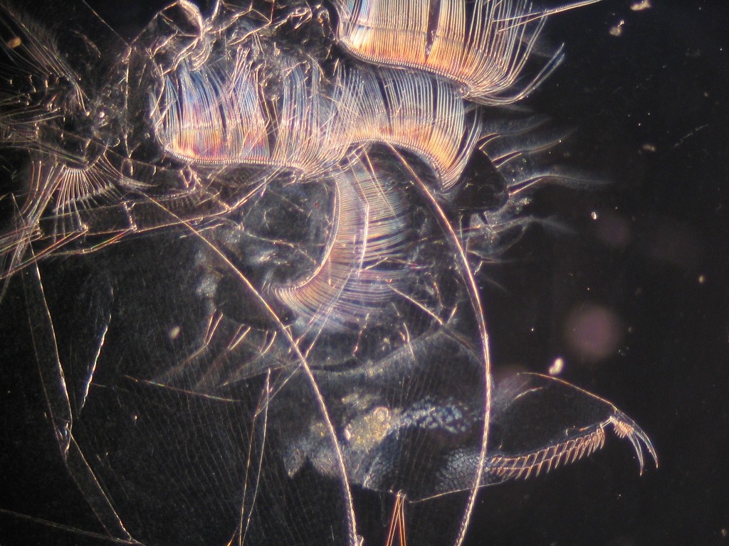
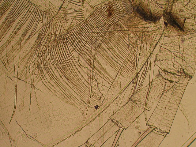
Daphnia pulex exo-skeleton, carapace and appendages
40x darkfield
reduced to 640x480
|
|
 |
|
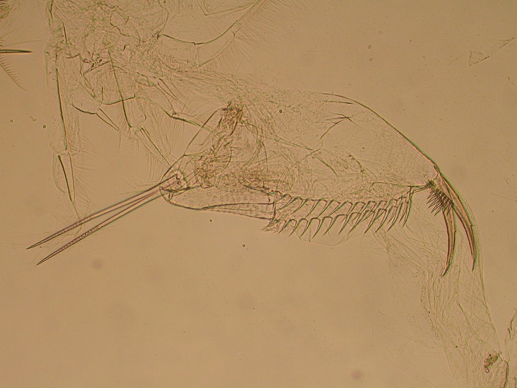
Daphnia pulex post-abdominal process
40x brightfield
reduced to 640x480
click on image for 1024x768 image |
|
|
|
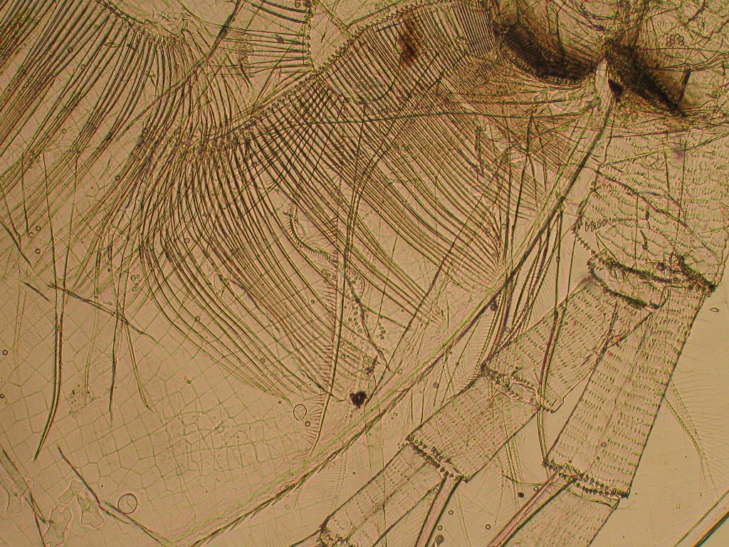
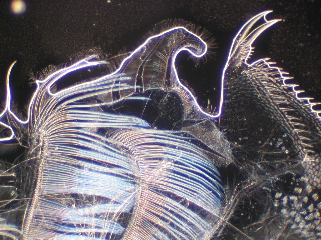
Daphnia pulex appendages and antenna
40x brightfield
reduced to 640x480
click on image for 1024x768 image |
|
|
|
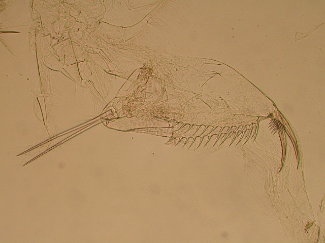
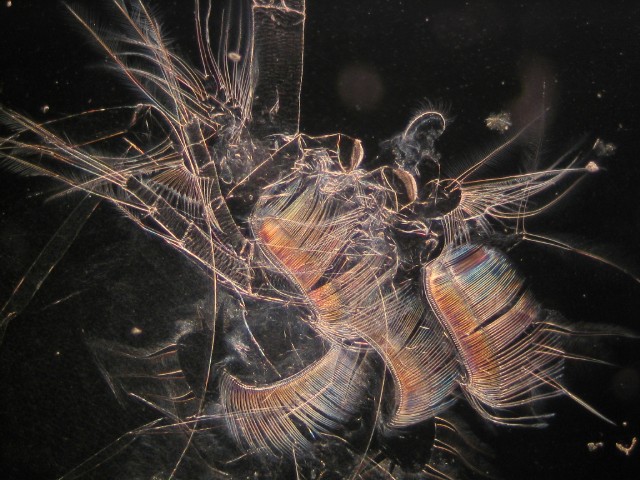
Daphnia pulex appendages and post-abdominal claw
Note: the neon effect is the water meniscus, the wet mount started to evaporaten due to the lamp heat.
40x darkfield
reduced to 640x480
click on image for 1024x768 image |
|
|
|
Daphnia pulex appendages
40x darkfield
reduced to 640x480
click on image for 1024x768 image |
Daphnia Anatomy Notes
It is helpful, when looking at a daphnia skeleton,
to remember that they are small distant relatives of lobsters - a shell with
legs and antenna attached. The daphnia has a head shell, which smoothly joins
the carapace, which attaches at the back of the 'neck'. The body hangs from
inside this carapace. The thorax has 4 to 6 pairs or swimming legs, which filter
food particles and move them toward the mouth. The body ends in the post-abdominal
claw. This claw is used to clean the thoracic legs, and is occasionally used
for movement.
When daphnia molt, they usually split at the
back of the 'head' into two parts. The 'head shield' or 'helmet' separates from
the back clam-shell like carapace, while the antenna and other body appendages
remain attached to the carapace. Occasionally these appendages remain within
the carapace, but usually they are suspended outside.
The long swimming secondary antenna are easily
identifiable, as they look the same as on a live daphnia. The post-abdominal
process retains its characteristic shape, noted by the prominent 'claw'. The
rest is mostly various other swimming appendages, prominent for all of their
fine filtering hairs. The two dark object are grinding surfaces of the mouth.
Image Note
Images were shot with the camera operated through
the computer. The 10x objective was used (100x microscope magnification), with
3x optical zoom on the camera. The camera flash and auto-focus light were off,
but all other settings were set to 'automatic'.
The original images were shot in large-fine mode (2048x1536 pixel).
To avoid sending 3 meg files over the web, the images have been reduced.
Darkfield
The (color) images follow standard darkfield
procedures.
Light Field
The light-field images ('monochrome') follow
standard procedures, but the light diaphragm was stopped way down. The shells
are so thin, that the light must be greatly reduced for any details to be visible.
Reducing the light to these low levels shifts the background color (from white
to sepia/brown); there has been no post-processing manipulation of the images.
Comments to the author Howard
Webb are welcomed.
Technical Details
Microscope: Bausch & Lomb monocular,
10x ocular, 4x, 10x and 40x Nikon objectives.
Camera: Canon A70
References
Daphnia
paleolimnology - daphnia skeleton (parts), extracted from lake sediments,
are often used to identify species of past populations.
Microscopy
UK Front Page
Micscape
Magazine
Article
Library
codebase="./" code="email.class" name="emailer" archive="email.jar" align="bottom" height="2" width="2">©
Microscopy UK or
their contributors.
Published in the
July 2004 edition of Micscape Magazine.
Please
report any
Web problems or offer general comments to the Micscape
Editor,
via
the contact on current Micscape Index.
Micscape
is the
on-line monthly magazine of the Microscopy UK web
site
at http://www.microscopy-uk.org.uk/