 |
| Fungus
and delamination in optics The problem and possible cures By Paul James (uk) |
I thought a few notes covering this potential problem would not go amiss following up on last month's article relating to physically damaged optics.
The initial reaction when discovering that your favourite objective has the dreaded delamination problem and or fungal growth within the optic body, is understandably going to be one of mild distress. Those observers using phase 'scopes are quite familar with the internal state of their objectives, since scrutinising the internal optical alignment of the annuli is required as a matter of phase contrast procedure. The majority of other observers who use brightfield etc., might only peep into the internal optical assembly of an objective using a loupe, and whilst this simple method has some merits it is subject to erroneous interpretations. The phase telescope can reveal the internal state of all the elements involved in objectives, and though this might seem like a great idea for those without one, its use for solely this purpose can cause unnecessary concern. This article has been written to allay some of the understandable feelings and also myths that have arisen through the microscopist community over the years.
An example of the problem
|
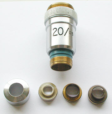
I use as a typical example of both fungal growth and *delamination discovered in a Vickers x20 objective whose imagery was definitely impaired and lacking in normal contrast. Discovering the fungal growth was not too surprising however as this specimen is about 40 years old. To younger generations that might seem like a lifetime! However, many fine optics can be found which are twice as old and are still perfectly sound. I once owned about a dozen old brass bodied objectives which were around 100 years old at the time and not one of them sported fungal growth or showed any sign of delamination. There is some credence to the notion however that certain manufacturers have issued objectives at various periods which have been definitely subject to delamination in the ensuing years. *( the process of the separation of glass elements bonded together with balsam cement. Usually the balsam dries or changes at the periphery of the lens combination and light transmission through this delaminated area is impaired ) .
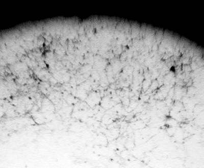
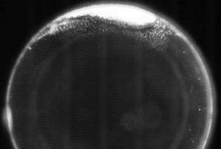
Phase telescope images of the Vickers objective
The first image below shows a portion of the internal surface of a glass element supporting fungal growth. In fact the whole surface was covered which accounted for the lowish contrast of this objective's imagery.
 |
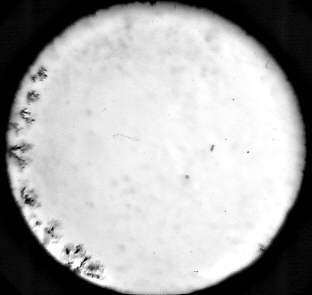 |
The 2nd image above shows the phase telescope's image of the peripheral boundary of two cemented elements which are in the process of delaminating. (The fungal growth can be seen blurred in the background).
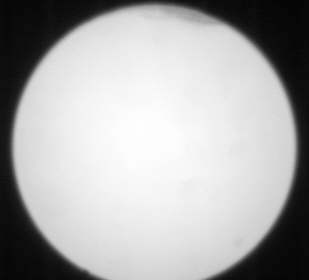
The two images below show some marginal delamination in a Wild achromat x40 objective. Top image is in brightfield and darkfield below. In this example the effects of this fault are not noticed in actual use, and providing the separation of the two elements remains static, which I believe it will, the objective will still project excellent imagery.
 |
 |
Fungal growths
As you might imagine the effect of fungal growth is to disperse light off axis and reduce contrast. In this example the scale of the growth was gross and it was apparent in the imagery. Fortunately fungal growths can be easily removed by simple and careful cleaning of the surface of the offended optic, but getting to it is the real problem, and it is a fact that many modern objectives are not easily disassembled. The Vickers shown above fortunately is constructed in a simple and straightforward way which requires no special tools to extract each housing from the main body. In fact the chromed bezel cap shown at left above traps and holds all the brass cages in place and as you can see is easily removed. Their position and orientation should be sketched on paper so reassembly will be straightforward and without problems.
Removing fungal growths
Most fungal growths that I have come across are easily swept away with the help of cotton buds and occasional use of lens cleaning fluid. When cleaning an optic in a brass cage I'd advise anyone to avoid lens cleaning fluids if they can because some of the fluid will inevitably percolate into the tiny gap between the metal and glass, which is undesirable in the long term. Since spores will inevitably remain somewhere inside after reassembly it is likely that the fungus will develop again. However, working in a warm dry environment whilst tackling the whole job might reduce the likelihood of renewed growth, by reducing the water vapour inside the optic's assembly.
Delamination......... Cures ??
This is a far more serious problem than fungal growth because 'major surgery' is required. By that I mean that the offending pair of elements that sandwich the balsam fault must be removed from the brass cage and then separated by warming to soften the balsam. The latter then has to be dissolved off the glass surfaces with appropriate solvent and then re balsamed together and finally placed into the cage etc.. That this is deemed an easily accomplished task is a fallacy, though the skills of re balsaming without dust or air inclusions are possessed by many with care, the vitally important remounting in the cage with both axial and radial precision without the use of reflective aligment in a spinning mandrel renders the task as being a hit or miss affair.
Delaminations, if peripheral as they usually are, are best left in my opinion since brightfield microscopy usually occasions substage iris closure anyway, so why bother chancing a precision repair in the first instance? Very often I believe that the skill in rectifying a problem is knowing when to leave well alone..............and some delamination faults must surely come into this category ?
I have rebalsamed a pair of lenses in a condenser and also a large format photographic lens with success. However they were relatively large in comparison to a microscope objective's glassware, and therefore an altogether easier proposition.
Cleaning glass
The art of cleaning glass surfaces has been documented before in Micscape, so I shan't make any comments here save to stress that grease left on any glass surface will have infinitely more image ruining properties than a typical sprinkling of fine dust that a normal homestead will provide! Dust appears to be instrumental in our almost manic desire to have clean surfaces on lenses of all kinds, and will always lead to unnecessary cleaning. It's often the cleaning that causes more problems than the dust, and it is so easy to introduce greasy streaks upon those tiny elements found inside a typical microscope objective.
Having removed the fungal growth and decided that the delamination was something that could be left, I reassembled the caged elements. I find the very best way of assessing the amount of dust ( and greasy marks ) on a lens surface is to view the lens against a bright light source, by orientating both the lens and eye such that a darkground effect ensures :-
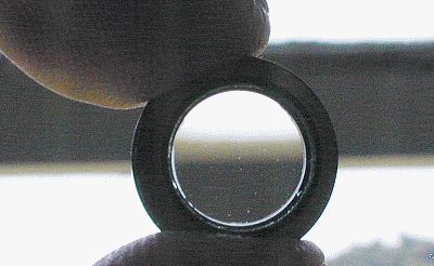 |
This is an extremely sensitive method of dust and grease smear detection and is even more effective when using bright small sources such as from halogen units. The main concern here is to be practical and rational about it. There is no point in "scrubbing" away to try to remove the final imperceptible specks and leave a number of lighting cleaning marks instead !!! A few specks will be totally inconsequential in their effects on imaging, so that when assembled the lenses might be marginally more 'dusty' internally but fungus free.
CONCLUSION
Despite the appearance of a suspect objective's internal glassware through the phase telescope, the critical issue with delamination and fungal growth is whether the job of trying to remove or cure the problem is a sensible one given the risks involved. If the lens is a fairly old one of moderate to low power and of conventional construction then the chance of its correct reassembly after removal of the fungus will be high, and with care worth the risk. High power apochromats with correction collars and the like are a different breed altogether, and should never been tampered with. Their intrinsic mechanical tolerances of construction are extremely high and are realistically beyond the scope of kitchen table surgery.
"Leave well alone" is a phrase that comes to mind on this subject, and if your objective still performs quite well despite a little peripheral delamination, then do leave it alone.
| All comments welcome by the author Paul James |
Microscopy
UK Front Page
Micscape
Magazine
Article
Library
Please report any Web problems or offer general comments to the Micscape Editor.
Micscape is the on-line monthly magazine of the Microscopy
UK web
site at Microscopy-UK