IMPROVING THE PERFORMANCE OF THE SINGLE LENS MICROSCOPE
By: Alvaro Amaro de Azevedo - (Brazil)
1. - Introduction
In the early days of microscopy, optical instruments were severely affected by numerous deficiencies such as chromatic and spherical aberrations yielding images plagued by distortions and artifacts due to their primitive optical design. Microscopes were lucky if they could yield about 20 to 30 times magnification. The first pioneers who have developed their research using early versions of microscopes were Robert Hooke (1635 – 1703), Marcello Malpighi (1628 – 1694), Jan Swammerdam (1637 – 1680) and Antony van Leeuwenhoek (1632 – 1750) to mention some of them. However for the mainstream of the new born scientific community, microscopes were not taken seriously as they were regarded nothing but mere toys. Only after the aberrations of optical systems started to be solved around the first decades of the nineteenth century, the microscope would become widespread among a variety of fields ranging from biology to metallurgy. Simple microscopes were a suitable option for microscopists of early times as their image were generally superior due to their simpler design. Although compound microscopes have undergone a remarkable evolution reaching a practically perfect performance, the same didn’t seem to have happened with simple microscopes as they have being gradually abandoned ever since the compound pieces have surpassed them in quality near two centuries ago.
2. – Limiting Factors of Simple Microscopes
My aim was to find out if it is possible, by developing the optics of the simple microscope, to achieve a level of performance comparable with that from modern day compound microscopes. I started from scratch, and by making an extensive research throughout the web, I found several useful sources of information. My first reference was "The Glass Sphere Microscope" from Giorgio Carboni. He gives a comprehensive introduction to this issue teaching how glass spheres can be made and used to create simple microscopes. I spent more than a year replicating and improving the design of this instrument. In a further reading of the patent 'Lenses and Uses Including Microscopes', the authors discuss the use of an algorithm to calculate lens apertures in order to optimize the resolution. They give a table where a list of ball lenses varying from 0.5 mm up to 9.0 mm are related to their optimal aperture and the corresponding resolution in microns. One can figure out that ball lenses when properly optimized with the right lens stop aperture can yield highly resolved images up to 250 times magnification. Above this, the balls have to be smaller and smaller and when the lens stop is added, the resulting image will be affected by diffraction due to the minute dimensions of the pin hole used as aperture. So, although greater magnifications can be achieved, no further increase of resolution will be possible limiting the efficacy of the simple microscope. In other words, as the magnification power surpasses 250 times there will be no further increase in the ability of the lens to supply more information about finer structures and this is called empty magnification. Ball lenses have a geometry that doesn’t permit them to be combined forming compound systems so I couldn’t go any further using this type of lenses.
In September 2005 I was given a copy of a paper named “The Microscopes from van Leeuwenhoek” by J. van Zuylen and this was a turning point in my homemade research and development of the simple microscope as from this point on, I could work with lenses more suitable for multiple elements systems. This article proposes how a miniature lens can be made using non-sophisticated hardware and techniques. The author himself made some lenses using this technique that according to him might be the same that Leeuwenhoek employed to make his own microscopes. In my previous article "The Challenge of Grinding Lenses for Single Lens Microscopes" I describe the process that has been used to produce small lenses. Ground lenses can perform as good as ball lens counterparts in the range of 50 to 300 times magnification however, smaller lenses seem to be affected by diffractive interference that reduces the resolution. So, if one would want to improve the resolution for obtaining higher magnification, we would have to abandon the single element design.
Ground lenses have an advantage however. They can be combined to create a compound lens system. I was given a copy of an article by Roger Baker entitled "The Homemade Microscope"* printed in 1991. In this article, the author makes an interesting conjecture stating that Leeuwenhoek’s microscopes might have had compound lenses to increase the resolution. He describes how he did manage to build his own model and gives all necessary details to those who want to replicate it. In fact, he describes an array of lenses that is called a Wollaston doublet, invented in 1812 by the notable Englishman William Hyde Wollaston. This doublet lens is comprised of a stack of two plano-convex lenses having their both flat sides facing the subject. The lens closer to the eye is larger and has longer focal length. Another characteristic of this array is that it gives a wider angle of view rather than the narrow field yielded from a single element lens By combining two small plano-convex lenses each having about 1.0 mm focal length, I was able to craft a Wollaston doublet, see pictures 06 and 07.
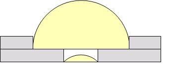
Figure 01 - Wollaston Double
The illustration represents the constructive aspect of the combined lens in the metallic frame. The performance of this doublet at higher magnification had superior resolving power than any element based on one single lens previously built. Nevertheless there was still a significant amount of spherical aberration, which made useful only the central part of the view field. The effect could be reasonably minimized by mounting the lenses facing each other’s curved side, see Figure 02.
Figure 02 - Aplanatic Doublet
3. - Experimental Setup
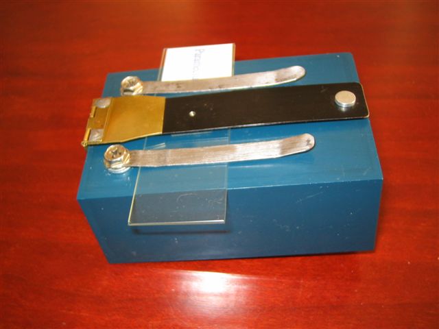
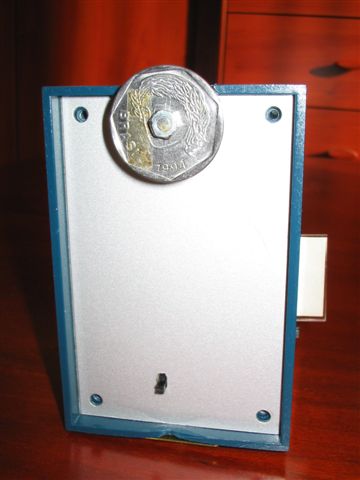
Another microscope was built, this time using a lens frame able to be lifted for the sake of centralizing the subject in the stage (see picture 01 to 05). Despite the versatility of this design, the construction is plagued with some jerkiness during focusing which makes it a little awkward to use.
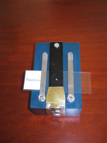
Picture 01 – Top View
Picture
02 – Side View
Picture 03 – Lens Frame Lifted
Picture
04 – Focusing Knob
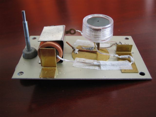
Picture 05 – Internal Illumination Setup
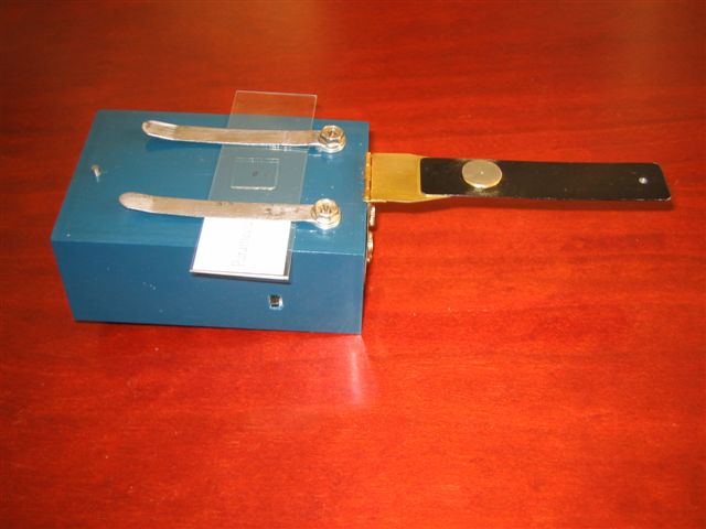
The Lens used in this microscope was mounted as illustrated below
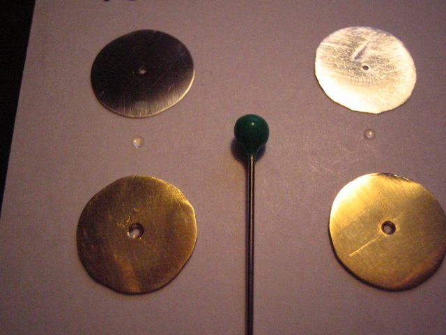
Picture 06 – Two Plano Convex Lenses and Frames
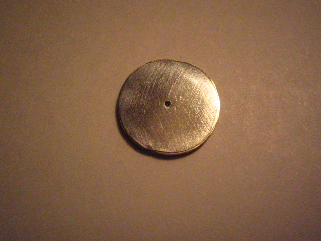
Picture
07 – Lens Once Assembled
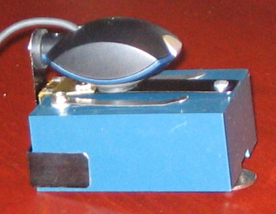
The microscope as showed above was coupled with a webcam so that images could be captured and recorded for further and more detailed observation, see picture 08.
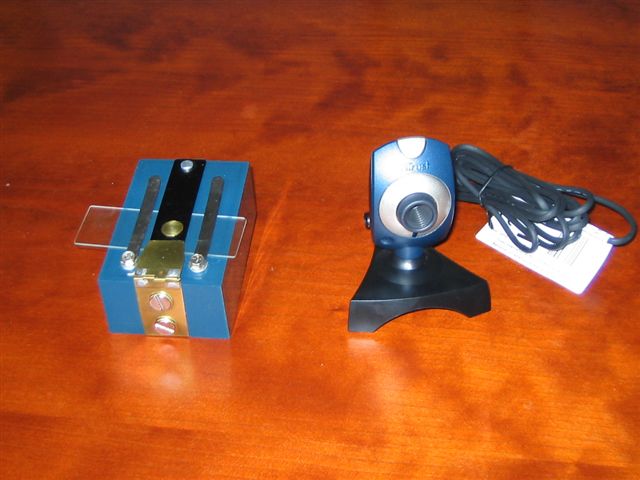
Picture 08 – Microscope and Webcam
Picture 08a – Microscope and Webcam
in use
4. - Final Notes
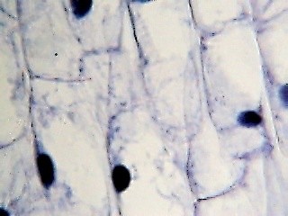
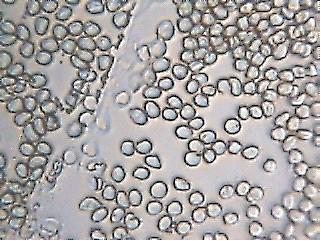
One may notice that the images in the gallery are flatter than my original ones obtained from one element single lens microscope, see comparison below.
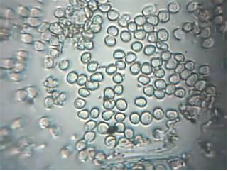
Blood cells image from a single lens Blood
cells image from a doublet lens
I came across a resolution problem. The webcam is very limited when compared with a digital camera that has a much superior number of pixels to resolve the image. It’s still not possible to use a digital camera as this cannot have its lens removed to be coupled with the microscope. However despite that, the images presented in this report indicate that reasonable quality can be obtained from such a crude resource.
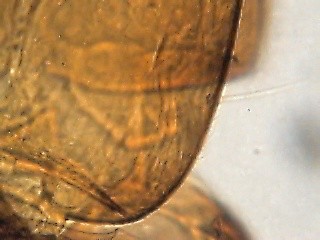
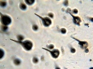
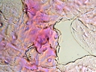
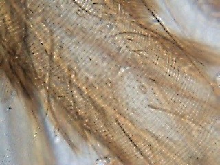
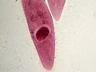
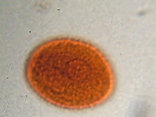
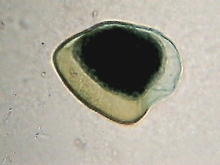
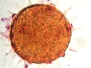
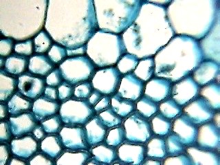
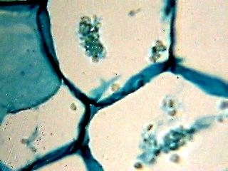
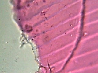
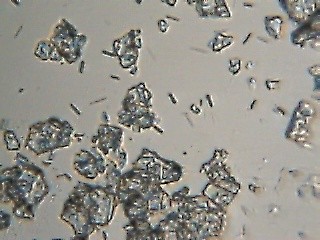
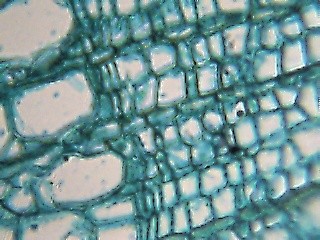
4. - Gallery of Images
|
|
|
|
|
|
|
|
|
|
|
|
|
|
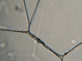
Spider
web silk
5. - Apologies:
For not being able to determine the magnification power as measurement of the focal length was not possible in this setup. The subjects being viewed appear to have the same size as when observed with a compound microscope set at 400 X.
For not being able to identify all the specimens illustrated in some images above despite my attempts.
6.- Other links / references
Comments to the author Alvaro A. de Azevedo are welcomed.
Published in the July
2006 edition of Micscape.
Please
report any Web problems or offer general comments to the
Micscape
Editor.
Micscape is the
on-line monthly magazine of the Microscopy UK
web
site at
Microscopy-UK
© Onview.net Ltd, Microscopy-UK, and all contributors 1995 onwards. All rights reserved. Main site is at www.microscopy-uk.org.uk with full mirror at www.microscopy-uk.net .