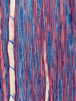
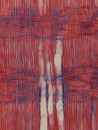
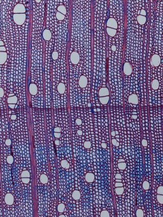
Left to right: tangential, radial and cross (transversal) sections of stained wood Acer (Acer pseudoplatanus),
a hardwood (broadleaved) growing in Europe. The sections were colored with safranin (red color) and Astrablue (blue color).
.
|
Wood under the microscope by Alan Crivellaro, Italy |
What wood is
Wood is the product of the metabolic and physiological activity of woody plants. Because of its function in live plants, wood must be mechanically resistant (it sustains the weight of the crown, leaves, water, wind, snow etc.) and at the same time it must be porous: photosynthesis in the leaves requires water and inorganic substances (sap) to pass through the wood from the ground. Both these functions, mechanical support and sap conduction, are supplied by cells. Wood is composed of cells which are characterized by a solid wall surrounding a lumen. The wood cells are fusiform and about 90% of the cells in the wood have a vertical orientation. The cell wall has a good mechanical resistance to traction and compression, and the cell lumen can be covered by the sap.
How to prepare wood for microscopic observation
Wood must be observed under the optical microscope in very thin slices, called sections. The sections can be easily obtained with a razor blade from wood recently cut out from a tree. If you need to observe the anatomical features of a piece of dry wood it must be boiled. Boiling it in water the cells walls will be softened; the boiling process can be repeated many times for very hard woods. Because of the orientation of the wood cells in the plant’s trunk the sections cannot be cut casually from a wood piece: the surfaces of every wood piece must be oriented following the anatomical wood constitution. It means that three plans of orientation must be detected. The most important is the so called transversal surface, it corresponds to the face of the trunk which you can see at the end of a cut plant. It can be easily recorded in a wood piece because of the visibility of the growth rings. The other two cutting plans for wood orientation are longitudinal: they follow the cells orientation in the trunk. The first type of longitudinal section, the so-called radial one, passes through the pith: the centre of the trunk. The second type, the tangential section does not cross the pith, but it is a tangent of the growth rings. For general observation purposes the wet sections can be placed on a glass slide with a drop of water, covered with the cover glass and observed under the microscope. For specific observation, the sections can be washed and stained.
What you can see
The principal anatomical features of wood can be easily recorded with a magnification of 25 to 50x.
The most important surface for wood observation is the transversal, because of the high number of observable characters. Anyway transversal, radial and tangential sections each have a specific appearance and characters to observe.
How to identify wood
Observing the diagnostic anatomical characters in different wood samples permits wood identification. As the face of every person is composed of two eyes, a nose and a mouth, but each person is different to another, so wood is composed of different kinds of cells but with different arrangements: each type of wood is identifiable from another. Through wood identification it is possible to obtain information about wood properties: it is sufficient to check in a book the characteristics of the identified wood.
Comments to the author, Alan Crivellaro , are welcomed.
Left
to right: tangential, radial and cross (transversal) sections of stained
wood Acer (Acer pseudoplatanus),
a hardwood (broadleaved) growing in Europe. The
sections were colored with safranin (red color) and Astrablue (blue
color).
.
Microscopy UK Front Page
Micscape Magazine
Article Library
Please report any Web problems or offer general comments to the Micscape Editor.
Micscape is the on-line monthly magazine of the Microscopy UK web site at Microscopy-UK.