|
Stentors
by
Howard Webb (St. Louis, MO,
USA)
|
Introduction
Stentors are a unicellular ciliate, noted for their trumpet
like shape (hence the name stentor, after the Greek herald of
the Trojan war). Stentors are one of the largest single celled
organisms, occasionally being several millimeters in length.
Where to Find
Stentors are usually found in the calm water of ponds and
lakes, usually near the surface attached to leaves or twigs.
While they are capable of free swimming, they are most often
noticed clustered together in small colonies. These particular
stentors were found in a cluster attached to the side of a
collecting jar. I had gathered a few twigs from shallow water
(I was actually looking for hydra), and they became apparent
after about a week. I used a small pipette to scrape the
colony from the side of the jar and transfered it to a slide
for observation.
Normally it takes a bit of hunting to find stentors, though I
have on occasion found them so thick that a sample is tinged
green with their presence.
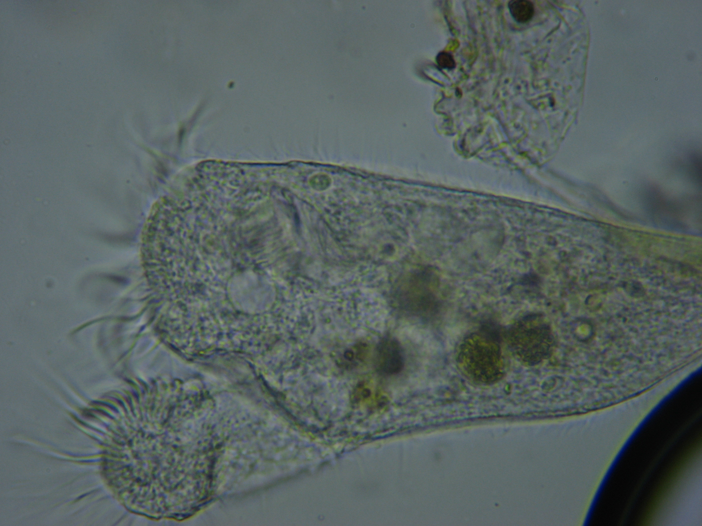
Anatomy
Most notable of the stentor is the 'crown' of cilia
surrounding the trumpet 'bell'. This crown is not a complete
circle. These cilia are used to create a current of water from
which it sweeps food. Every little while, the stentor will
close up the cilia crown and contact, bringing the food within
its cell structure.
The crown is not the only cilia on the stentor, its whole body
is covered with shorter cilia, which are used for locomotion
when free swimming. When moving, the stentor is contracted
into an oval or pear shape.
Being single celled, there are no separate parts which make up a
"mouth" or other organs. For digestion, the cell wall
envelops the food, and separates to form a round bubble like
"vacuole" within the cell. After the nutrition from the
food is extracted, this vacuole moves to the outer cell wall
and 'pops', evacuating the remaining contents. Since
stentors have a cell density higher than the water in which
they live, osmotic pressure causes water to transport into the
cell. The stentor cell actively collects this excess water
into a vacuole, and expels it; thereby maintaining the
internal fluid density.
Behaviour
Stentors, like most ciliates, are filter feeders; passively
eating whatever happens to be swept in their direction. They
normally eat bacteria and algae, though large stentors are
reported to opportunistically eat rotifers or anything else
that they can catch.
Images
|

|
|
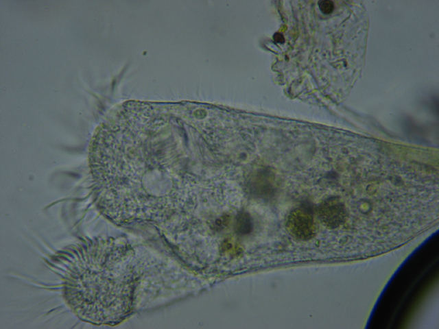
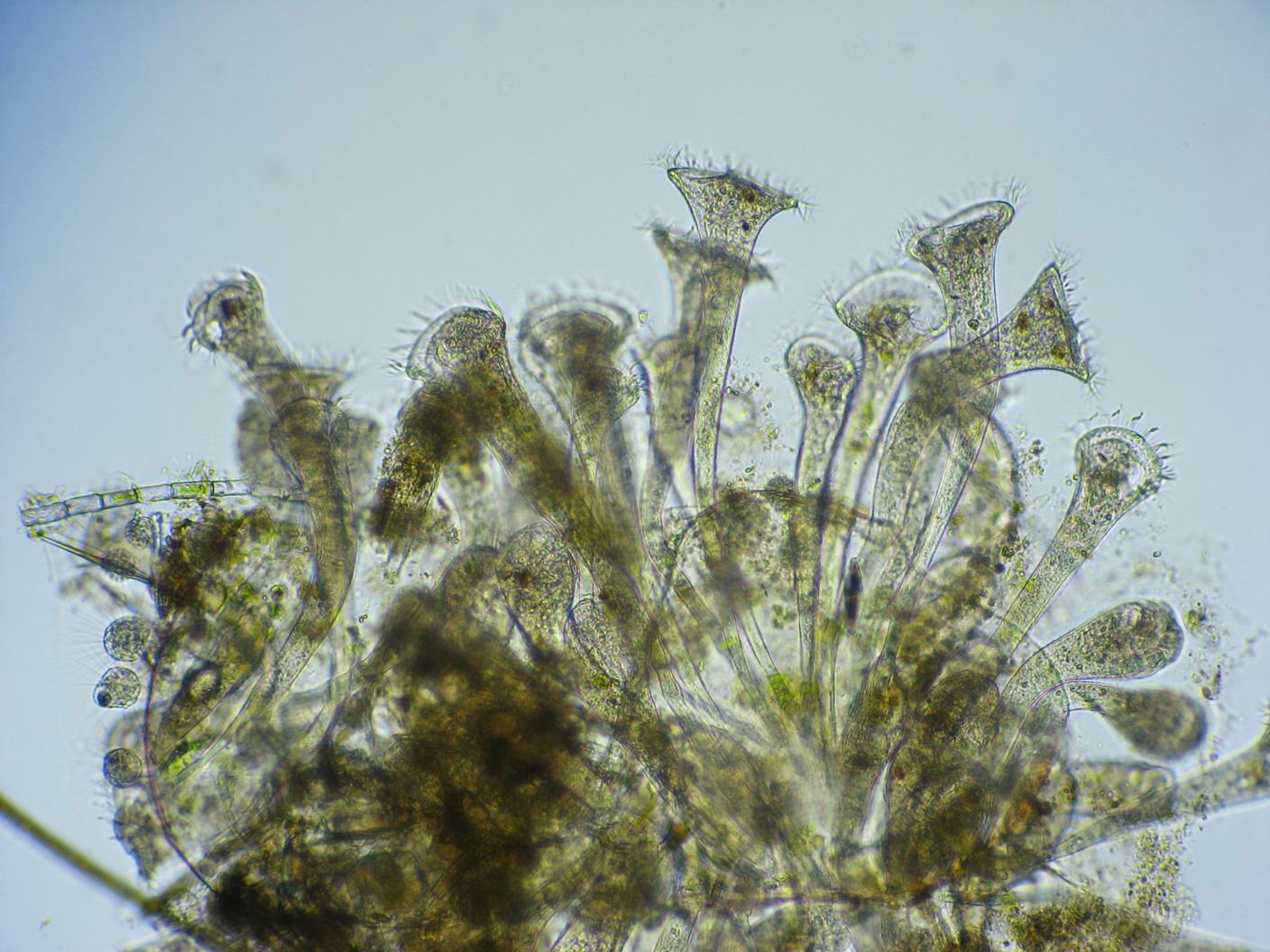
Unlike some vorticella, where the whole colony
is connected as a single organism, a colony of
stentors is a group of individual organisms
which just happen to be located together.
This colony looked like a spot of
mold beginning to form on the side of the
collecting bottle. I pushed it loose with a
pipette, and transferred it to a microscope
slide.
|
Stentors
|
|
|
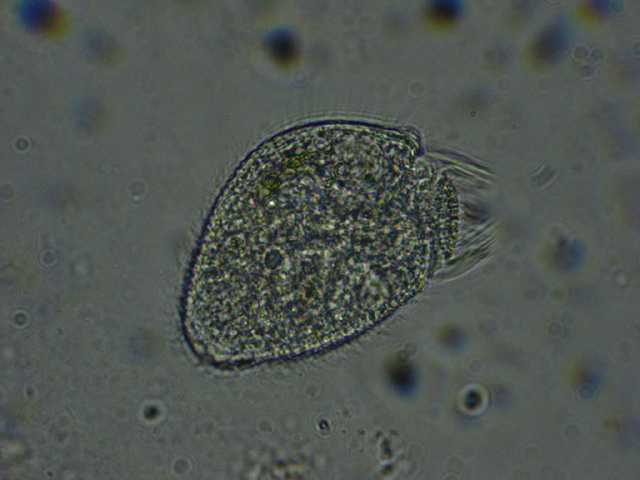
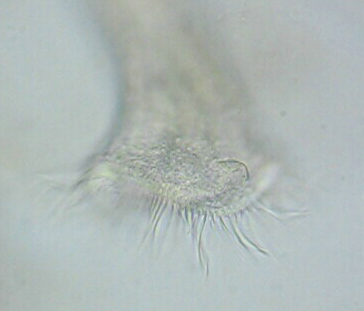
When stentors are
traveling, they are not in the typical trumpet
shape, but contract into a more oval shape.
The cilia on the trumpet bell closes up, and
the cilia on the body are used for locomotion.
The illuminator was moved down to give more
contrast (and also shows up more background
artifacts - compare to the next photo).
|
|
|
|
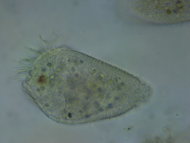
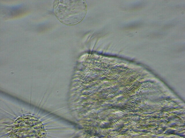
Another stentor in motion.
|
|
|
|
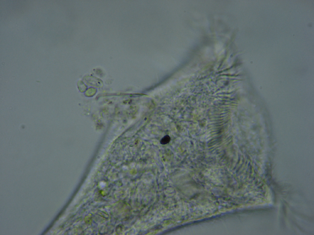
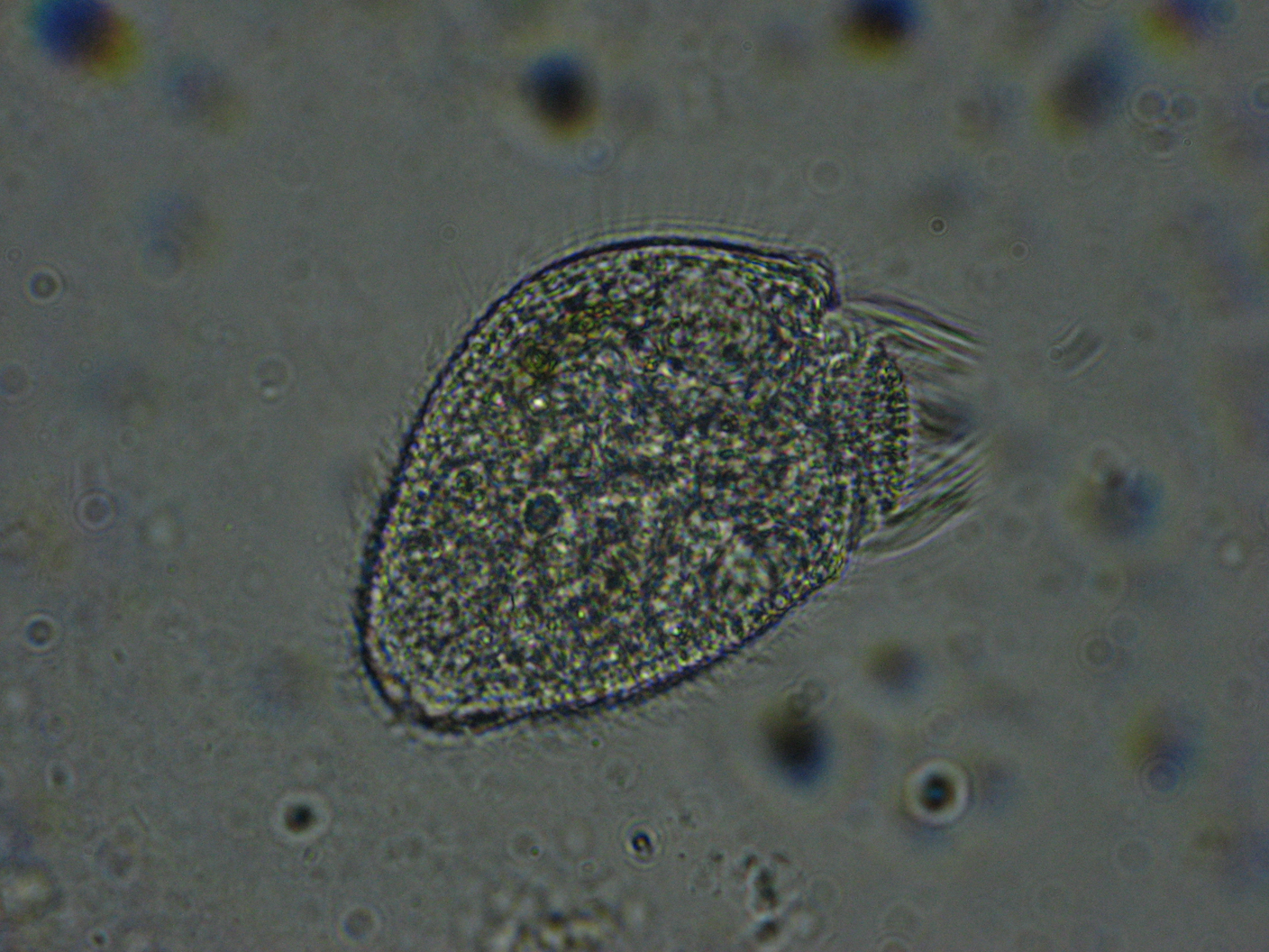
Stentors have a rather complex reproductive
cycle. Here a stentor undergoing division.
|
|
|
|
Food vacuole being evacuated.
|
|
|
|
Movie of the cilia around the bell of the
stentor. The bell is more than a
simple round shape.
|
|
|
|
Movie showing water current produced by cilia,
and contractile vacuole
shrinking.
|
Technical Details
Environmental Conditions:
Water temperature:23C
Depth: collected near the
shore, at less than 6 inches depth
Secchi visibility: 2 meters
Location: quarry at
Whitecliff Park, Crestwood, MO (lat: 38.5561, long:
-90.3688 (NAD83/WGS84)). The park contains an old, abandoned
quarry, which is probably one of the deeper bodies of water in
the area. The quarry is generally closed to the public, though
the city has been kind enough to give me access.
Microscope: Bausch & Lomb monocular, 10x ocular,
4x, 10x and 40x objectives.
Illumination: Luxeon K2 LED
Camera: Canon A540 (6 megapixel)
Software: Photoshop Elements, VirtualDub
References
Vance Tartar, The Biology of
Stentor, Oxford, New York, Pergammon Press,
1961
This is a classic work. Most of the text describes
studies of slicing and dicing stentors, and observing their
regeneration; though there is a thorough coverage of their
anatomy and behavior, as well as culture techniques.
Comments to the author
are welcomed.
© Microscopy UK or their
contributors.
Published in the July 2007
edition of Micscape Magazine.
Please report any Web problems
or offer general comments to the
Micscape
Editor,
via the contact on current Micscape Index.
Micscape is the on-line monthly
magazine of the Microscopy UK web
site at
http://www.microscopy-uk.org.uk/

