The fascinating mold
Empusa (Entomophtora) muscae.
Some data, old and new.
INTRODUCTION
It is widely known that the mold Penicillium notatum, the producer of penicillin, has saved millions of lives. Penicillium is a benefactor of humankind.
The name of the mold Tolypocladium inflatum is hardly known to the world. However, the name of one of its metabolic products, Cyclosporin A, has been known for decades to thousands of patients as a potent immunosuppressant effective in preventing rejection after organ transplantation. Tolypocladium inflatum is also a benefactor of humankind.
We shall now turn to a mold with a far more modest claim to human gratitude than either Penicillium or Tolypocladium, but one that may come readily to the attention of many amateur microscopists almost anywhere. It is a fly-eating mold; it is Empusa muscae, later renamed Entomophtora muscae. Chance may bring it in our very own houses.
THE MOLD THAT FLIES
Our interest in Empusa goes back years. In 1999 (1), one of us wrote:
“When I was a teenager using my first microscope, I noticed a dead fly on the window glass of my “laboratory.” Two facts stimulated my curiosity; one was that the unfortunate critter had remained attached to the glass. The second was that a circle of whitish dust surrounded the body. The fly’s body was covered with a dust-like material, but the material forming the ring around it, whatever it was, did not contact the body directly. Instead, there was a separation of a few millimeters between the body and the ring. I assumed then that the ring could not be an extension of molds feeding on the carcass, but the mystery remained unsolved. But not for long. An antique book dealer offered a copy of L. Trabut’s Précis de Botanique Medicale (2). I bought it. Its price was $5.00 Argentinean pesos, about $2.50 USA in 1948. It was a daring investment for a teenager, but it was worth of it. There, at p. 600 was a long paragraph with the key to the mystery of the white fly.”
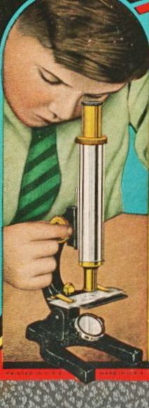
Figure 1. Young man and his microscope.
Figure 2. Frontispiece of Trabut’s book. Pablo Aguilar, likely the first owner of this copy, signed it on March 9, (18)99. The life of this volume expands now over three centuries.
What follows is an extract of Trabut's eloquent description, followed by a loose translation of it:
“Empuse muscae. - Vers la fin de l’été les mouches atteintes par ce parasite meurent avec un abdomen volumineux distendue a surface blanche, farineuse; …. Un examen au microscope du contenue de l’abdomen montre a thalle dissocié, cest-à-dire de cellules arrondies indépendant et burgeonnant.”
[Now comes the punch line.] “Pour producire des spores, chacunne des cellules isolées s’est allongée en un filament qui a percé la peau de la mouche et donné un spore. La spore ainsi formée est projectée a une assez grande distance comme un bouchon de Champagne; …”
[Isn’t this a beautiful description?]
Which translates like: “Towards the end of summer the flies attacked by this parasite die with a voluminous, distended abdomen [covered by] a surface white flour-like. A microscopic examination of the contents of the abdomen reveals a dissociated thallus, that is to say [one] formed by independent, round, budding cells. To produce spores each of those single cells elongate into a filament that pierces the skin of the fly and produces a spore. The spore formed this way is projected at quite a large distance as the cork of a champagne bottle.”
Next we will review Empusa information available after that provided by Trabut in 1898.
Chronologically, our next stop is a 1951 book by Clyde Christensen (3). Christensen provides information on Empusa under the subtitle “Fungus Parasites of Insects.” Reading Christensen one becomes immediately aware of the considerable amount of information that was gathered in the half a century that elapsed between the publication of this book and that of Trabut’s. When Christensen wrote his book it was known that the spores that form a halo around the fly carcass are endowed with very peculiar survival properties. If they are able to directly infect another fly their objective has been reached and the cycle continues. However, should infection of a new fly have not taken place within some time, then the spores germinate on whatever surface they have landed and produce a short stalk that produces a second generation of spores. Should those new spores also fail to infect a new fly, then they also form a spore-producing stalk. These tertiary spores represent the last chance for Empusa to propagate. Presumably by then all the nutritional reserves are used and the tertiary spores face the stark choice of finding a host or dying. The story sounds attractive; but it is true? Regardless, Christensen observes that the effect of Empusa infestation in reducing the fly populations is limited.
Contemporary with Christensen’s book was the publication of the second, posthumous edition of Henrici’s book (4), a classic of the mycological literature. Henrici gives a description of the disease that goes no further than Trabut’s, but he also provides a microscopic illustration of the infected tissue, and a photograph of a dead, infected fly. The illustrations are reproduced below as figures 3 and 4.
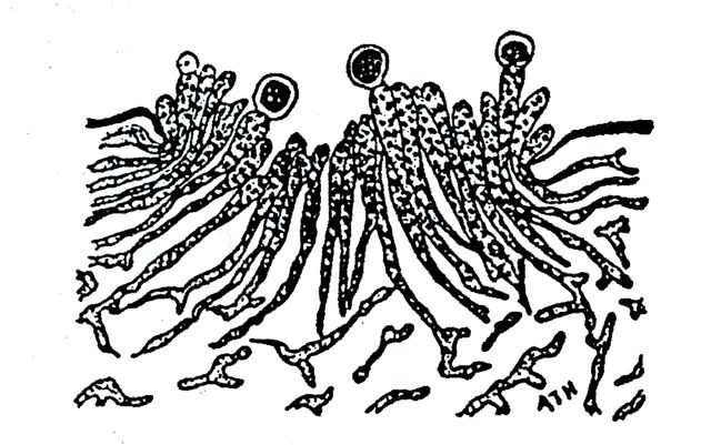
Figure 3. A camera lucida drawing by Henrici showing a stained section through the body wall of a fly infected with Empusa muscae. The preparation shows clearly the development of the conidiospores, the spore-producing filaments, and the conidia, or spores. The thick lines at both sides near the top of the illustration represent the body surface of the infected fly. It is easy to appreciate that most of the infecting fungus grows inside the body.
Copied from Reference (4).
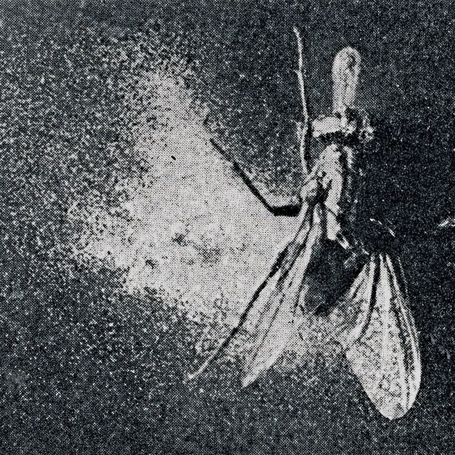
Figure 4. Henrici’s photograph of a fly dead as a result of Empusa’s infection. The picture is one a few available in the classic literature illustrating the end point of the infection. It presents however, an atypical example of asymmetrical distribution of the spores, instead of the usual ring-like formation surrounding the body without contacting it.
Our final book reference will be one published exactly a century after Trabut’s (5). This book written by George Hudler became highly popular introduction to mycology. It summarizes in a vivid, highly readable style, the knowledge on Entomophthera muscae, as he calls it, following modern nomenclature.
Taken together, this century-long accumulation of knowledge describes Empusa as a fly killer; it agrees on the fact that late summer or early autumn is the time of the year when the action of Empusa becomes evident. The authors also identify related species of molds that attack other insects, such as aphids, grasshoppers, crickets, and others.
Of course, no search of bibliographic sources would be complete these days without an on-line search, even a brief one. Not unexpectedly, such search provided additional information, both from the present days and vintage. Of the latter a most informative is a 1923 article by Bessie Goldstein (6) that refers to the first study of Empusa by Cohn in 1885. Cohn provided an uncommon observation of the last minutes in the lives of infected flies. These died in convulsions (“unter schweren Kämfen”).
Most articles from the recent literature are too technical for discussion here, but we shall make an exception with that by Six and Mulliens (7). In studying E. muscae infestation in California dairies, they confirmed with numerical data the old observation that infection is an autumnal event, September-November in California. They also correlated the infection level with the weekly temperature changes. Remarkably, they documented a site where “infection at a site exceeded 70%.” Bravo for California!
Bibliographic research is both a source of information and an intellectual pleasure, but as observational microscopists we wish to see the object under consideration, not only to read about it. Thus, we present in the following four illustrations from a commercially obtained slide of Empusa (figs. 5-7).
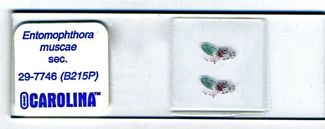
Figure 5. Two consecutive sections of a fly infected with E. muscae. Paraffin embedding. Trichromic stain.
The abdomen is filled with a green-staining material. The next two pictures show details of the abdominal infestation.
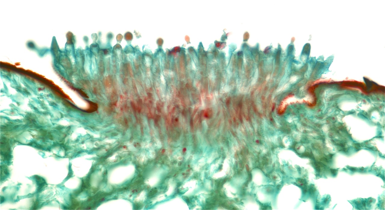
Figure 6. Photomicrograph of spore-forming hyphae erupting through the abdominal cuticle. Compare with Henrici’s drawing in figure 3. Field width 550 µm.
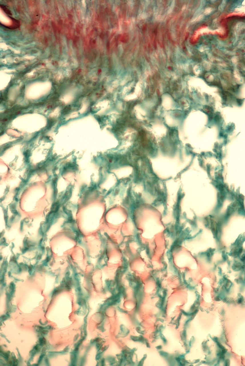
Figure 7. Vertical view of the field shown in figure 6. The pink walled, empty structures seen at the bottom half of the field are the fly’s trachea, or air-conducting ducts; they are immersed in a mass of fungal hyphae. Field height 550 µm.
The time of this writing is months away from the Empusa season in the Northern hemisphere. We hope that some of us will be successful this coming fall in spotting infected flies, and in seeing Empusa under the microscope.
Good hunting!
Comments to the authors are welcomed.
REFERENCES
(1) del Cerro, Manuel (1999) 5. The Amateur Microscopist. Journal of the Microscope Historical Society, 7: 19-22.
(2) Trabut, L. (1898) Précis de Botanique Médicale. Paris, Masson et Cie., 739 pp.
(3) Christensen, Clyde M. (1951) The Molds and Man. An Introduction to the Fungi. Minnesota University Press, pp. 157-159.
(4) Skinner, Ch. E., Chester W. Emmons, and Henry M. Tsuchiya (1947) Henrici’s Molds, Yeasts, and Actinomycetes.
A Handbook for Students of Bacteriology. John Wiley & Sons, New York, p. 82.
(5) Hudler, George W. (1998) Magical Mushroom, Mischievous Molds. Princeton University Press. Princeton, NJ, pp 210-211.
(6) Goldstein, Bessie (1923) Resting spores of Empusa Muscae. Bulletin of the Torrey Botanical Club, 317-328.
(7) Six, Diana L. and Bradley A. Mullens (1996) Seasonal Prevalence of Entomophthora muscae and Introduction of Entomophthora schizophorae (Zygomycotina: Entomophthorales) in Musca domestica (Diptera: Muscidae) Populations on California Dairies. Biological Control, 6: 315-323.
Published in the July 2008 edition of Micscape.
Please report any Web problems or offer general comments to the Micscape Editor .
Micscape is the on-line monthly magazine of the Microscopy UK web site at Microscopy-UK
© Onview.net Ltd, Microscopy-UK, and all contributors 1995
onwards. All rights reserved.
Main site is at
www.microscopy-uk.org.uk
with full mirror
at
www.microscopy-uk.net
.