|
|
A
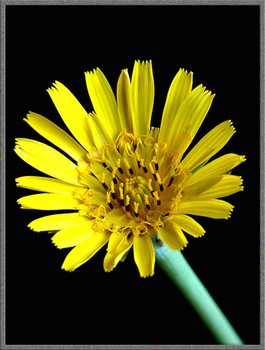
Close-up View of the Wildflower (Tragopogon dubius) |
|
|
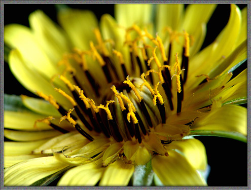
A
Close-up View of the Wildflower (Tragopogon dubius) |
A
casual glance at this bright yellow wildflower might leave the observer
with the impression that it is a dandelion on steroids! It is in
fact a member of the same family, (aster), but there are many
differences that contribute to Yellow Goats-Beards uniqueness.
The most obvious difference is
size. Goats-Beard can grow up to a metre in height, and the
single composite flower-head
containing both ray and disk flowers can have a diameter up
to seven centimetres. This large flower-head opens and twists
slightly towards the sun each morning. In the early afternoon,
the flower-head closes again. The English poet, dramatist and
essayist Abraham Cowley (1618 - 1667) wrote the following about the
Goats-Beard plant.
The propensity for early closing
resulted in old country names of Noon-flower and
Jack-go-to-bed-at-noon being given to the plant. The genus name
Tragopogon
comes from the Greek words tragos, meaning
goat, and pogon
meaning beard. This is in reference to the seed-head of the
plant (which will be shown later). The species name dubius can be
translated as doubtful, and may refer to the fact that, due to
hybridization with other similar species, identification may be
uncertain.
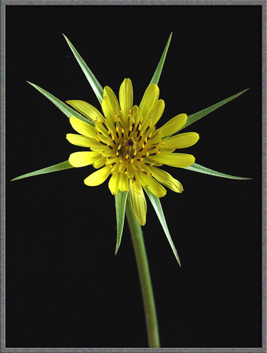
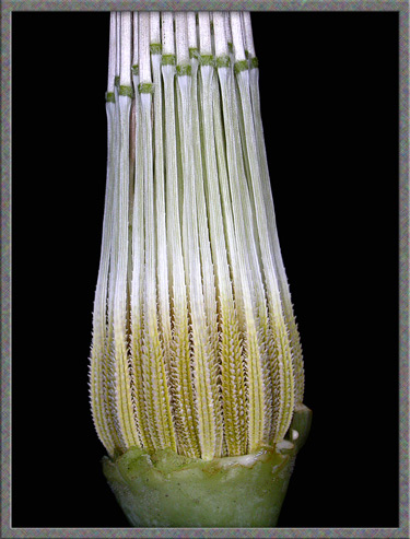
One of the most important
characteristics of Yellow Goats-Beard is the ring of sharply pointed
modified leaves called bracts
which are arranged in a radial pattern beneath the flower-head.
In this species these bracts must be longer than the petals. The
first photograph in the article, and the two images below show these
bracts. Note that the number of bracts is variable, but most
plants have about ten.
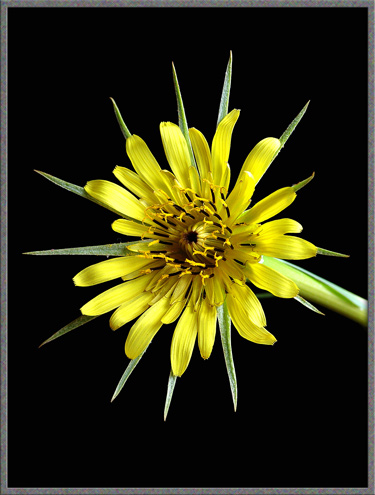
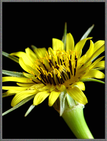
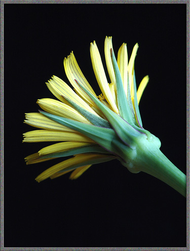
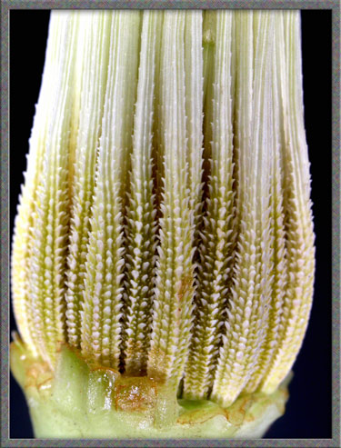
A swollen growth at the base of the
flower-head is another characteristic of Yellow Goats-Beard. It
can be seen in the right-hand image above.
The first flowers to bloom in the
composite head are the outer ray flowers to which the yellow petals,
called ligules, belong.
The pale green, yellow tipped columns at the centre of the head are
immature stamens.
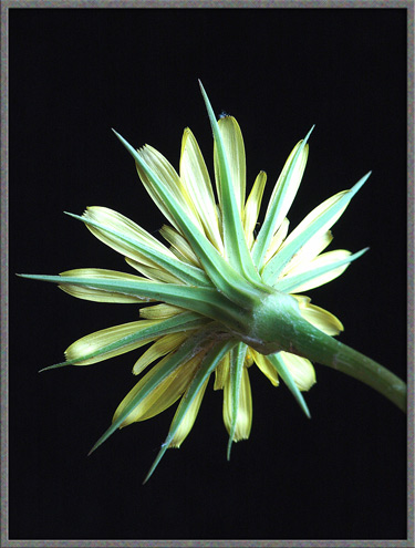
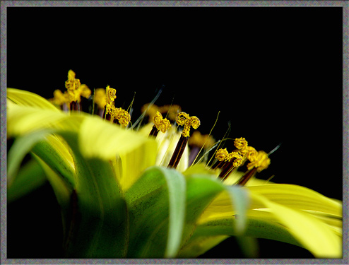
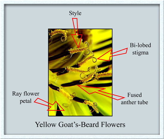
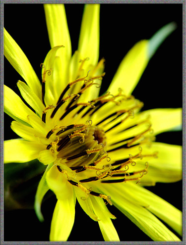
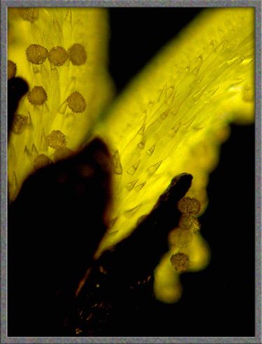
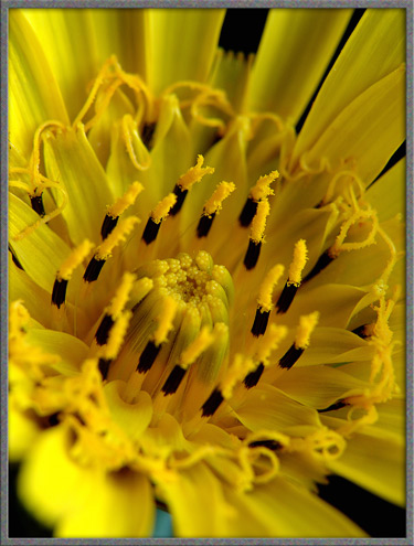
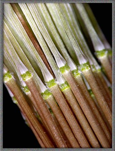
The three images below show side
views of the outer ray flowers. The dark, almost black columns
are actually tubes formed by fused anthers
(the male pollen producing structures). Growing up through each
of these tubes is the style, which supports a two-lobed stigma (the female pollen accepting
structure). It has been my experience that the beautifully curled
lobes of the stigma exist for a very short time, and they soon
straighten out to a straggly ribbon form. All three images show
that the stigmas surface is coated with tiny pollen grains.
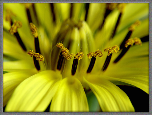
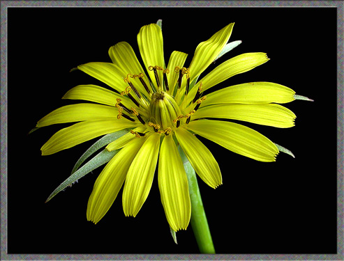
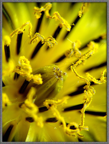
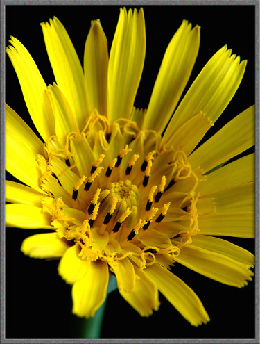
The unkempt appearance of the
stigma lobes after a period of time can be seen in the following image.
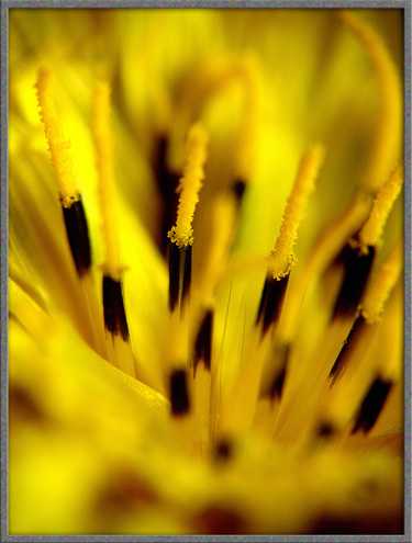
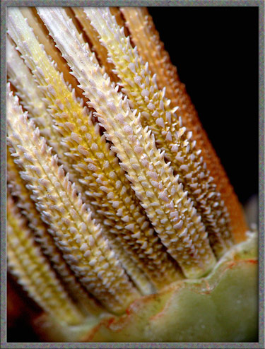
A closer look reveals more
detail. In particular, notice that each of the anther columns
forming the central structure in the image, is divided at its tip by
fine black lines, into five sections. These are the five anthers
which are fused to form a particular column. Given time, the
stigma and style will grow up through this tube and beyond it.
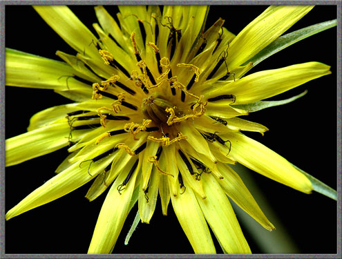
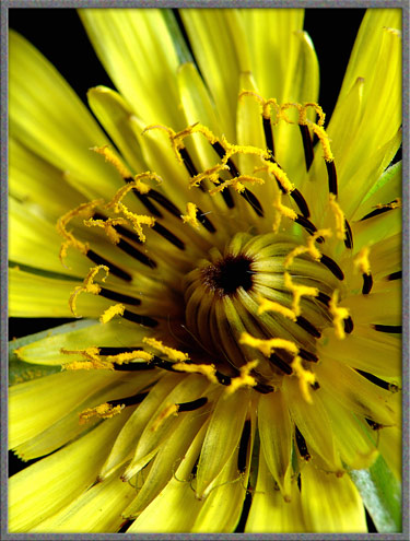
As the flower-head matures, the
central columns straighten slightly to reveal a dark centre.
Compare the two images below with the one above, in which no dark
centre is visible.
Two
further images of a flower-head are shown below. Note the swollen
base in the first image.
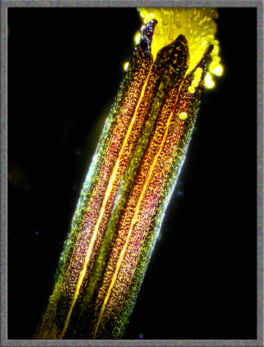
Under the microscope, the details
of a fused column of anthers can be seen. Note that three of the
five anthers are visible in the photomicrograph, each with a triangular
top. The yellow column protruding from the end is the style.
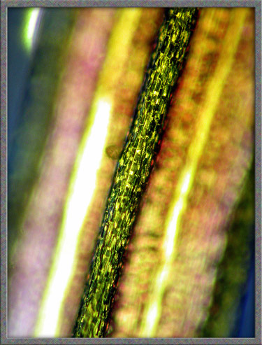
The highly magnified central
longitudinal ridge of one of the anthers is shown below.
In the following photomicrograph,
the bi-lobed stigma has just pushed out of the anther tube. As
time passes, it will grow out still farther, revealing the style that
supports it.
A much higher magnification image
of the exit point of the style is shown below. Note the hair-like
protuberances on the styles surface that tend to capture any pollen
that come into contact with them.
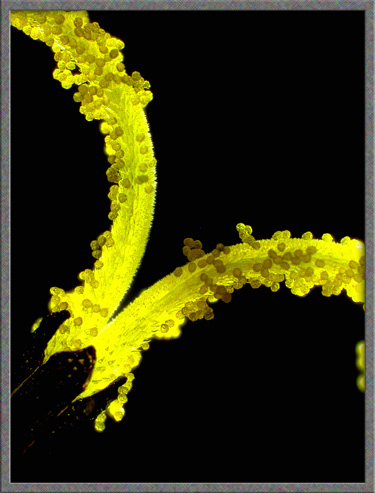
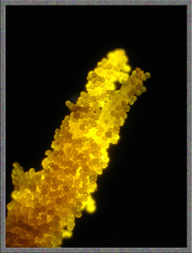
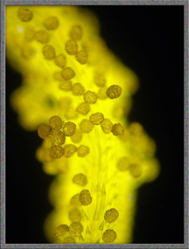
Two further images reveal the many
pollen grains which have become stuck to the surface of stigma and
style.
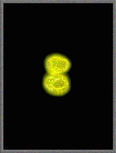
The surface of a pollen grain
appears to be pock-marked by many tiny depressions.
A Second Species
(Meadow Goats-Beard ?)
The flower-head in the two images
below looks remarkably like Yellow Goats-Beard, but the pointed green
bracts are shorter than those of that species. This is probably
Meadow Goats-Beard, Tragopogon pratensis.
I say probably, because although pratensis does
have shorter bracts, it is not supposed to have the swelling beneath
the flower-head - and this one clearly does. Welcome to the
nightmare of identifying Tragopogon species! One possibility is
that all of the many Tragopogon species tend to form hybrids when
unknowing insects carry the pollen of one plant to another of a
different species. The resulting hybrids have a mixture of the
properties of both plants. This may bring joy to a plant
taxonomists heart, but not to mine!
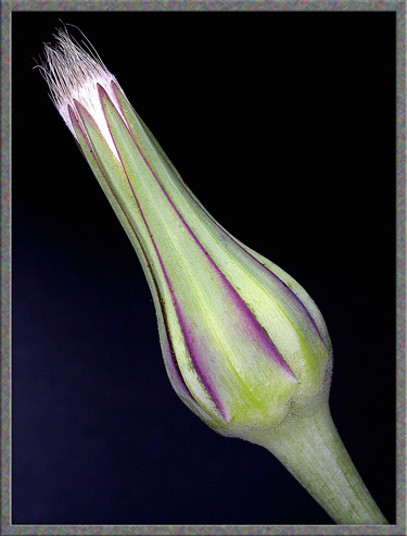
The swelling at the base of a
flower-head is particularly noticeable while still in bud form.
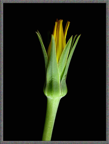
The three photographs below
demonstrate how similar the Meadow Goats-Beard flower-head is to that
of the Yellow Goats-Beards.
Back to Yellow
Goats-Beard
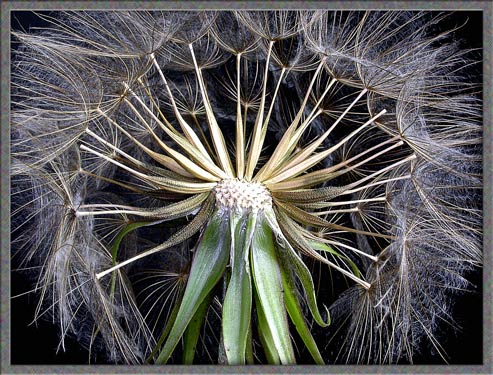
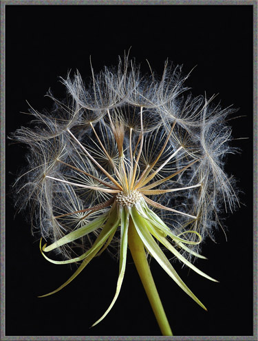
Once the flower-head has finished
blooming, a remarkable thing happens. The bracts close up around
the fertilized flowers, completely hiding them from view.
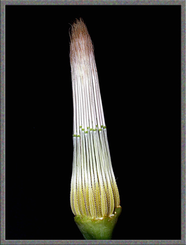
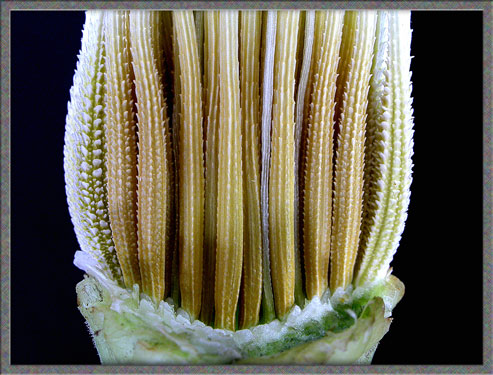
If the bracts are cut away, it is
possible to see the developing seeds and pappae, composed of the hairs that
will eventually form the dandelion-like parachute. Note, in the
image on the right, that the spiked seeds are beige in colour.
The white columns beyond the narrow green rings are composed of the
fine white threads which will eventually form the parachute.
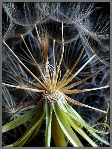
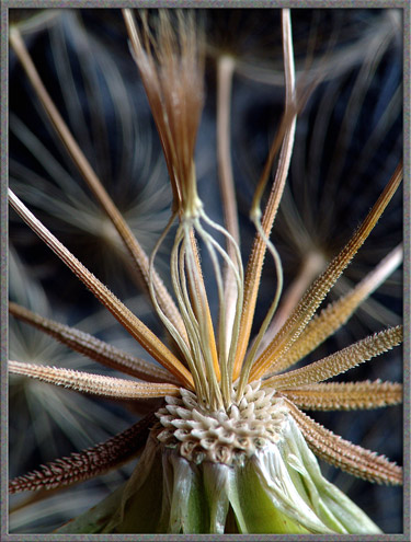
The higher magnification
photographs which follow reveal details of the developing seeds and the
green rings which are the point of attachment of the pappae.
As can be seen in the two images
below, the seeds, called achenes
are covered in spikes which form on the many longitudinal ridges.
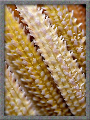
It is
interesting to note that if several of the outer achenes are removed,
those visible in the inner core have greatly reduced spikes on their
surfaces.
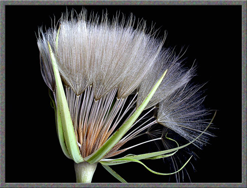
Once the seeds have sufficiently
matured, the bracts re-open and the pappae on their long stalks begin
to dry out and open into parachutes.
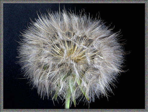
When this process is complete, the
startlingly large (up to 10 centimetre!) globe-shaped plumed head is
revealed. These balls of parachutes completely dwarf those of the
largest dandelion.
A
gust of wind is all that is needed to dislodge seeds from the white pad
to which they are attached. (For this photograph, I acted the
part of the wind in order to remove the front-most achenes.)
While searching in the field with
my trusty magnifier for a photogenic specimen, I noticed the seed-head
below. Several of the central seeds and pappae have failed to
grow properly and have formed an interesting and unique basket-shaped
cage. In the last image, note the strange shape of the
depressions in the pad to which the seeds are attached.
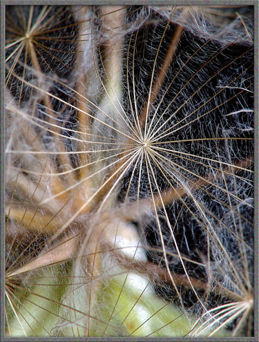
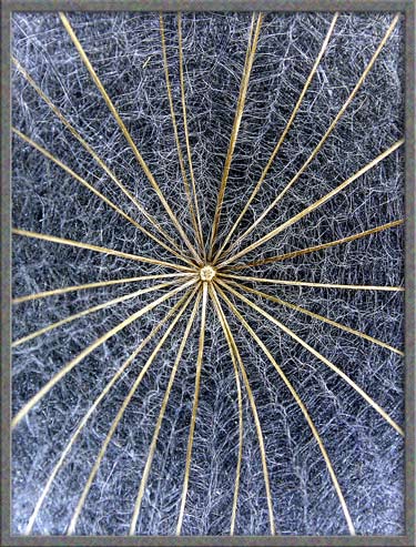
The parachute of a Goats-Beard
seed is almost flat and the surface is perpendicular to the column that
attaches it to the seed. Strong (beige) radial arms attached to
the central column hold fine (white) fibers that completely fill the
space between the arms.
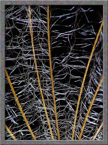
Under a higher magnification, it
can be seen that these fibers form a tangled web between each pair of
radial arms.
When I first became interested in
the macro-photography of wildflowers two summers ago, Yellow
Goats-Beard seed heads were abundant. Although it was only
August, I could find no flowers. (To be truthful, I did find
several very misshapen examples that were too ugly to
photograph.) With less than perfect patience, I had to wait until
this past summer to obtain images of flower-heads. Strangely,
this summer, which was much cooler and wetter than the previous one,
proved to be ideal for Yellow Goats-Beard. Flowers bloomed right
up to the middle of September! It was worth the wait. This
plant is one of my top ten favourite wildflowers.
Photographic Equipment
The photographs in the article were
taken with an eight megapixel Sony CyberShot DSC-F 828 equipped with
achromatic close-up lenses (Nikon 5T, 6T, Sony VCL-M3358, and shorter
focal length achromat) used singly or in combination. The lenses screw
into the 58 mm filter threads of the camera lens. (These produce
a magnification of from 0.5X to 10X for a 4x6 inch image.) Still
higher magnifications were obtained by using a macro coupler (which has
two male threads) to attach a reversed 50 mm focal length f 1.4 Olympus
SLR lens to the F 828. (The magnification here is about 14X for a
4x6 inch image.) The photomicrographs were taken with a Leitz SM-Pol
microscope (using a dark ground condenser), and the Coolpix
4500.
Note:
Since the photographs in this article were obtained over a period of
two years, my Sony DSC-F 717 and Nikon 4500 were used to take the
earlier photographs. They can be distinguished by the fact that
the gray frames around them are of slightly greater width.
References
The following references have been
found to be valuable in the identification of wildflowers, and they are
also a good source of information about them.
A Flower Garden of
Macroscopic Delights
A complete graphical index of all
of my flower articles can be found here.
The Colourful World of
Chemical Crystals
A complete graphical index of all
of my crystal articles can be found here.
Published in the July
2009 edition of Micscape.
Please report any Web problems or
offer general comments to the Micscape
Editor.
Micscape is the on-line monthly magazine
of the Microscopy UK web
site at Microscopy-UK
© Onview.net Ltd, Microscopy-UK, and all contributors 1995 onwards. All rights reserved. Main site is at www.microscopy-uk.org.uk with full mirror at www.microscopy-uk.net .