Cross-Eyed 3D Microscope Image Gallery
Part 1
by Mol Smith July. 2014
|
|
|
|
| I thought it would be fun to create some 3D images which you can view through cross-eyed viewing. Big disclaimer here "If you get stuck looking like a cross-eyed Jack, or you get eye strain, don't blame me." Some of these have come from our 3D SEM microscope where Dennis Kunkel kindly gave permission for me to convert some of his magnificent images into 3D, and others have been created by me in 3D from photos and images from contributors to Micscape Magazine. So, you have to stare at each pair of images, cross-eyed. A third image appears between them. Focus on this one until it becomes clearly focused and in 3D. |
 |
|
|
|
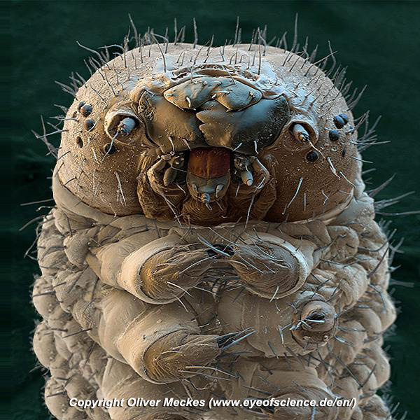
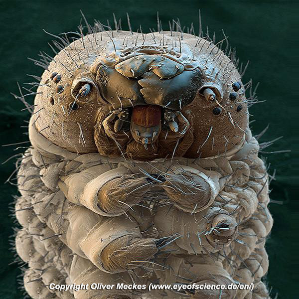
This is the head of a silkworm, close-up. It is not a worm but a larva, the caterpillar stage of a moth. The silk from the silkworm's cocoon is a single, continuous thread. It is made of a protein that is secreted from two salivary glands in the caterpillar's head. Image is copyright of Oliver Meckes www.eyeofscience.de/en/ [All rights reserved]. View in our 3D Microscope?
|
||
|
|
 |
|
|
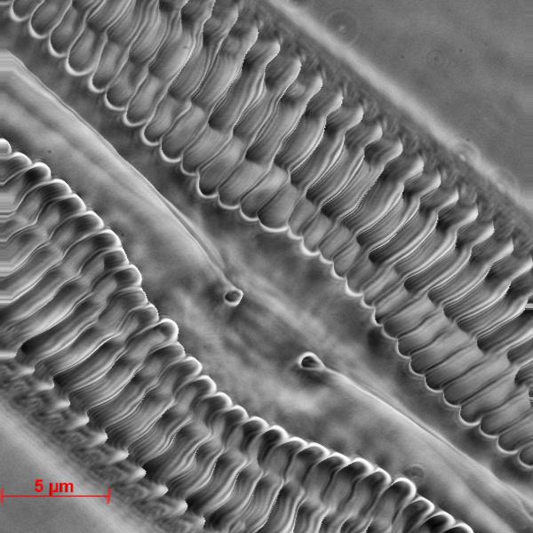
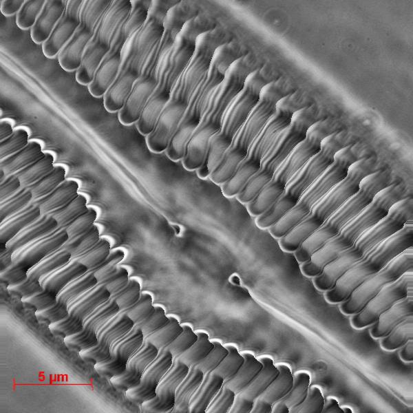
Diatoms are a major group of algae (microscopic plants), and are among the most common types of phytoplankton. Most diatoms are unicellular, although they can exist as colonies in the shape of filaments or ribbons. Diatoms are producers within the food chain. A unique feature of diatom cells is that they are encased within a cell wall made of silica (hydrated silicon dioxide) called a frustule. View in our 3D Microscope? |
||
 |
|
|
|
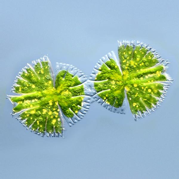
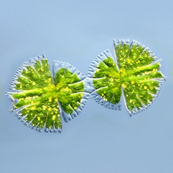
Desmids are microscopic plants living in water. They multiply by fission, asexual reproduction. Here we see a cell division of Micrasterias fimbriata. Desmids cell division of Micrasterias fimbriata copyright Wim van Egmond. View in our 3D Microscope? |
||
Have you gone CROSS-EYED yet?
No? Ok - more...
 |
|
|
|
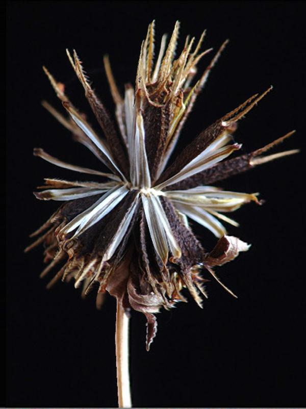
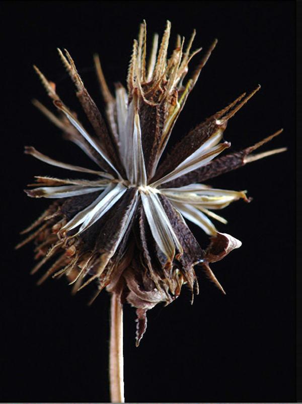
Devil’s Beggar-Ticks - Bidens frondosus |
||
|
|
|
|
|
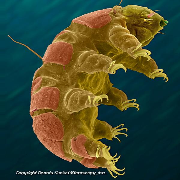
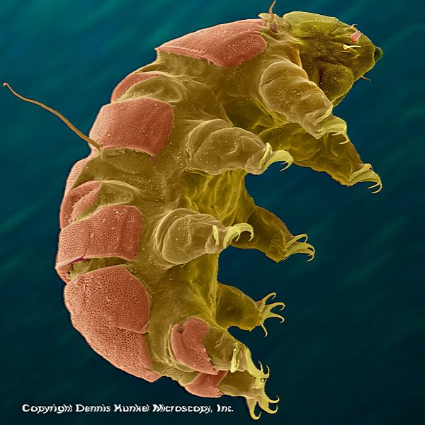
What about a Waterbear? I did this one so it's a hologram, in so far as much as it stands forward of your screen instead of behind it.
Micscape Article |
||
Did you enjoy that? Why not go cross-eyed again. I'll do some more. Look out for them.
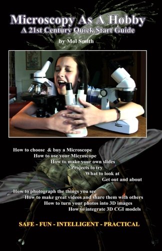
Want to find out how to make these from single microscopy photographs?
I explain how in my book: here!
mol