|
More Chitons by Richard L. Howey, Wyoming, USA |
In the last article on chitons, I said that in this next article I would look at some of the micro-detail. Well, as often happens, I ambushed myself. I found some sources for some additional specimens and so you will have to wait until the next article for the micro-stuff (maybe), because this time, I am going to share some more macro-views of chitons with you. As I look at them more and more closely, I am much taken with the patterning on the valves and am still marveling at their general architecture. So, this will essentially be a gallery of some additional species of chitons and also I want to raise the issue of variation within a species–an interesting problem.
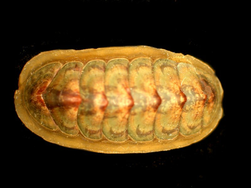
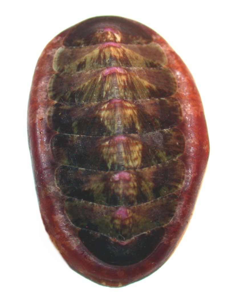
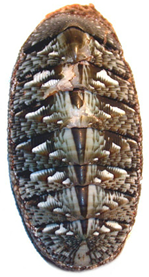
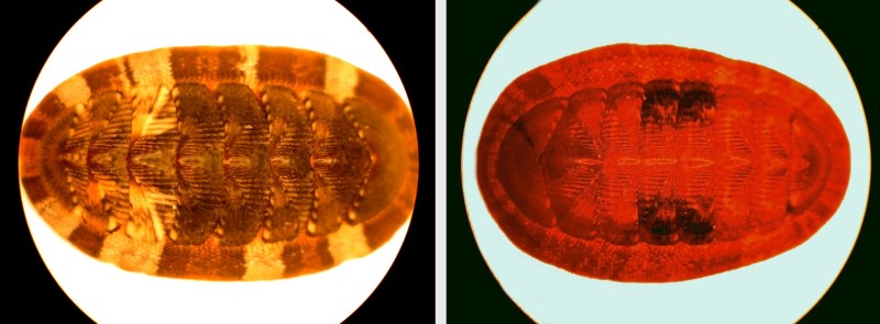
The taxonomy of these organisms is maddeningly complex and one can easily go astray by relying on size, color, and patterning, and counting the valves is no help because they all have eight. We looked at eyes last time, but remember, some groups don’t have any eyes at all and others have thousands (literally!) Let’s begin by considering a species called Tonicia lebruni. If you Google it, you will find a bewildering group of organisms that are all classified as Tonicia lebruni. For example, there is one in which the valves (or plates) are a rich green and the girdle (that outer edge of tissue which surrounds the valves) is lemon yellow. Then there is another specimen in which the central portion of the valves is copper-colored and then shades into a greenish “patina” toward the edges. Yet another is a pinkish-rose color and a fourth is a light lime-green color with pink in the center of the valves. Here is an image of the specimen which I have and you can see that it is yet different, combining colorations of several other types. This one is from Tierra del Fuego.
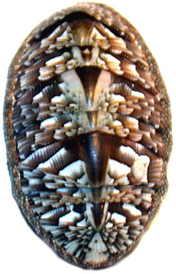
If now, we return to the Google page of images and look at species other than lebruni, but still in the genus Tonicia, there are some stunning variations, especially this one by Yuri Hooker.
Here the patterning and the pastel pinks and greens, make this small creature into an astonishingly lovely creature.
Some chitons appear to be rather hirsute as you can see here in this specimen of Chaetopleura lurida. Taxonomically these are designated as bristle hairs. This specimen is from Baja California, Mexico.
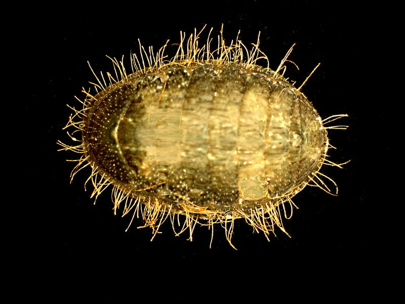
You will also notice that on the anterior valve (left) there are lots of rows of eyes and additional ones on the edges of other valves as well.
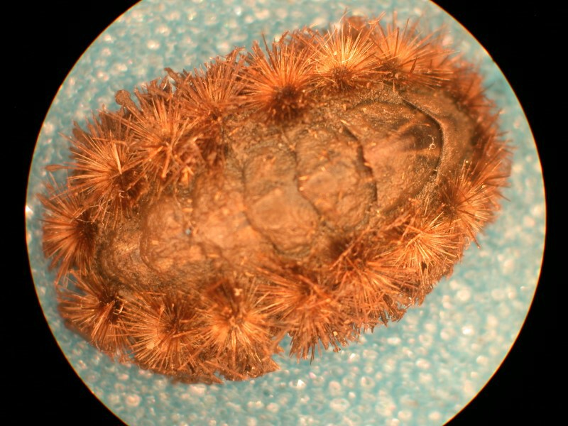
However, an even fuzzier chiton is to be found in Acanthochiton garnoti. These bushy bursts are called sutural tufts. This specimen is from South Africa.
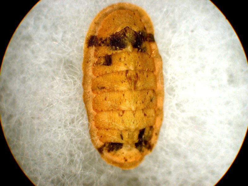
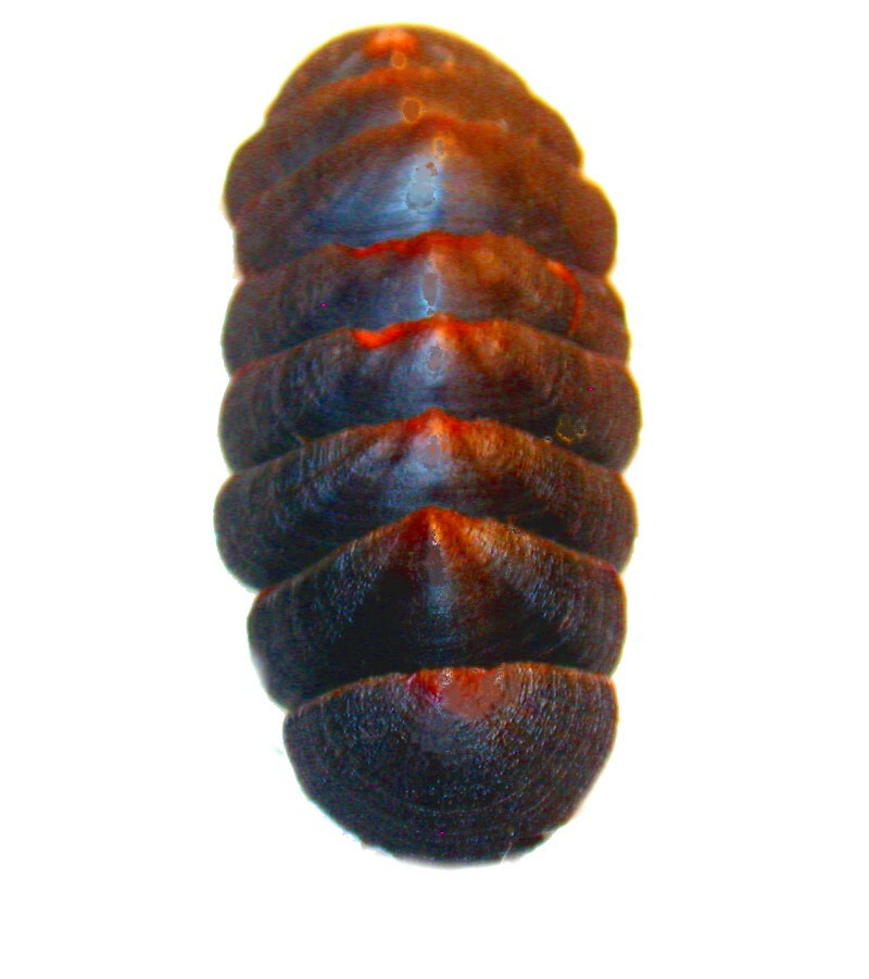
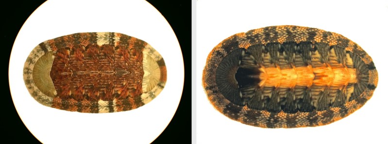
Next we have a specimen with no hairs, a light orange and black surface on which the valves and the girdle are very clearly demarcated. This is from British Columbia, Canada.
The central area of the valves is generally the highest part of the arch on the valve and is frequently patterned in a fashion quite in contrast with the rest of the valve plate. We can see this clearly in the specimen below of Callochiton dentatus which was collected in East London.
We have already seen instances of considerable variation in the girdle and here’s another one. In this specimen of Chiton japonica (from Japan) the girdle looks like it is composed of thousands of tiny bits of brown grit.
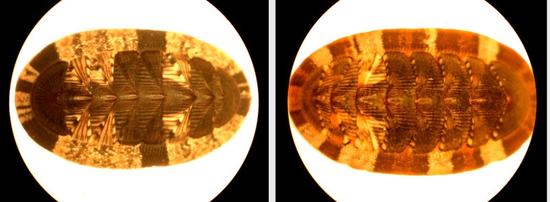
However, in another species C. tulipa (again collected from East London), the girdle appears almost as a pattern of cross-hatching and the central arches of the valves look quite like isosceles triangles.
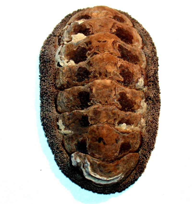
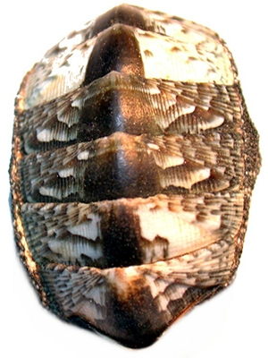
The majority of chitons have a body type described as “oval” or “elongated oval”, but some are described as “broad oval” and the specimen of Chiton tuberculatus below from St. Croix is a good example.
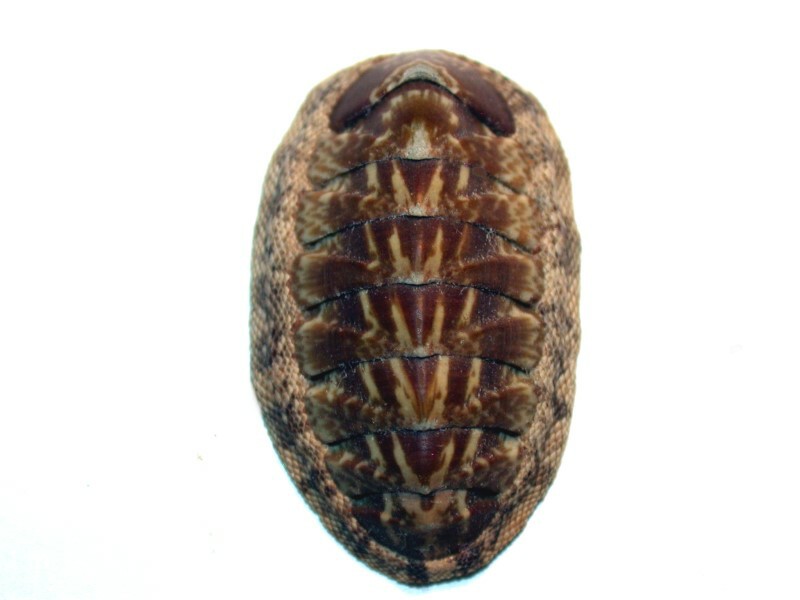
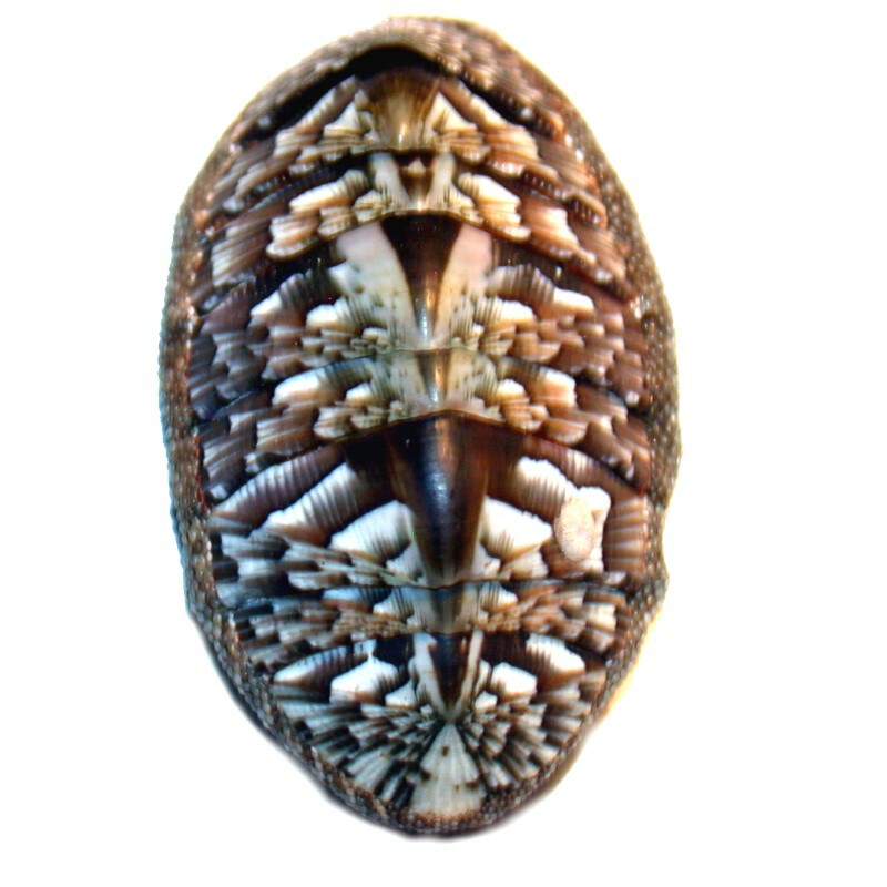
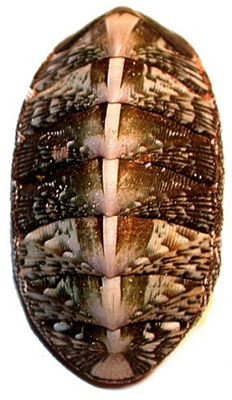
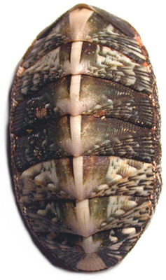
There are also species which have intricate patterns across the entire surface of the valves and have the look of an elaborate set of inlays. One can imagine using a variety of fine woods and creating a beautiful oval coffee table based on the patterns in Chiton densiliratus (from Leyte Island, Philippines).
Some of the specimens which I acquired recently came packaged in small groups in a plastic baggy with no identification other than “Chiton species”. For example, these 3 specimens came packaged together.
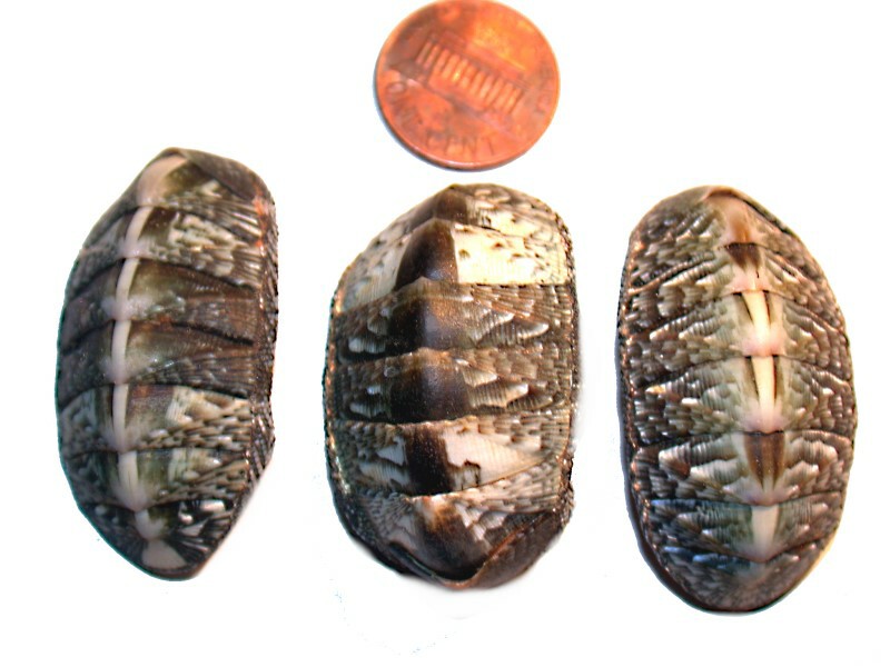
If we look at the 3 of them comparatively, we can, I think, conclude that it is likely that specimens 1 and 3 are of the same species, but that the middle one is of a different species (although very likely, the same genus). This is based on the difference in body type “broad oval” as against “oval” and the difference is in the patterning particularly along the central arch of the valves.
Another dealer sent 8 specimens in one plastic baggy and again the only listing was “Chiton species”. Now this clearly means that there are some people out there who are even lazier than I am.
Here’s a picture of all 8 of them.
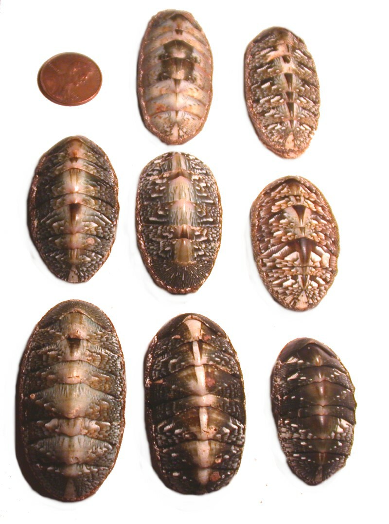
This time we can get a bit of aid from an identified specimen which we viewed above, namely, Chiton densiliratus (remember the inlaid coffee table specimen). Even in this picture of all of them together, the one on top right and the one on the middle right are strikingly similar to our C. densiliratus and this becomes even more evident if we look at closeup images of them. The one on the left is the one we know to be C. densiliratus, and the other 2 are the ones we were just discussing.
In addition, the center specimen in the middle row might also be the same species. As I mentioned earlier, there can be considerable variation in the patterning in the same species. The question as to whether or not the other 5 specimens are of this species is not so easy to answer and would depend upon a much closer examination and a better grounding in the taxonomy of these wonderful creatures than I have.
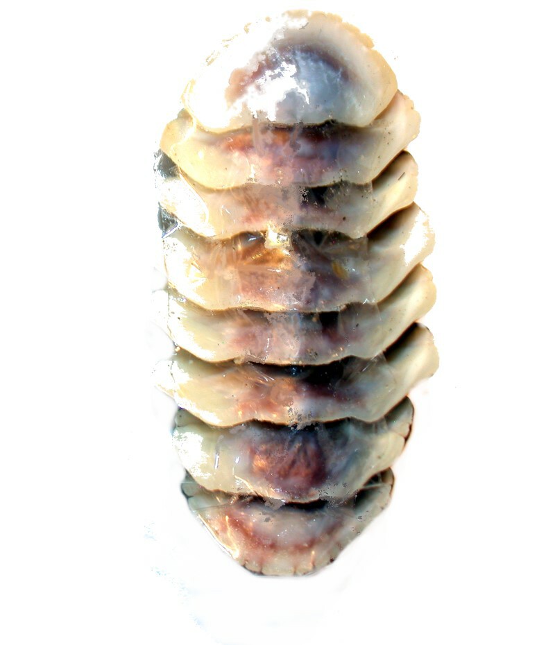
Another dealer sent 2 specimens which had only the valves and he ingeniously used a clear tape on the ventral side to hold the valves in position. First, I’ll show you a dorsal view of a specimen of Acathopleura spinosa (from Coron Island, Philippines).
And here is the underside and you can see the light reflections off of the tape.
The species problem got even more difficult when I looked at a group of specimens which were shipped from Italy, but all of which were listed as coming from Sardinia. There are 9 specimens and all are listed as Chiton olivaceus. Here are 6 stitched together in 3 images to allow for a side-by-side comparison. Two things struck me immediately: 1) the general morphology seems virtually identical and 2) of course, the very significant variation in general appearance.
I did a search for images of this species and found the following comparative plate with the comment at the bottom “Highly variable in patterns and colors”.
So, these are very likely all of the same species and they demonstrate the extraordinary capability of Nature to constantly present us with puzzles.
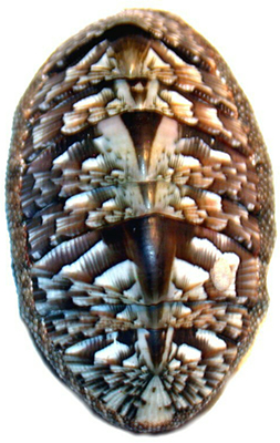
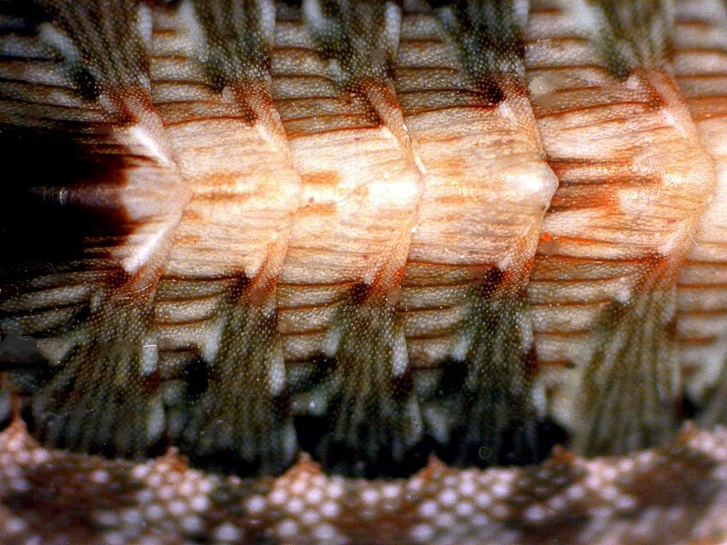
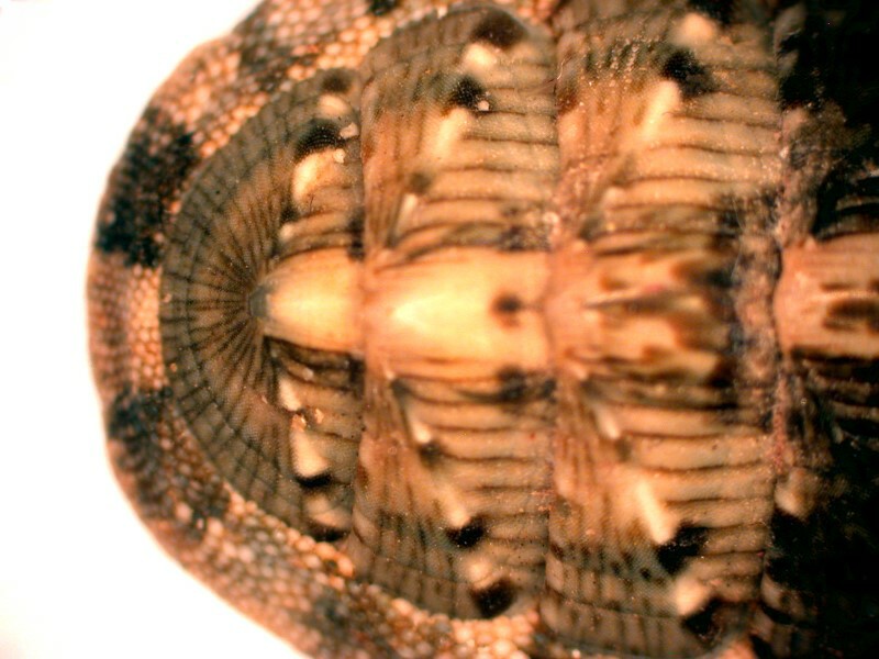
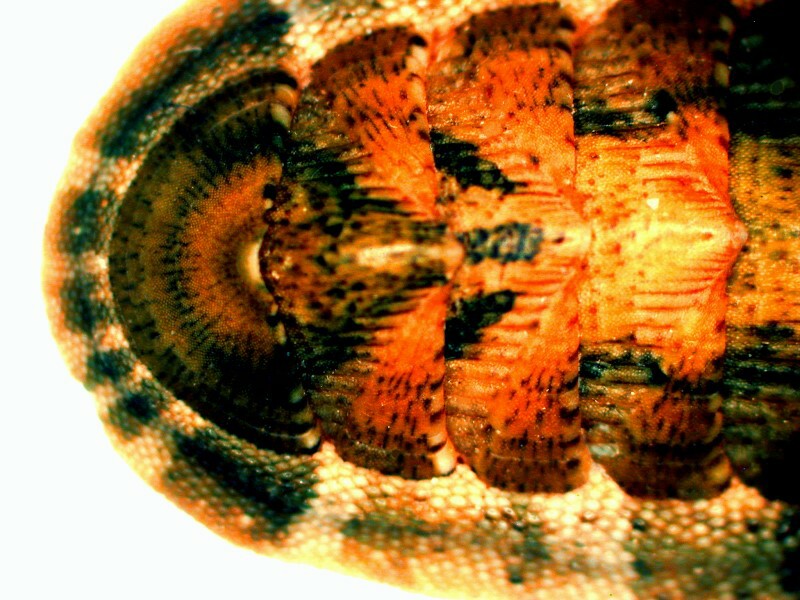
However, before we wind this up, I would like for us to take a closeup look at a few of these Sardinian chitons. The first image is along the central ridge of the valves and from the problem of focus out on the edge at the girdle we realize that the valves are arches and the center is the high point. Furthermore, this image reveals to us the granular crystalline nature of the valves.
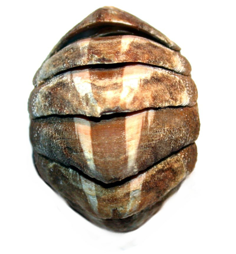
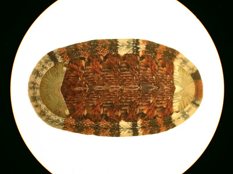
In the next images, I want to take a closer look at the anterior or “head” valve which in this species has the appearance of a rather elegant fan.
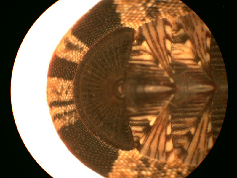
And finally, another view of the entire chiton which shows us that both the anterior and the posterior valves have this fan-like shape and it is not the case that in all species there is such a resemblance.
I hope this brief tour of a few examples of chitons has proved of interest and in the next part, I hope to take up an examination of some of the intriguing micro-structures which are found in and on chitons.
All comments to the author Richard Howey are welcomed.
Editor's note: Visit Richard Howey's new website at http://rhowey.googlepages.com/home where he plans to share aspects of his wide interests.
Microscopy UK Front
Page
Micscape
Magazine
Article
Library
Published in the July 2016 edition of Micscape Magazine.
Please report any Web problems or offer general comments to the Micscape Editor .
Micscape is the on-line monthly magazine of the Microscopy UK website at Microscopy-UK .
©
Onview.net Ltd, Microscopy-UK, and all contributors 1995
onwards. All rights reserved.
Main site is at
www.microscopy-uk.org.uk .