| |
Mosses and liverworts, simple
plants?
|
by Jan Parmentier with photographs by the author
|
| |
|
|
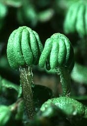
Mosses (Bryophyta) are quite interesting, simple green
land plants with leaves and a stem and always without
roots. In many mosses, the leaves are only one cell
thick, except for the midribs, which are sometimes
present. So the leaves are easy objects for the
microscope. The plant is normally attached to the ground
by delicate, colourless or brown threads, the rhizoids.There
are two major groups in the Bryophyta: Mosses (Musci) and
Liverworts (Hepaticae). See Footnote.
Most mosses are found in areas with a humid and a cold
to moderate warm climate. In the tropics, mosses are
found especially in the mountains. In Europe, the south
western part of Ireland is a paradise for mosses.
Mosses can reproduce asexually, by means of small
clusters of cells or plates of tissue which break away
and germinate to become new plants. Especially liverworts
do this.
|
| The normal, sexual method of reproduction
however, involves special organs, the antheridia and the
archegonia. These organs are the interesting parts for
the microscopist who is interested in the biology of
mosses. In the Musci the antheridium is the male organ, a
delicate sac in which the male gametes are formed. It has
a greyish or brown colour and an ovoid or globose form.
It is a spectacular sight to see the male gametes, with
two flagella, escape under the microscope from the
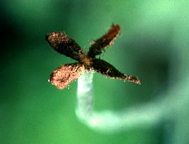
antheridium. These antheridia are normally accompanied by
numerous short filaments of cells, the paraphyses (see
right image). The archegonium is easy to recognize,
with a shape like a little bottle or flask. So look
carefully with a hand lens among wet patches of mosses,
archegonia and antheridia are often found in special cups
of leaves.
|
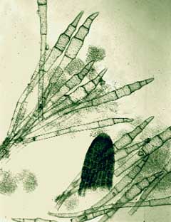
paraphysis with archegonium
|
 |
I always try to study first some of the
common microscopical objects in detail, especially their
biology and then try to determine the names of the
species that are more difficult to find. So I studied
very common mosses to see some details of their
reproduction. The male gametes, escaping from the
antheridia, need water to reach the egg in the
archegonium. After fertilization, the egg develops in
most cases into a spore-containing capsule on a stalk
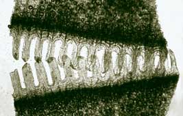
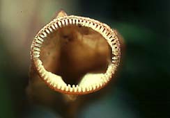
called a seta. Capsule and seta together form the so
called sporophyte. At maturity, the capsule sheds the
spores as a fine dust. The spores can be held back in wet
weather by a mechanism whereby the teeth of the capsule
close it.
Mosses can have one or two rows of teeth, (images left
and below), an important aspect for the determination.
|
|

|
| In the leafy
liverworts, the antheridia generally occur in a
packet-like swelling, the androecium, which
develops on the lower portion of a modified leaf.
The sporophyte develops from the archegonium. The
seta is very delicate, often white and glassy,
grows very fast and perishes after the shedding
of the spores. The capsule is black, often
globose or ellipsoid. |
 |
|
|
|
|
|
| |
|
|
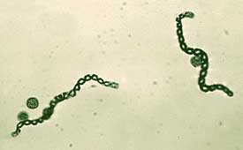
In most species the capsule (sporangium) peels open
in four sections (image above), exposing the spores and
the elaters, cells which have helically arranged moisture
absorbing wall thickenings. (Image below).
|
| |
|
|
|
| |
|
|
These cells are sensitive to slight changes in
humidity, causing a twisting action that aids in
dispersing the spores. The elaters are initially attached
at both ends to the sporangium. Upon drying, one end of
each elater snaps loose from the center of the
sporangium, spreading the spores.
|
| |
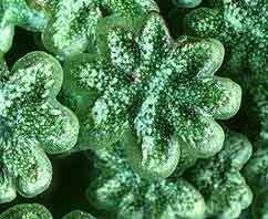
Marchantia polymorpha, a more complicated liverwort, is
common in flowerpots in green houses, on moist bricks in
gardens and on badly drained soils. On its leaves we can
see small cups, (gemma cups) with small oval pieces of
tissue, which can be spread by rain drops and become new
plants |
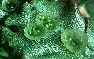
gemma cups
|
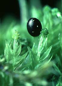
| This dioecious liverwort is
known immediately by the male and female
"umbrellas". These umbrellas carry the male
and female receptacles. The numerous sporogonia develop
on the underside of the umbrellas; each capsule contains
spores and elaters.
|
the male umbrella
|
|
the female umbrellas
|
You may call mosses simple plants, but important biological
processes are easily studied by looking carefully at these
sometimes fascinating plants.
Footnote: This is the classification adopted
until recently and found in many books available on mosses and
liverworts. The Phylum Bryophyta (mosses, liverworts and
hornworts) has recently been split by taxonomists into three
separate phyla: Bryophyta (mosses), Hepatophyta (liverworts) and
Anthocerophyta (hornworts) e.g. see 'Margulis and Schwartz' in
'Further Reading' below. Return to article.
Further reading:
E.V.Watson, British Mosses and Liverworts. 2nd
ed. Cambridge UP 1978.
A.J.E.Smith, The Moss Flora of Britain and
Ireland, Cambridge UP, 1978.
H.N.Dixon, The Students Handbook of British
Mosses, 3rd ed. Reprint Wheldon and Wesley 1970.
S.M.MacVicar, The Students Handbook of British
Hepatics, 2nd ed. Reprint Wheldon and
Wesly 1971.
D. Aichele, H.-W. Schwegler , Unsere Moos- und
Farnpflanzen, Kosmos, Stuttgart 1984
W.D. Margadant, H.During, Beknopte flora van de
Nederlandse Blad- en Levermossen,
KNNV 1982, Thieme, Zutphen.
P.H.Raven, R.F.Evert, S.E.Eichhorn, Biology of
Plants, 5th ed., Worth Publishers, New York 1992.
L. Margulis, K.V. Schwartz, Five Kingdoms: An
Illustrated Guide to the Phyla of Life on Earth, 3rd ed., W. H.
Freeman, 1998. See Micscape Review.
Comments to the author Jan Parmentier are
welcomed.
Prepared for the Web by Wim van Egmond
All Material Copyright: © Jan Parmentier
First published in July 1998
Micscape Magazine.
Please report any Web problems
or offer general comments to the Micscape Editor,
via the contact on current Micscape Index.
Micscape is the on-line monthly
magazine of the Microscopy UK web
site at Microscopy-UK
WIDTH=1
© Onview.net Ltd, Microscopy-UK, and all contributors 1995 onwards. All rights
reserved. Main site is at www.microscopy-uk.org.uk with full mirror at www.microscopy-uk.net.