|
THE DOUBLE LIFE OF OBELIA by Wim van Egmond |
|

|
|
| I must have been five years
old when my grandfather showed me a book about prehistoric life illustrated with the
paintings of Z. Burian. The first picture I saw had such impact that it formed the start
of a lifelong interest in natural history. The painting showed life in the Cambrian seas,
the Cambrian period starting 650 million years ago being the era of the early crustaceans,
the trilobites. This wonderful impression of ancient life depicted several trilobites
walking around on their many segmented legs. Above them floated a group of jellyfish.
Trilobites are long extinct but jellyfish still swarm the oceans. Observing lifeforms such as jellyfish gives you a glimpse of this wonderful past. Jellyfish belong to a group called the Cnidaria (nettle animals) or coelenterates. They are not so difficult to study and they have a very interesting lifecycle. To study them alive the best thing to do is try and capture them with a plankton net or try and grab the polyp stages from the growth you can find on rocks and on the poles in a harbour. They can be kept alive for many days when put in a glass container. One of the most interesting aspects about jellyfish is the fact that they have two life phases. There is a medusan and a hydroid stage. The medusa is free swimming and reproduces sexually. Their eggs grow to form ciliated larvae that attach themself to a surface. Then the hydroid stage starts to grow into a colony of polyps. These polyps multiply asexually, forming buds. |
|
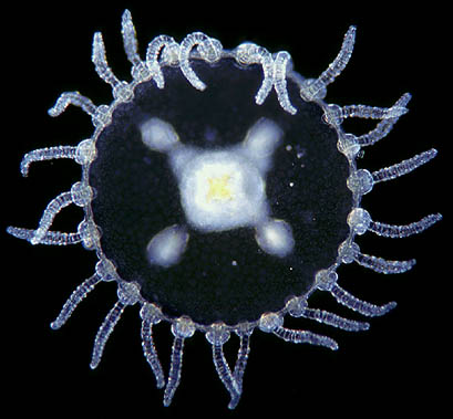
The medusa of Obelia. At the centre is the manubrium, it's feeding opening. Around it lie four gonads, male or female reproductory organs. The tentacles are based on a muscular band called the velum. This is the swimming organ. At the base of the tentacles little vesicles can be found, the statoliths ,which serve as organs for equilibration. |
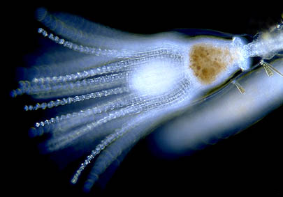
| The Cnidaria can be devided into three classes: the Hydrozoa (hydroids), the Anthozoa (corals and anemones) and the Scyphozoa (true jellyfish). Obelia belongs to the Hydrozoa. The Hydrozoa differ from true jellyfish not only in size. (Like many hydrozoa Obelia grows not much larger than a few millimetres.) Some organs, like the muscular band for locomotion, the velum, are only present in the hydrozoan medusae. One of the things that can be noticed is that for the Hydrozoa the polyp stage seems more important than for the true jellyfish. For the Scyphozoa the polyp stage is not more than a budding stalk and often the polyp stage does not occur at all. Obelia and it's kin on the other hand forms a long pink-white branching colony more than ten centimetres long.They develope many tentacles for capturing prey. The tentacles of Obelia are housed in transparent cups for protection. When touched or exposed to the microscope's light the tentacles will withdraw and hide in the cups. |
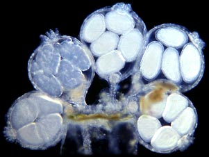
| After the colony of polyps is fully grown, reproductive polyps are formed. These are budding of little medusae. The image shows a reproductive polyp of Gonothyraea. The hydroids are very similar to Obelia, but the reproductive stages are very different. |
|
| The individual polyps are all connected by the branching
stalk. You can see under the microscope how they share their food via a fluid running
through the stalk. The living inside of the stalk contains cilia that make a current so
that the fluid can move in two directions. The next time you go to the ocean don't forget to take a little jar with you. There are many fascinating creatures that can be found in coastal waters. Hydrozoans like Obelia are almost always present and fun to watch. You don't even need a microscope, just a good handlens will do. With a bit of concentration you can travel back in time and imagine life in the Cambrian seas, half a billion years ago. |
|
|
Comments to the author Wim van Egmond are welcomed.
visit Wim's Homepage
© Wim van Egmond 1998
First published in July 1998 Micscape Magazine.
Please report any Web problems or offer
general comments to the Micscape
Editor,
via the contact on current Micscape Index.
Micscape is the on-line monthly magazine of
the Microscopy UK web
site at Microscopy-UK