Having read Dave Walker's interesting article on diatoms as test subjects, and being new to the hobby, I thought I'd see what my microscope could do. So I acquired a diatom test slide from Klaus Kemp and after a bit of experimenting came up with the attached pictures of Pleurosigma angulatum and Amphipleura pellucida. The images are:-
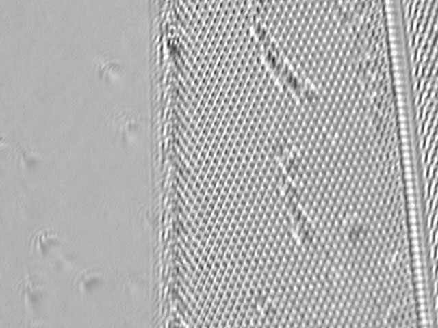
1. P. angulatum: A flat-fielded (compensation for varying illumination across the image) and slightly contrast stretched but otherwise raw image.
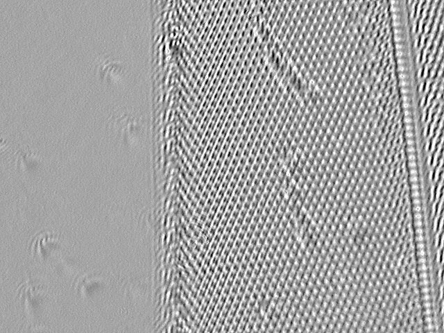
The variation in focal plane seems to be showing the surface pits near the bottom right and the underlying slots towards the top and left. This may be my imagination, but I hope to do some more work on how the images we see relate to the true 3d structure of these tiny transparent objects at high magnification, and with oblique illumination.2. P. angulatum: A lightly unsharp masked (sharpened) version of the same image.
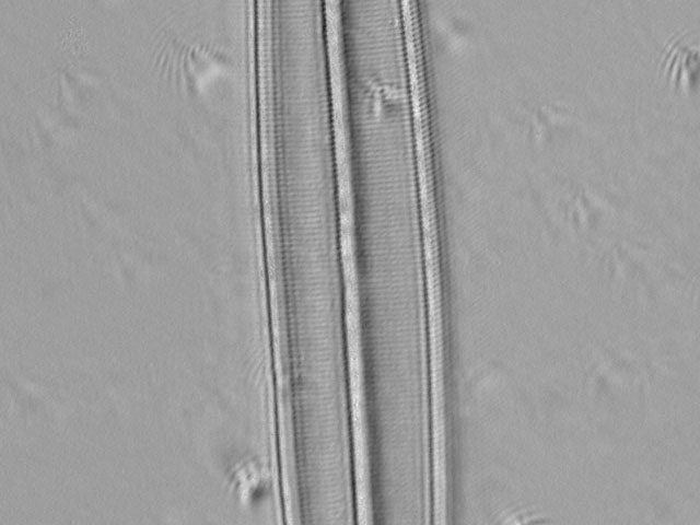
3. A. pellucida: A flat fielded and contrast stretched but otherwise raw image.
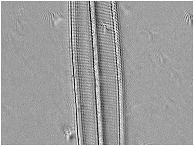
Both vertical and horizontal rows of dots can be discerned. All the images are approximately 40 microns x 30 microns.4. A. pellucida: A lightly unsharp masked version of the same image.
My microscope is a Gillett & Sibert binocular. One eyepiece holder is replaced by a lensless CCD USB webcam which puts its CCD chip close to the focal plane of the objective (so that the camera and remaining eyepiece have almost matched focus). The objective is a Leitz-Wetzlar EF N.A. 1.25 oil, which had a long hard life in a working lab, but still seems to be OK optically. Illumination for these images was by a single blue LED run at 3V pointing upwards from below the slide, and held in place by a lump of plasticine so that its quite narrow beam can be angled towards the sample at a suitable oblique illumination angle. The odd specks of dust on the images show a sort of interference bow-wave and wake effect which indicate the direction of illumination. I could not get such good images using the standard Köhler illumination. The contrast of the A. pellucida image was very low and the only internal structure seen visually was the four broader longitudinal humps either side of the 'spine' into which the smaller rows of dots seem to be grouped. Different angles of illumination would show visually either the lateral or longitudinal fine structure separately, but not both at the same time.
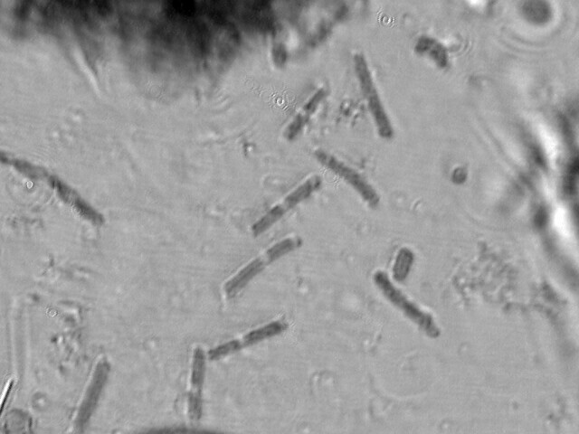
Finally, to show that the same setup also works for 'normal' specimens, here is an image from a prepared Brunel stained and mounted slide.