 |
|
|
Digital Photography for the Field Mycologist by Paul F Hamlyn, UK
|
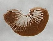
Accurate identification of the larger fungi often involves a two-stage process. Observations on fresh fruit bodies in the field are followed by detailed microscopic examination at home. Generally there is insufficient time to examine all specimens collected under the microscope while they are still in a fresh condition following a days foraying. However, this is not of concern since most material can be dried and then reconstituted for examination at a later date. The advent of the digital camera has provided a new opportunity for the field mycologist to record both the whole fruiting body and important microscopic structures such as spores in a form that can be readily shared with other mycologists via email and Internet based discussion groups.
The Camera
After spending several months reading numerous review articles on digital cameras and evaluating the pros and cons of various alternatives I eventually decided to invest in a Nikon Coolpix 5000. Its small and compact design would be advantageous on field trips and the camera reportedly has a good macro focus ability making it ideal for close-up work. In addition, the Coolpix 5000 has a 5.0 megapixels CCD capable of high-resolution photography. The flip-out and twist LCD monitor has proved extremely useful when the camera is attached to a microscope. Since the cameras lens systems cannot be removed another important feature is that there is a screw thread in the lens housing which can be used to attach a microscope adapter via two coupling rings.
Unilink Microscope Adapter
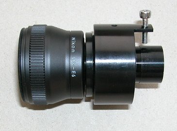
The Unilink from Brunel Microscopes is an adapter with a built in wide field lens system that will attach a digital camera with a lens housing screw thread to virtually any make of microscope. It has a clamping device to hold the camera steady whilst on top of the microscope. The Unilink has a standard 37mm screw thread which using step rings can be converted to fit the screw thread of any digital camera. In the case of the Coolpix 5000 I required a Nikon UR-E6 step-down ring (provides a 28 mm thread) and a 28 to 37mm step-up ring obtained from Brunel Microscopes. The complete assembly ready to attach the camera to the microscope is shown below. I am using a Brunel SP10 with trinocular head and therefore do not need to remove an eyepiece to fit the camera to the microscope. An electronic remote release cable (Nikon MC-EU1) is used to prevent camera vibrations and the following camera settings are employed when the camera is attached to the microscope:
|
|
|
Results
In the case of photomicroscopy vignetting (darkening of the image around the edge of the picture) or circular images are sometimes experienced but these can usually be avoided by using zoom to fill the field of view. In addition, because the fungal structures that I am interested in are very small an image processing program (e.g. Paint Shop Pro) can be used to crop out the required portion of the picture. The 'ring artifact' problem reported with some Coolpix models was not evident at least under the conditions that I have been using. Overall I have found the Coolpix 5000 to be a versatile camera for both fieldwork and microscopy. Although a bit pricey when it was first brought out the Coolpix 5000 has been around for over 2 years now so it should be possible to get a good second-hand deal.
|
|
|
|
Spore Prints (for gilled fungi)
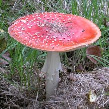
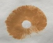
Spore colour is an important characteristic for identification purposes and can be readily observed by taking a spore print. Remove the stem from a mature and fresh mushroom and place the cap, or a section of the cap, gills downward on a piece of paper. Cover the cap with a small glass or tub placed upside down to maintain humidity and leave overnight or for a couple of days at most. To describe colours accurately it is necessary to use a reference colour chart showing various shades of cream, brown etc. After noting the colour a spore print is an ideal source of mature spores for subsequent microscopic examination.
 |
 |
 |
||
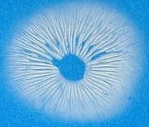
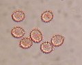
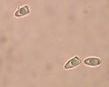
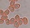
Fungal Spores
Spore size, shape, ornamentation and chemical staining reactions can all be important in the identification of fungi. For example, species belonging to the genus Laccaria are characterised by a white spore print and under the microscope the spores are usually spiny in appearance (a). Lepiota also have white spores but some species (e.g. Lepiota cristata) have spores that are distinctively bullet-shaped (b) whereas Entoloma have pink to pinkish brown spores that are typically angular in shape (c). Hygrophorus is yet another white-spored genus but in this case the spores are ellipsoid in shape (d).
| (a) | (b) | (c) | (d) | |||
| |
||||||
 |
 |
 |
 |
|||
All comments to the author Paul F Hamlyn are welcomed.
Useful References
Largent, D.L., Johnson D. & Watling R. (1987). How to Identify Mushrooms to Genus III: Microscopic Features. Eureka: Mad River Press.
Guides for the Amateur Mycologist No. 2. Guide to Identification with a Microscope by Jack Marriott. (£2.50 from the British Mycological Society).
Brand B., Henrici A. & Leonard P. (2001). Guide to the Literature for the Identification of British Basidiomycetes. (£5.50 from the British Mycological Society).
Web Resources
You can learn more about identifying fungi by joining a local fungus recording group. If you live in the UK visit http://fungus.org.uk/ to find out if there is one near you.
Details of mycological clubs in North America are available at:
http://www.namyco.org/clubs/index.html
A detailed review of the Nikon Coolpix 5000 by Phil Askey (January 2002) is available at:
http://www.dpreview.com/reviews/nikoncp5000/
Microscopy UK Front Page