|
MICROSCOPIC
FAUNA
some lifestyles
part 1
Walter
Dioni
Cancún, México
|
Except
when noted otherwise, the included images
are personal pictures obtained with a digital camera of 0.4 Mpx.
integrated into
my National Optical DC3-163-P microscope equipped with planachromatic
optics.
(Ocular 10x, Objectives: x4 (NA 0.10), x10 (NA 0.25), x40 (NA 0.65) and
x100
HI (NA 1.25)). The original ones have been captured at 640 x 480 px.
and
reduced or trimmed as was necessary to include them in this work. All
picture
formatting work was made in Photo Paint. In each picture's legend
the objective upon which it was taken is indicated, just as a
suggestion of the power
used. The widest
illustration in this page is 960 pixels, half width pictures, are
therefore 460
pixels, which correspond to the real size of a 640 px. picture.
|
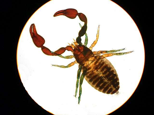
| Pseudoscorpion,
arthropod from the litter, mounted in PVA-G; excellent photomicrography
kindly communicated
by Mike André, one of my North American correspondents. |
The amateur microscopist likes
to explore the micro-world
of fresh or
marine waters. The diversity and beauty of the species that populate
them are
enough to captivate their interest for many hours that generally
would be extended to days, months, or years.
This is the world of free living creatures, which move at
will in the medium
in which they live, even though they often crawl or glide over
live or
inanimate substrata. To explore those diverse habitats can be an
infinite
occupation, and it is normally the first task that the amateur assigns
himself.
A few examples of free living aquatic
organisms are included below.
|
|
|
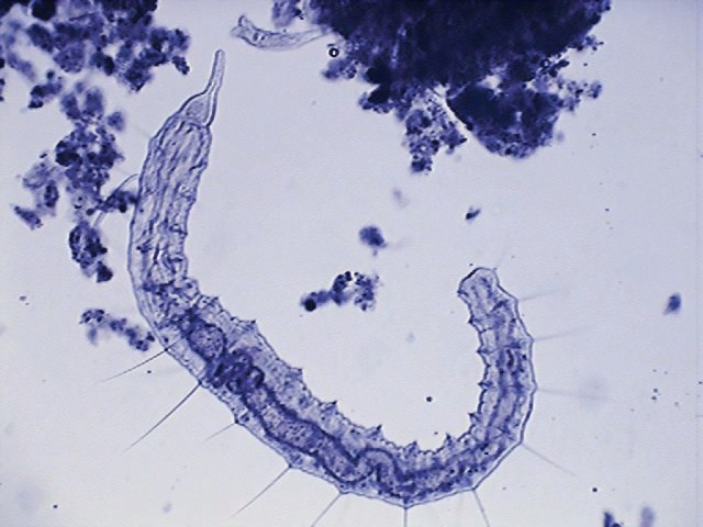
Naidid
oligocheta of free life - x4, blue toning
|
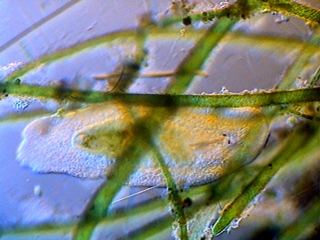
Macrostomum, a turbellaria of free life - x4
Oblique Illumination
|
|
|
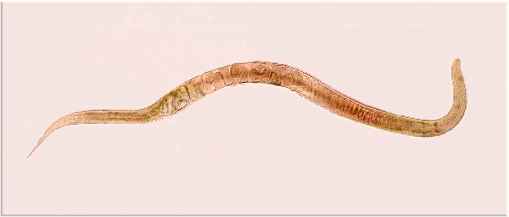
Marine
Nematode, ovoviviparous, of the psammon of a beach of "Isla
mujeres", Cancún; the image is a mosaic composed with photos
taken with
the objective 10x, original size 1933 x 825 pixels.
|
|
|
|
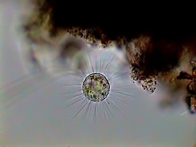
A
heliozoan, x100, bright field
|
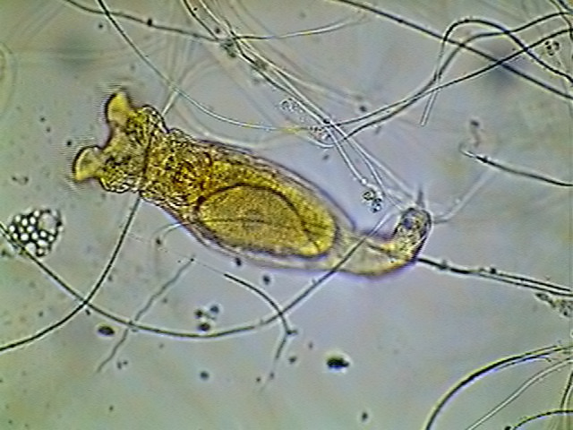
Rotifer of the genus Philodina,
x40, Rheinberg filter, blue
center, yellow periphery
|
|
|
|
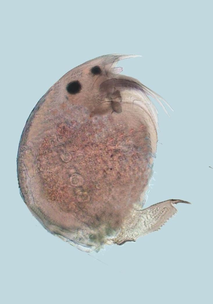
A Cladocera of the
family Chydoridae, mosaic of 8 images, x10, bright
field. It lives between the floating plants.
|
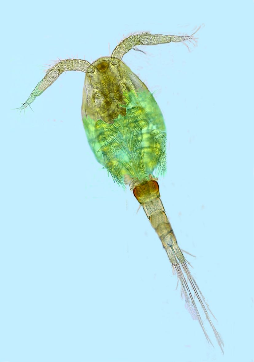
Copepod of the
Cyclopidae family, mosaic of 12 images, x 10, common
inhabitant of plankton.
|
But living species
have developed diverse styles of life, in addition to free
life.
Some
(many) of those aquatic species live fixed to a substrate,
they are sedentary, adhering to stones and other animated or inanimate
substrates.
|
|
|
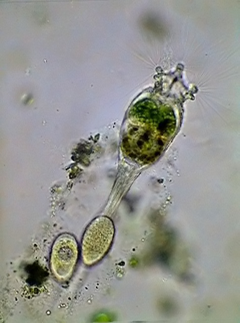
| Collotheca,
rotifer sessile, x10, bright field, from
a
pool in a plant garden at Cancún.
|
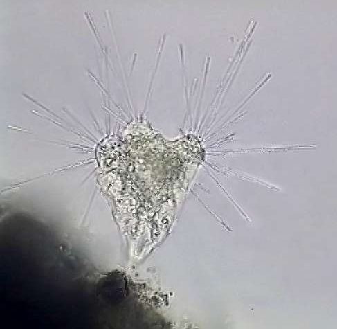
Tokophrya,
protozoa, with sucking tentacles, x100, bright field. Three images
amalgamated
with CombineZ 3.0, from a brackish water aquarium, Cancún.
|
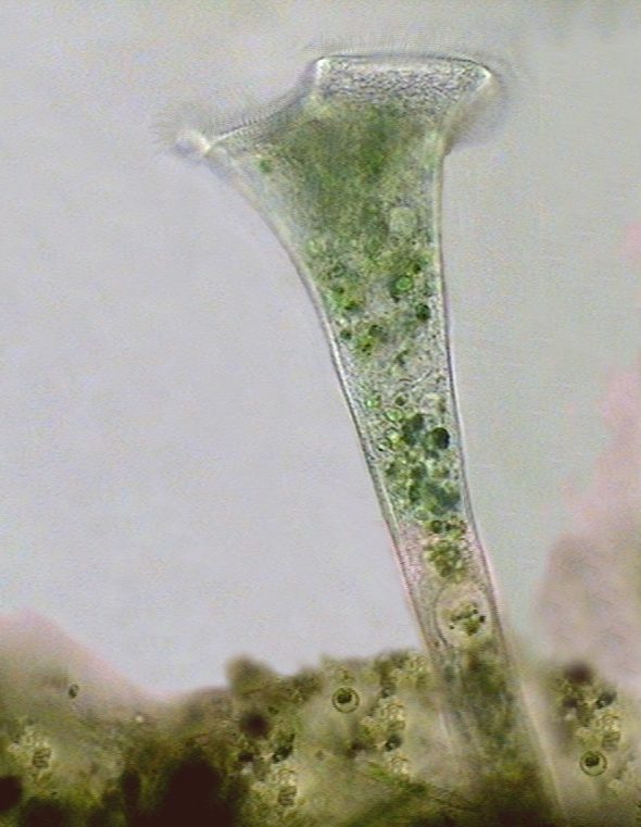
Stentor
muelleri,
x40, bright field from a brackish water aquarium, Cancún. |
Others are even
associated to other species
(algae
for example) without causing any damage to them. Although they cannot
be
considered "free", they are yet independent beings. They need the live
substrate only for a support.
There
are the epiphytes (i.e: they live on plants).
|
|
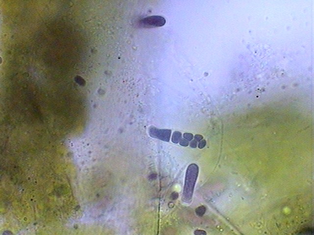
Cyanobacteria, of the
Chamaesiphonaceae family, epiphytic
on algae of the genus Cladophora,
in the photos there are several stages
of the
reproductive cycle, x100 HI, bright field, from one brackish water
aquarium,
Cancún. The color is the real one the pigment this
cyanophycea had.
|
and the epizoics (those
that live on animals).
|
|
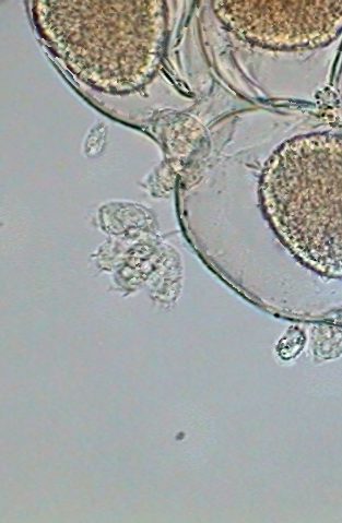
Micro-peritrichs,
fixed on the egg package of a female of a Diaptomid from Durango, Mx. fixed with GALA,
mounted in PVA-G. Bright field, x100 HI.
|
MUTUALISM - Of these last
associations of species there are some which imply that the “living
support" is not only a substrate, but that shows the development of
dependency relationships (denominated "mutualism") between the
associate species.
Commensalism (one
of the forms of mutualism) is a type of relationship in which
one
of
the members
are more or less passive, whereas the other benefits from the relation
without
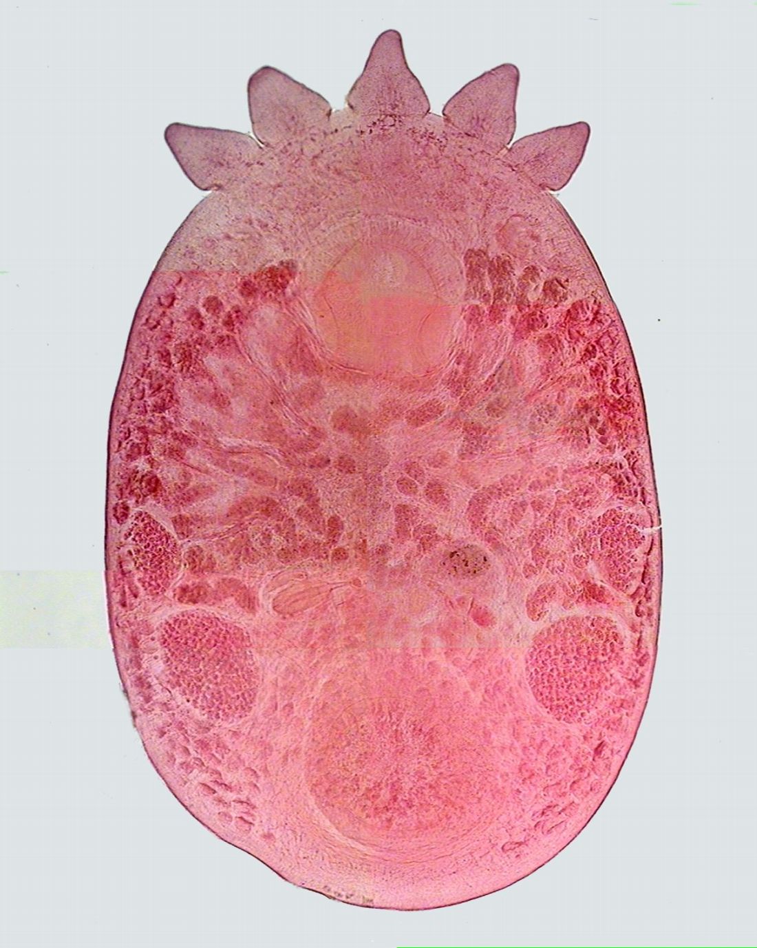
damaging its associate. The following image, typical of a “commensal”
must
necessarily be large so that its structure is well appraised.
|
|
Temnocephala
pignalberiae, Dioni,
1967 – Commensal on crabs from the Paraná River. Mosaic of
6 images, bright field x4.
Preparation made by Prof. Cristina Damborenea (Univ.de la Plata, Rep.
Argentina). This and other Temnocephala
will be described in a special article.
|
Symbiosis implies that both organisms
benefit from each other by the intimate relation established between
them. The most
well known and complete example are the lichens, among the plants
and
termites among the animals. Lichens are classified in genus and
species
although in fact each “species” is an intimate and indissoluble
relationship
between a species of alga and another one of fungus.
|
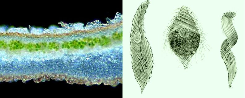
Left.- hand made
section of a lichen, trimmed from a picture posted to the "Forum
Mikroscopia", by Advocat Andre
(Aphylla) on 01/08/05. He describes an upper layer of hypha, underwhich
there is a green layer of algae continued by the bluish medula and a
lower grayish layer both of which are also hyphae. I have never seen
such a clear illustration of the structure of a lichen. Right. - magnificent
drawings,
made by J. Leydi
of termite symbionts. He didn't know of planachromatics, apochromatics,
photomicrographic cameras, and so on. Surf the web for drawings by Joseph Leydi to see his
impressive plates of protista.
|
A more spectacular
symbiosis, with probably the older origins,
although it has been accepted only recently (after 1980), is the one of
all living cells with its mitochondria. According to the
concepts accepted today (although sometimes still discussed)
mitochondria are
protobacteria (prokaryotic
organisms, that is to say, without nucleus) with aerobic breathing,
that introduced itself into eukaryotic cells (those with nucleus but
which were anaerobic)
obtaining food and protection, and offering them true power factories
and the
possibility of using the oxygen which by that time began to dominate
the
atmospheric
gases.
In the following
elementary
references, you can see in addition to a summary of the modern theories
some
illustrations of the structure of mitochondria.
HTTP:they //en.wikipedia.org/wiki/Endosymbiotic_theory
Today it is unthinkable for the
cellular operation of all living beings
to occur without these symbionts.
PARASITISM (another form of mutualism)
There
is nevertheless a habitat that the amateurs explore exceptionally
and if he does, it is generally when he is already an advanced
microscopist. It
is no less interesting and it can be really fascinating. It is the
world of the
animals that choose
to live at the expense of other
living species: they are the PARASITES.
Each species of plant or animal
has several parasites, generally
specific, which is to say that they only live on that species, and
normally in
a well defined niche (place and environment).
Apart from my picture of
gregarines below (fig 1), the presented few examples are
only to suggest the enormous variety of parasitic species, and have
been
obtained and
modified from the Internet. It must be greatly acknowledged the
contribution of
those who share their images, and have contributed effectively to the
extension
of knowledge of this form of microscopic invertebrate life.
It
must be clear that numerous vegetable parasites exist; but they are
mostly out of the sphere of knowledge of this author.
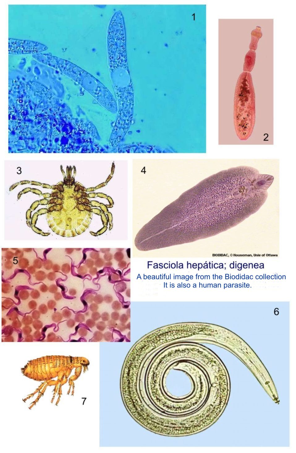
|
1- gregarines
(protozoa, apicomplexa) from the intestine of a naidid, x100 HI,
oblique illumination, teinted blue. 2 - Echinococcus granulosus, a cestode
that parasitises dogs as an adult. 3 - an acarina parasite of domestic
dogs and cats. 5 - a blood parasite, a flagellate of the genus Trypanosoma. 6 - a nematode
parasite on plants. 7 - one of the well known fleas.
|
This
is the first article in a short series that
tries to introduce the microscopists
with a spirit of adventure to the investigation of this specialized
habitat
that just as the outer world offers a great diversity, and demands a
variety of
techniques to be investigated.
I
could have selected as a subject, a frog or toad, with the certainty
to find more parasite diversity.
In
them it is easy to locate trypanosomes
in the blood, apicomplexa
(previously called esporozoa) in the
gall bladder, a variety of trematodes
(Digenea) (different species according
to the location) in the mouth, stomach, thin intestine and rectum, and
also in the
urinary bladder, and even in the lungs, where they are also located
specific nematodes. If
this is not enough they also have cestodes in its intestines.
And have even external parasites
like red acarii that
adheres to their skin.
But
surely, although this choice would be most beneficial, probably,
and for several reasons, it is not the best one for a first experience
of
amateur microscopists into the investigation of those who live hidden
in
the inner
world.
I
have then chosen a bug that only very few people consider a friendly
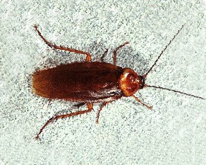
pet: the cockroach. The added picture shows a Periplaneta
americana, the most common cockroach in México taken with
the Cannon Power Shot A300. Natural size.
The
second and
third parts of this series are dedicated to this interesting insect and
to the
even more interesting parasites that are associated with it.
Comments to the author,
Walter
Dioni , are welcomed.
© Microscopy UK or
their
contributors.
Published in the June 2005 edition of
Micscape.
Please report
any
Web problems or offer general comments
to the Micscape
Editor.
Micscape is the on-line
monthly
magazine of the Microscopy
UK web
site at Microscopy-UK
©
Onview.net Ltd, Microscopy-UK, and all contributors 1995 onwards. All
rights
reserved. Main site is at www.microscopy-uk.org.uk with full mirror at www.microscopy-uk.net.
|


