I'm sure plenty of you will have seen advertised on eBay digital eyepiece cameras designed for microscopes. This is my brief review of two such cameras.
The Cameras
The two camera's themselves are shown below:
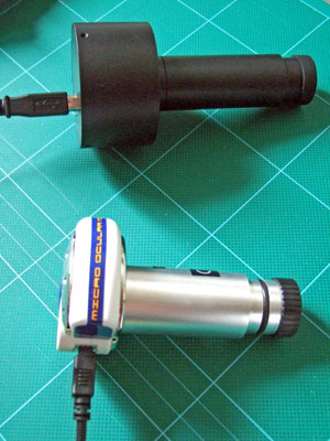
The upper most is a 5 megapixel camera - the DCM500. The lower was is a 1.3 megapixel camera - model number MD-130.
Although not specified I believe both are made in China. Click here to view the The specs for the DCM500 can be found on the manufacturer's web site.
I had problems finding specs for the actual MD-130 camera, the closest match can be viewed by clicking here. However this is not the same camera and not only looks different but has different software. Editor's note April 2008. A reader kindly suggested this MD-130 specifications link.
My set up
I have a Leica CME microscope which I also bought off eBay! I also have the Leica darkfield slider. I'm very new to microscopy and still a huge amount to learn about even using the microscope! So please excuse the rather poor quality of slides and common subject matter.
Initial Impressions
The 1.3 megapixel MD-130
Installation
The camera didn't come with the software the seller claimed it did on the eBay sale. In fact it had a basic driver and some software designed for a web cam.
Installation proved tricky as the manual ( in Word document format on the CD-ROM ) didn't match the actual camera and software supplied. Undaunted I inserted the CD ROM and prepared for install. A menu box popped up with 2 options as regards my camera's frame rate. As the manual was for a different camera I had no idea which of the 2 options to choose and went for a random choice! This turned out to be wrong and after an uninstall and re-install I chose the right option!
So not exactly smooth so far.
In Use
Once I got over the install and the lack of microscope specific software I actually found the images from the camera were not too bad. Unlikely to win awards in photographic magazines but perfectly acceptable for keeping track of things viewed under the microscope. I use it to record what I've seen, then use the image later when trying to identify the subject matter.
The ability to take videos of items under the microscope is particularly fun and quite useful. Still pictures don't tell the whole story and actually seeing how things move is so much more useful. Again the image quality won't be used by the BBC Planet Earth team any time soon but is perfectly acceptable.
The camera and software are very easy to use and generally little adjustment is needed. Having seen something interesting under the microscope, it only takes a minute to get the camera attached, software running and start taking photos and videos.
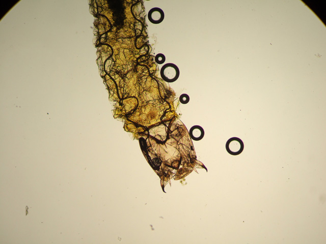
Sample Images
As I'm fairly new to microscopy, the slides are poorly prepared and far too many bubbles got in!
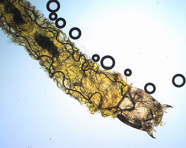
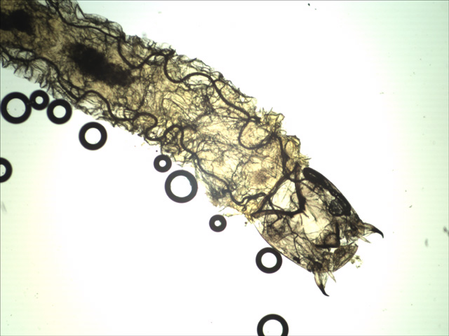
Larva slide 40x - View Larger Image
Image above taken straight - no adjustments to camera settings. Microscope needed re-focusing but otherwise no changes.
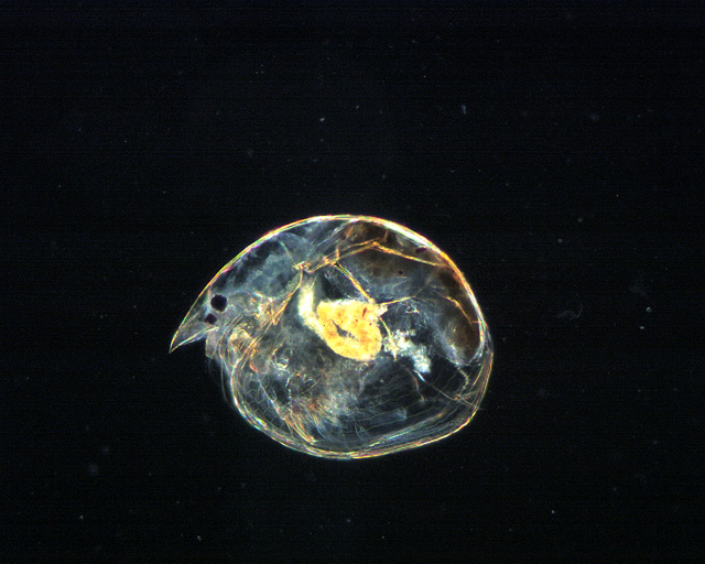
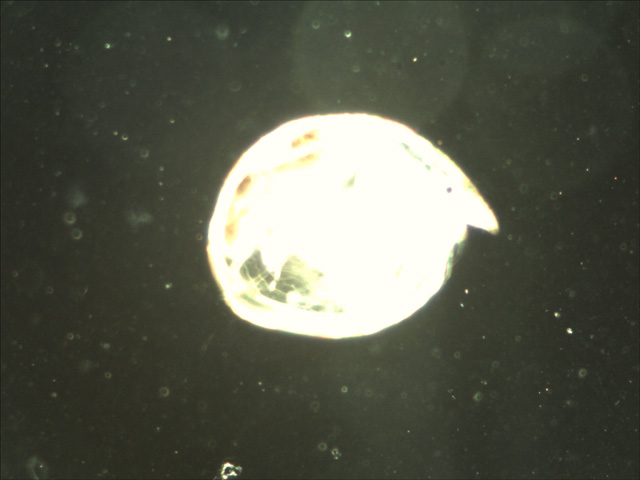
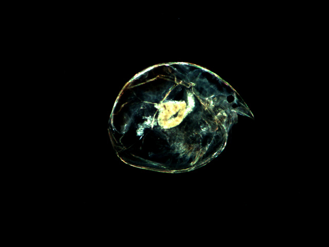
Water flea 100x darkfield. View Larger Image View PhotoShop Adjusted Image
Some small adjustments made in exposure settings on camera. I've also tweaked a version in Photoshop - helping enhance details.
Videos
Both videos (mpeg format) are quite large and in original size and compression. Right clicking and choosing save works best as at least you get a download progress indicator!
Brachionus_Calyciflorus 400x - 9mb
(Editor's note: the videos are excellent and well worth the download time.)
The 5 megapixel DCM500
Installation
Installation went much more smoothly. This time I got the software designed for the actual microscope camera. A good start and within minutes I was ready to go!
In Use
There were 3 main issues with the actual camera:
1) Very significant and for me unacceptable levels of vignetting.
2) The camera's driver software appeared to make it virtually impossible to get a decent image. Attempts to change exposure and white balance settings were futile.
3) Live image update was slow - very, very slow. Any changes to the image or settings took half a second to reflect in the image. This may not sound long but made focusing, white balance and exposure setting very tricky.
Having set up the microscope and installed the digital eyepiece I launched the software and used the default image settings ( exposure and white balance being the most vital ).
On default settings the images looked awful! Exposure and white balance were all wrong ( see images ).
So I turned to the camera's settings menu and proceeded to play with the exposure and white balance settings. There's a choice of auto settings where you specify a preference ( darker, lighter etc ) and the software tried to match that. I found the tools to be very much a blunt instrument - either too dark or too light but never just right. Also while I might get the exposure acceptable it'll cause the white balance to change. So I'd adjust the white balance to barely acceptable which caused the exposure to change - arrrrggghh!
There are manual settings and these worked better though not 100%. Exposure was acceptable to use but manually adjusted white balance by moving 3 sliders ( red, green, blue ) was a massive challenge.
I doggedly persisted and was determined to get a decent image. After a few very frustrating hours I gave up. The images you see in the next section are the best I could manage after a LOT of fiddling with both microscope and camera settings.
Sample images
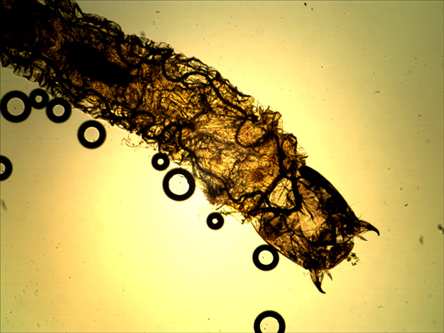
Larva 40x - default camera settings. View Larger Image
Larva 40x - after a LOT of fiddling with camera and microscope settings. View Larger Image
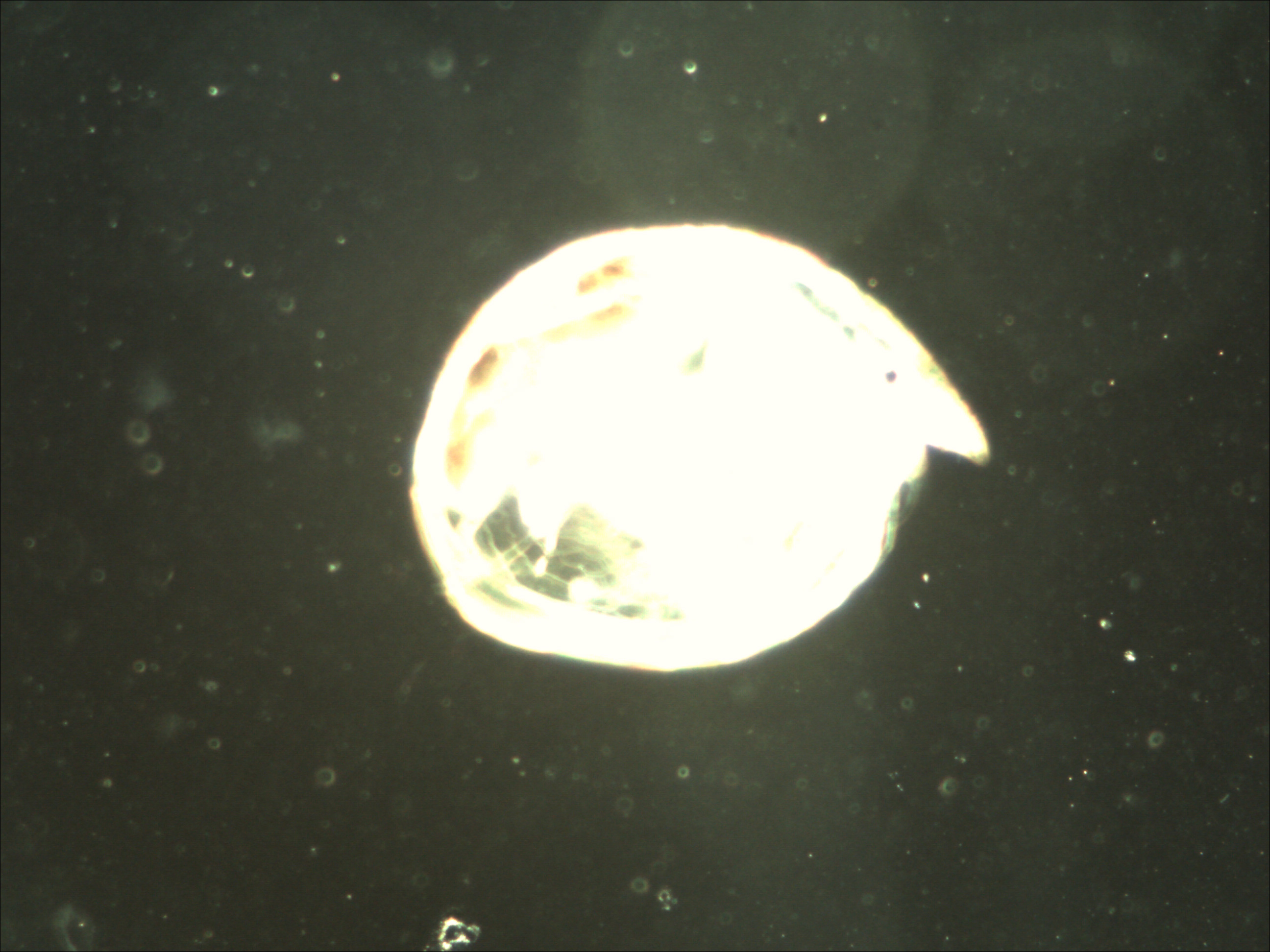
Water flea darkfield 100x. Default "auto" settings. View Larger Image
Water flea darkfield 100x. After a LOT of adjustments. View Larger Image
Videos
I did do some test videos but found that:
1) On maximum setting the frames per second rate of 2 is way too slow for a video.
2) Changing the video size to a smaller size resulted in poor image quality and lots of pixellation.
None of the videos are worth uploading.
A "Normal camera"
For comparison here's a shot taken with a compact digital camera via the normal microscope eyepiece:
Larva taken using Fuji compact digital camera through eyepiece.
Conclusion
The 5 megapixel eBay camera was a total waste of money. Very hard to adjust to get a good image. 2 frames per second video is too slow. I'm currently trying to get my money back but this being eBay it's not looking hopeful! I wasted £200 on this pile of junk and I only hope anyone contemplating buying one of these sees the review first!
The 1.3 megapixel camera is actually not too bad. Easy to use, reasonable speed of refresh in videos and often requiring little adjustment to produce acceptable images. I still have this camera and use it regularly. Image quality is far from outstanding but it'll do until something better comes along.
Note added by author March 2010: A lot of eBay sellers are providing the camera with no support or software, making the camera unusable. Check first with the seller to see if it comes with software for your operating system. Unfortunately I no longer have or use this camera and don't have any software I can provide.
Editor's note March 2010: Owners of TWAIN compliant cameras without software or find the software unsuitable may wish to try the freeware Micam software by Marien van Westen. It is designed for microscopy stills / video capture and includes measurement and documentation features.
All comments to the author Paul Wilton are welcomed.