Water Immersion Phase Contrast
Robert Pavlis, Girard, Kansas USA
In 1953 Frits Zernike was awarded the Nobel prize in physics for the development of phase contrast microscope objectives. When transparent objects are immersed in a transparent medium they normally are visible only because the path of light is deviated at the interface between the medium and the object by refractive index differences. High numerical aperture objectives are essential for high power, high resolution microscopy. Such objectives take in a very wide cone of light. This results in almost all of the deviated light being "scooped up" by the lens, as well as light that passes straight through. The result is that as numerical aperature is increased, transparent objects become less and less visible.
Phase contrast objectives require that the subject be illuminated by a hollow cone of light. That light that passes through the cone is passed through an annular neutral density filter in the back of the objective that also throws the light passing through it out of phase from light that has been deviated by the specimen, so that when the deviated and non-deviated rays combine they combine with strong phase differences that result in constructive or destructive interference.
Oil immersion lenses depend on having the specimen and all the space between the specimen and the objective being immersed in an clear material having the same index of refraction as the lowest element of the objective. The velocity of light is diminished by being in this medium, which shortens its wavelength, and quite dramatically increases resolution.
There is an optical problem here which is not addressed as often and as well as it should be: As stated above oil immersion lenses require that the specimen and all the space between the specimen and the objective have a constant index of refraction equal to the refractive index of the lower element of the objective. Phase contrast is primary used to observe small living microorganisms. Small living microorganisms do not ordinarily survive in media other than water. The index of refraction of water is about 1.33. The index of refraction required for the space between objectives and the specimen is normally about 1.518. When light passes from one index of refraction it is deviated according to Snell's law.
Suppose an aqueous preparation of bacteria is placed on a microscope slide, with a standard cover slip over the bacteria laden water, and standard immersion oil is placed between the cover slip and the objective. Unless the bacteria are directly in contact with the cover slip, the light first passes past them, and then hits the cover slip. When it does so, its path is deviated. This introduces severe spherical aberration!
Standard oil immersion and phase contrast observation at high powers are thus rather incompatible things.
There is an obvious solution to this problem: Design the objective so that it is corrected for WATER immersion, rather than air or oil. There really are two rather different ways to do this: (1) Design a lens so that it produces an image without spherical aberration when all the space between the lower lens element and the specimen are immersed in water. (2) Design a lens so that it produces an image without spherical aberration when there is a glass cover slip between the specimen and the object with water above and below the cover slip.
Microscope manufacturers have produced objectives of both of these designs. The disadvantage of the first type is that the living specimens are not confined. They can move any place between the slide surface and the objective surface. The disadvantage of the second type is that cover slips tend to vary in thickness, requiring that a spherical aberration collar be incorporated into the design.
Unfortunately although microscope manufacturers have tended to produce many phase contrast objectives and also many water immersion objectives, they have produced very few phase contrast water immersion objectives. Recently this situation has changed, but the phase contrast water immersion objectives that are produced today generally are sold for truly amazingly high prices. These objectives are generally worth literally more than their weight in gold.
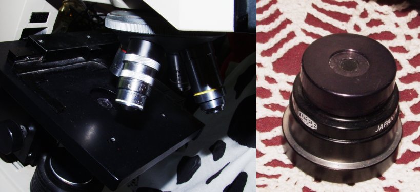
LOMO once produced a limited number of apochromatic 1.23 na 70X water immersion phase contrast objectives. From time to time these lenses appear for sale, often from vendors in Russia or other countries that were in the Soviet Union. These lenses are based on an ancient Zeiss design, but they have phase annuli.
I obtained one of these lenses some time ago. As is really essential for such a lens, it has a correction collar to compensate for different thicknesses of cover slips. Although it has substantial field curvature, the images are extremely sharp, even very small bacteria like E. Coli stand out sharply, dramatically better than with oil immersion phase contrast.
This lens is designed for the old RMS 31mm parfocality standard. With some modern microscopes it may be necessary to use some sort of lens extender to bring it close enough to the specimen to focus. The phase annulus of this lens is of a very odd size. Standard LOMO, Zeiss, Leitz, Nikon, Olympus, and Wild phase contrast condensers do not work with it!
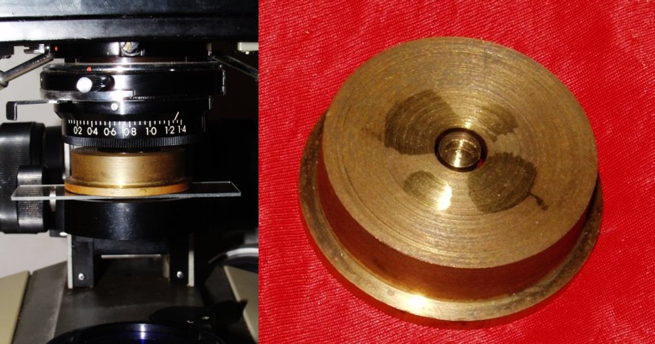
There are four obvious solutions to the phase annulus problem: (1) replace one of the rings in a standard condenser with a new ring whose diameter matches that of this LOMO lens. This is extremely difficult to do! (2) obtain a variable annulus phase condenser. The best is certainly the Leitz Heine condenser. They are expensive and hard to locate. Olympus has made a somewhat crude variable annulus condenser that they supplied with some of their CH microscopes. I have one of these, it works quite well. (3) Fabricate a phase ring out of aluminium or brass to fit beneath the condenser of a standard microscope. I tried to make an attachment to the bottom of the lens, but this turned out to be unusable because of centring problems. I found that the best solution was to make a stop that fits on top of a glass plate that can be moved until it is centred. I made a phase annulus for another microscope's condenser system that turned out to be a bad optical design for it. Amazingly enough, however, it was perfect for this lens!!! (4) Try to locate one of the original LOMO phase condensers used with these.
There is, by the way, one wonderful use of oil immersion phase contrast lenses: They are ideal for crystallographic and geological specimens.
The reality is that water immersion is about the only good way that has ever been designed that provides really good images of living bacteria. They are otherwise difficult to observe for two fundamental reasons:
(1) Bacteria are smalltheir dimensions typically run between 0.5 and 5 microns. Their dimensions are similar to the wavelength of visible light (about 0.38 to 0.72 microns).
(2) Most bacteria are colourless. However, their index of refraction is somewhat greater than water.
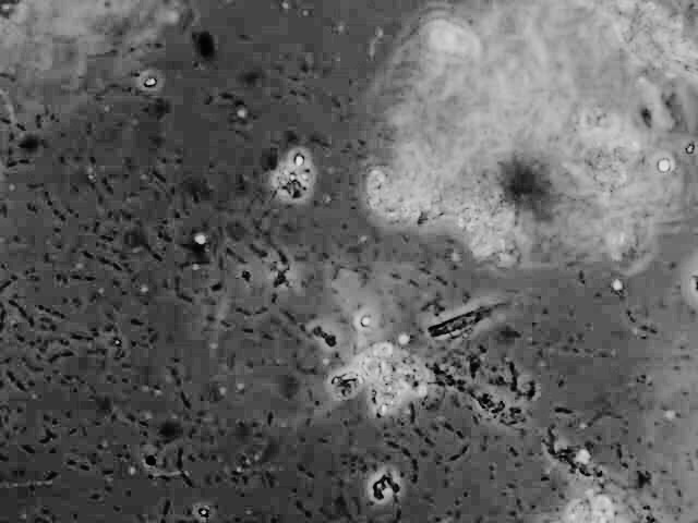
Observing mobile bacteria with this lens is truly amazing! If the preparation contains food particles they will move around it in great swarms! It is amazing that a so called "simple" cell is able to move in such an organised fashion.
Non motile bacteria that are not attached to the slide or coverslip are also in motion from the so called "Brownian motion". Bacteria are so small that they are bounced around by statistical variation in the number of collisions they undergo with water molecules!
The image below shows an aqueous preparation using this lens.
Another way to observe living bacteria is with dark field illumination. Dark field condensers project a hollow cone of light onto the subject so that the objective cannot take in the edge of this cone. Oil immersion dark field condensers typically have an "inner aperature" of about 1.1 or so. This generally permits their use with any dry objective, and it also permits their use with oil immersion objectives that are equiped with a special iris.
All comments to the author Robert Pavlis are welcomed.
Microscopy UK Front Page
Micscape Magazine
Article
Library
Please report any Web problems or offer general comments to the Micscape Editor .
Micscape is the on-line monthly magazine of the Microscopy UK website at Microscopy-UK