 Insight into the fascinating world of magmatic crystals –
Light and electron microscopy of accessory
zircon
By Robert Sturm
Elsbethen, Austria
Introduction
ircon
may be regarded as a remarkable and, from the geoscientific
point of view, as a very interesting mineral worthwhile for extensive
investigations, because it represents one of the very few mineral phases
occurring in a wide spectrum of magmatic,
metamorphic, and sedimentary rocks. Zircon thereby belongs to the so-called
accessory minerals which in contrast to the so-called main minerals are
characterized by a rather low abundance. In a granitic
rock sample with a mass of 10 kilograms, the respective mass of zircon crystals
amounts to about 1 gram. Since zircon mainly occurs as a mineral phase being
included into the main minerals, its size exceeds 1 mm only in exceptional
cases. Normally, a single crystal has a
length between 50 and 300 µm and a width between 20 and 80 µm. Besides its
ubiquitous occurrence, accessory zircon is marked by some further
characteristics enhancing its scientific value significantly. First, the
mineral has the unique ability to survive several cycles of erosion,
sedimentary transport, diagenesis, and metamorphism,
thus representing an excellent protolithic indicator
containing information of the original rock, within which it was formed.
Second, zircon contains minor but measurable amounts of U and Th and therefore may be subject to any dating procedures
yielding ages of crystallization, magma cooling as well as element
redistribution within the mineral phase. By using this dating technique it
could be shown that some zircon crystals have an age of more than 1 billion
years (1,000,000,000 years) and thus were involved into several cycles of orogenesis (formation of mountain belts). A third reason
underlining the fascination and scientific importance of zircon concerns its
way of crystallizing out of the magma. Only in very few cases, crystal growth
may be evaluated as continuous, with nearly equal amounts of chemical components
being added over the whole duration of crystallization. In most cases, however,
crystal growth is carried out in a discontinuous, more stepwise fashion due to
more or less rapid changes of chemical or thermal conditions in the direct
vicinity of the crystallizing mineral phase. As a consequence
of this ‘step-by-step’ growth, respective growth bands or zones are
established which sometimes can be already observed under the light microscope
but are best visualized with electron microscopic techniques (e.g.
backscattered electron imaging or cathodoluminescence).
By chemically investigating the growth zones of a zircon crystal, important information
on chemical and physical changes of the magma during the cooling process may be
obtained. What is the chemical composition of accessory zircon? In the ideal
case the mineral with the chemical formula ZrSiO4
consists of 67.1 % ZrO2 and 32.9 % SiO2. In natural zircon about 50 more elements
occur in the crystal structure, from which Hf, Y, P,
U, Th, La, and Rare Earth Elements are most
prominent. As will be shown in detail in the next chapter, the morphology of
zircon crystals is marked by two types of prisms and three types of pyramids,
being combined in several different ways (Speer, 1980).
Light Microscopy
efore
studying zircon crystals under the light microscope they have to be separated
from their host rocks and prepared according to well defined procedures.
Successful extraction of accessory zircon from the host rock includes rock
crushing with a hammer, milling, sieving, floatation (coarse separation of
single mineral phases according to their specific weight), magnetic separation,
and, finally, separation using so-called heavy liquids (Unfortunately, these
liquids are uniformly classified as extremely hazardous!). After this
time-consuming procedure, grains have to be prepared for light microscopy which
is best carried out by placing some zircon crystals on a glass specimen, adding
some Canada balsam or resin with high light fraction, and covering the crystal-liquid
mixture with a thin cover slip.
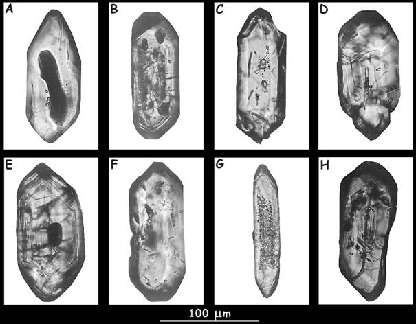 Figure
1. Light
microscopy of zircon crystals.
Figure
1. Light
microscopy of zircon crystals.
With
this rather simple microscopic technique it is already possible to visualize
the extern morphology of single zircon grains, respective growth zones (see crystals
B, E, and H), and mineral inclusions of different size.
°°°°°°°°°°°°°°°°°°°°°°°°°°°°°°°°°°°°°°°°°°°°°°°°°°°°°°°°°°°°°°°°°°°°°°
As exhibited in Figure 1, light microscopy
enables the detailed study of the outer or external morphology of accessory
zircon. While some grains are characterized by steep pyramids (e.g. crystals A
and G), others show a predominance of flat pyramids (crystals B, D, E, F, and
H). Most crystals separated from granitic rocks
contain both types of pyramids, being marked by a more or less remarkable
difference in size. It has been found that in granites with high contents of
Al, termed ‘peraluminous’, steep pyramids dominate
over flat pyramids, whereas in granites with high contents of Ca, K, and Na,
termed ‘calcalkalic’, the opposite case may be
observed (e.g. Vavra, 1994; Sturm, 1999). Besides the
pyramidal morphology also the prism morphology plays an important role
concerning the crystal shape of accessory zircon. Differing between the two
prism faces is indeed not easy under the light microscope but can be realized
in the following way: 1) The crystal is positioned on the prism {100}, if the
angle of the two pyramidal faces crossing on the top amounts to 96° (grains B,
D, E, and F in Figure 1). 2) The crystal, on the other hand, is positioned on
the prism {110}, if this angle has a value greater than 112° (grains A, C, and
G; see also Figure 2).
Another typical characteristic of
accessory zircon being already noticeable under the light microscope is its
growth zoning which in numerous cases is represented by a set of more or less
concentric growth shells (grains B, E, and H). If the growth zones are too weak
for an appropriate visualization under the light microscope,
electron-microscopic techniques have to be applied (see below). Zircon is normally characterized by a wide
spectrum of inclusions ranging from magmatic fluids
to mineral phases, from which apatite occurring as small needles (grains C, D,
and F) is most prominent. With light microscopy these inclusions can be
categorized and quantified.
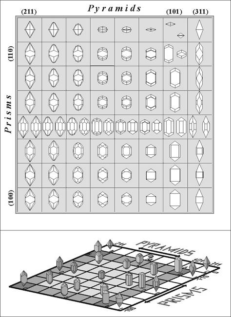 Figure 2. Typology diagram introduced by Pupin and Turco (1972) for the systematic classification of the
zircon morphology.
Figure 2. Typology diagram introduced by Pupin and Turco (1972) for the systematic classification of the
zircon morphology.
A
statistical evaluation of the average zircon morphology within a zircon
population consisting of all zircon crystals separated from a specific magmatic, metamorphic or sedimentary rock was outlined by Pupin and Turco (1972) as well as
Pupin (1980). Within this two-dimensional
classification scheme, eight rows and eight columns defining 64 crystal shapes
are given. Crystals
of the first row containing no prism faces and crystals of the 8th
column containing the very steep pyramid {311} do not occur with high frequency
in natural rocks. From row 2 to row 8 the ratio of size between the two main
prisms {100} and {110} is continuously changed. While in row 2 zircon crystals
exclusively develop the {110} prism, in row 8 only the {100} prism occurs. In
row 3 to 7 intermediate forms can be observed. The same arrangement is given
for the pyramids, i.e. in column 1 only the {211} pyramid is developed, whereas
in column 7 the {101} pyramid is not accompanied by a further pyramidal form.
In columns 2 to 6 the ratio of size between the two pyramids is continuously
changed with {211} becoming smaller and {101} becoming larger. In column 4 the
two pyramids are equally sized. A very specific crystal type is that in column
4 and row 5, because it develops not only pyramids but also prisms with the
same size.
To give
a further impression of the various crystal shapes resulting from the
combination of the prism and pyramid forms, another, let’s say, 3-dimensional
Typology diagram is illustrated below. From the examples drawn in this diagram,
the high variability of zircon morphology can be well estimated. The diagram
also shows a subdivision into several subtypes (A, B, C, etc.), which, indeed,
is important for a scientific analysis of a specific zircon population, but
would exceed the aim of this small contribution.
°°°°°°°°°°°°°°°°°°°°°°°°°°°°°°°°°°°°°°°°°°°°°°°°°°°°°°°°°°°°°°°°°°°°°°°°°°°°°°°°°°°°°°°°°°°°°°°°
ith electron
microscopy further detailed information on single zircon crystals can be
obtained. While scanning electron microscopy (SEM)
mainly serves for the investigation of the crystal surface, which is important,
if zircon underwent e.g. an extensive mechanical metamorphosis, electron
microprobe analysis of oriented crystal sections provides essential information
on crystal growth and chemistry. For SEM single
zircon crystals are picked out of the separated zircon fraction and are
afterwards mounted on a glass slide using resin (e.g. Epon).
After coating of the crystals with carbon (This is necessary for the leakage of
electrons hitting the sample.) SEM procedure can be
started. Respective results of this very impressive microscopic technique are
presented in Figure 3.
Figure 3. Scanning electron microscopy (SEM)
of selected zircon crystals.
The
morphology of single crystals is very well recognizable, whereby most grains
show a clear predominance of {211} over {101} and of {110} over {100}. Crystal morphology mainly
composed of {211} and {110} can be frequently separated from granitic rocks characterized by an enhanced concentration
of aluminium (Al) and a lower content of calcium (Ca). Such rocks are usually
termed ‘peraluminous’.
Regarding
the crystal surface, significant differences among the exhibited zircon grains
can be determined. Crystals A-G are marked by a well
developed surface which only shows some small scratches and roughening. The
pyramidal tops are not mechanically damaged. Another picture is given for
crystals H-J, where roughening and scratches become more remarkably intensified
and are added by so-called corrosion pits. Cracks running over
the crystal faces (grain I) give evidence for an increased mechanical influence
due to brittle or ductile shearing.
°°°°°°°°°°°°°°°°°°°°°°°°°°°°°°°°°°°°°°°°°°°°°°°°°°°°°°°°°°°°°°°°°°°°°°°°°°°°°
For obtaining information on the growth
development of individual crystals, electron microprobe analysis (EMPA) has to be regarded as a preferential technique. An
appropriate application of this technique, however, requires an extensive and
time-consuming preparation of single zircon grains. A scientific study of the
pyramidal growth is only enabled, if individual crystals are sectioned parallel
to their crystallographic c-axes, defining the axes running trough the
pyramidal tops (‘longitudinal section’, Figure 4, 5). Prism growth, on the other
side, can only be investigated in detail, if respective crystals are sectioned
perpendicular to their crystallographic c-axes (‘cross section’, Figure 4, 5).
Good results with the microprobe are guaranteed by using a
accelerating voltage of 15 to 20 kilovolts (kV) and a electron beam current of
30 to 40 nanoamperes (nA).
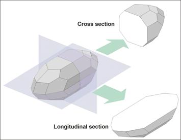 Figure 4. Preparation of zircon
crystals for backscattered electron imaging and chemical analysis using the
electron microprobe.
Figure 4. Preparation of zircon
crystals for backscattered electron imaging and chemical analysis using the
electron microprobe.
A
selected crystal can be sectioned either parallel or perpendicular to its
crystallographic c-axis, resulting in so-called longitudinal or cross sections.
As demonstrated in the following figure, longitudinal sections are most
appropriate for the study of the pyramidal growth development, whereas cross sections
are used for the investigation of prism growth. Before starting crystal
preparation, selected grains have to be mounted on a glass slide and embedded
in a layer of resin. Afterwards starts the grinding and polishing procedure
(Sturm, 1999). For obtaining best results, crystals have to be perfectly cut in
the middle (see the cutting planes in the sketch), because otherwise numerous
effects may complicate growth analysis immensely. Surfaces of the crystals must
be polished perfectly to guarantee an ideal interaction with the electron beam.
°°°°°°°°°°°°°°°°°°°°°°°°°°°°°°°°°°°°°°°°°°°°°°°°°°°°°°°°°°°°°°°°°°°°°°°°°°°°°°°°°°°°°°°°°°°°°°°°°°°°°°°°°°°°°°°°°°°°°°°°
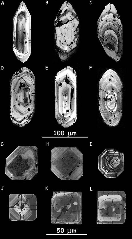 Figure 5. Backscattered electron imaging of zircon
crystals sectioned parallel and perpendicular to their main crystallographic
axes.
Figure 5. Backscattered electron imaging of zircon
crystals sectioned parallel and perpendicular to their main crystallographic
axes.
Crystals
A-F are sectioned parallel to their crystallographic
c-axes, so that respective sections provide useful information on the pyramidal
growth. As can be observed for most crystal, ratio of size between the two
pyramids {211} and {101} is not constant over the whole growth period but
changes continuously. This may be primarily regarded as a result of magma
chemistry changing permanently during crystallization. Most impressive in this
case is crystal C, where early growth stages are characterized by nearly
equally sized pyramids, while the outer or extern morphology (marked by the
outline of the crystal) shows a clear predominance of the steep pyramid {211}.
Crystals
G-L are sectioned perpendicular to their
crystallographic c-axes. Respective sections give an impressive insight into
the prism growth. Similar to the development of the pyramids, also prism growth
may not be classified as constant or static in many cases. Concerning crystals G-J, ratio of size
between the prisms is subject to a continuous change, resulting in
morphological differences between early and late growth stages.
°°°°°°°°°°°°°°°°°°°°°°°°°°°°°°°°°°°°°°°°°°°°°°°°°°°°°°°°°°°°°°°°°°°°°°°°°°°°°°°°°°°°°°°°°°°°°°°°°°°
For chemical analysis crystal sections
prepared for backscattered electron imaging are investigated by electron
microprobe analysis (EMPA). With this chemical
analysis method besides main chemical components of zircon (zirconium (Zr) and silicon (Si) also
chemical components represented with lower concentrations can be detected (see
introduction). The main question standing behind the measurement of chemical
profiles like those provided in Figure 6 is, whether there can be detected a
relationship between crystal chemistry and crystal growth or not.
Figure 6. Chemical profiles of the elements hafnium (Hf),
yttrium (Y), and uranium (U) measured on a cross section of a selected zircon
crystal.
The
profiles should demonstrate that magmatic element
concentrations are not constant over the whole growth period. Some elements
like U show a somewhat oscillating concentration, i.e. growth zones with low
U-content are followed by growth zones with higher U-content and vice versa. As
could be found out in the past, concentration of certain elements in the magma
is an essential factor controlling prism growth of accessory zircon.
Conclusions
rom the
brief study presented here it can be concluded that magmatic
crystals and particularly accessory zircon represent extremely interesting
objects for geological and mineralogical research. With the help of traditional
light microscopy on the one side and modern electron microscopy on the other
side, numerous results concerning magmatic mineral
growth and its control by environmental factors may be obtained. In future,
questions regarding the relationship between magma chemistry and zircon
morphology will be subject to a detailed review and to further investigations.
References
- Pupin,
J. P. (1980): Zircon and granite
petrology. Contrib. Mineral. Petrol. 73, 207-220.
- Pupin, J. P. & Turco, H. (1972): Une typologie originale du zircon
accessoire. Bull. Soc. Fr. Minéral Cristallogr. 95,
348-359.
- Speer,
G. (1980): Zircon. In: Ribbe, P. H. (ed.), Orthosilicates, MSA,
Washington DC, 67-112.
- Sturm, R. (1999): Longitudinal and cross section of zircon :
a new method for the investigation of morphological evolutional trends. Schweiz.
Mineral. Petrogr. Mitt.
79, 309-316.
- Vavra,
G. (1994): Systematics of internal zircon morphology in major Variscan granitoid types. Contrib. Mineral. Petrol. 117,
331-344.
Any
comments on the study or questions concerning mineralogical research are very
welcome by the author Robert
Sturm.