The continuing popularity of the dahlia
is not surprising. Dahlias come in an amazing variety of colours,
shapes and sizes. This member of the Aster family, (Asteraceae), reproduces
vegetatively. Onions, hyacinths, and tulips produce bulbs, crocuses produce a bulb-like
structure called a corm, but
dahlias (and potatoes) produce tubers.
Dahlias are named after the Swedish
18th century botanist Anders
Dahl. Sixteenth century Spanish conquistadors came upon
the ancestors of modern dahlias while conquering the Aztecs in the
mountains of Mexico and Guatemala. (All thirty-five species of
the genus Dahlia are found in
this relatively small region.) Several hundred years later, seeds
and tubers were located at the Royal Botanical Gardens in Madrid,
Spain, from which they were distributed throughout western
Europe. Since then, plant breeders have been actively breeding
dahlias to produce thousands of hybrids. The tremendous variety
of these cultivars is due to the fact that most plants have two sets of
homologous chromosomes, whereas dahlias have eight sets! Dahlias
are therefore referred to as octoploids.
Several dahlia hybrids are
investigated in this article, however photomicrographs of a flowers
reproductive structures are shown only for the last hybrid (near the
end of the article).
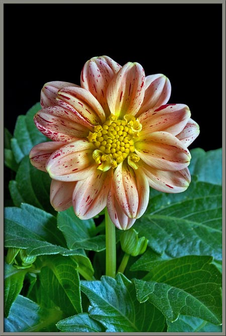
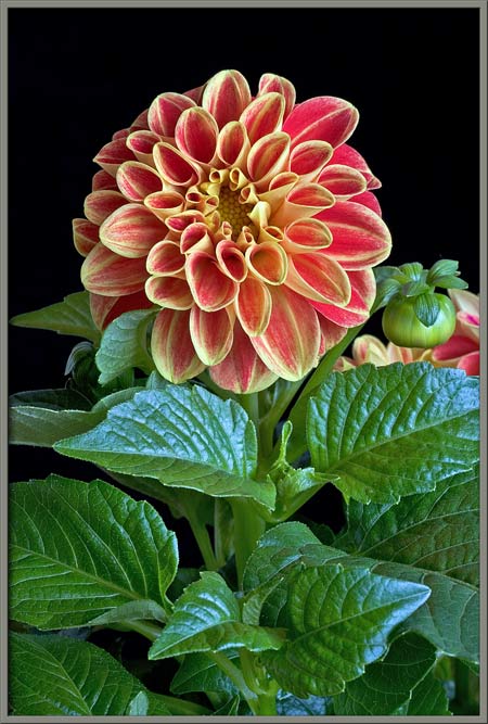
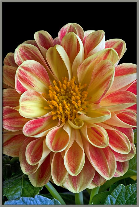
Dahlia
hortensis Patricia
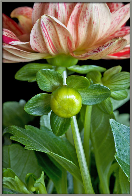
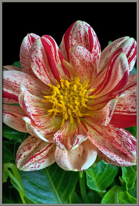
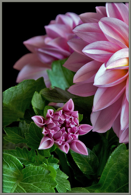
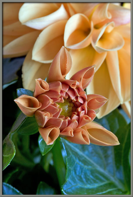
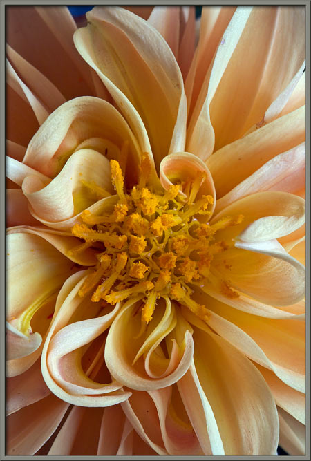
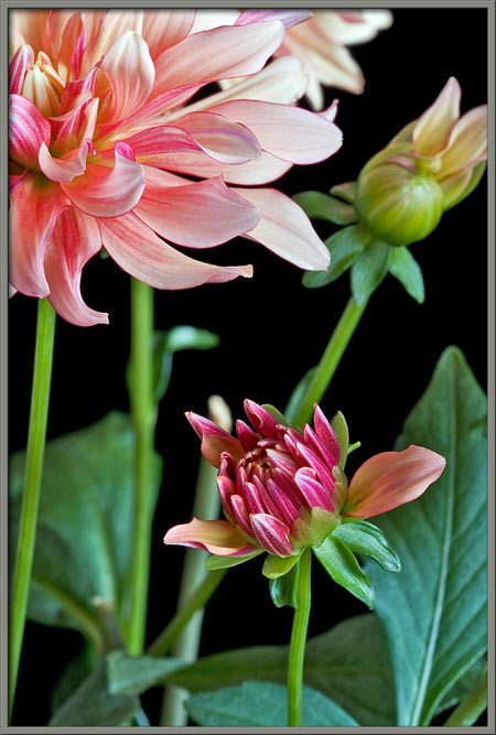
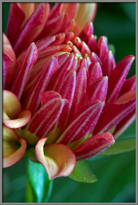
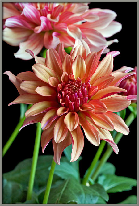
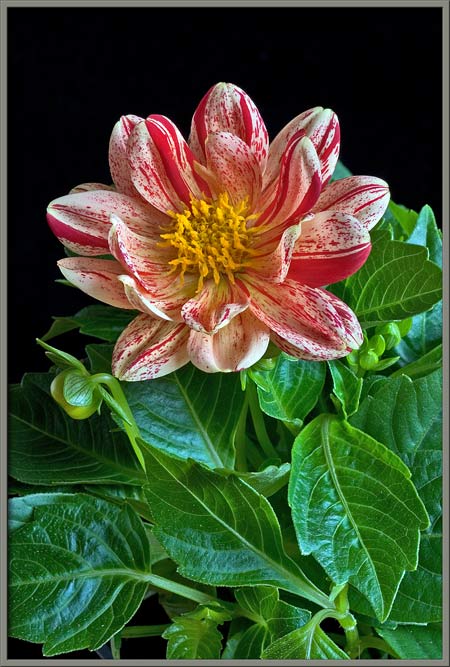
The first image in the article
shows this spectacularly coloured cultivar. Notice in the image
below that a dahlia bud is ringed by five oval, bright green
leaflets. Its hard to believe that the flower shown in the image
on the right had such an unspectacular origin.
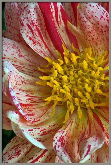
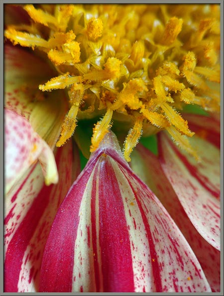
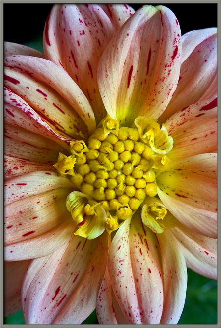
Closer views of the centre of a
flower show many columnar, pollen encrusted pistils emerging from the
disk flowers of the bloom. Careful inspection of the right-hand
image reveals the five pointed, star-like shape made by the fused
petals of each disk flower.
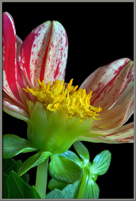
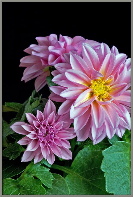
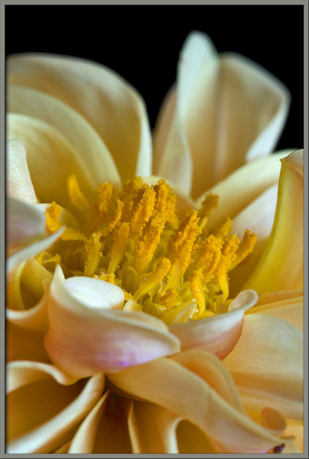
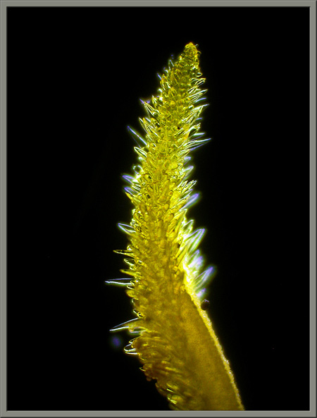
Several petals (better called ray
flowers) have been removed to better show the yellow forest of pistils
(stigmas and styles) at the centre of a flowerhead. A bloom is
cupped by a ring of light green sepals (modified leaves) that make up
the flowers involucre.
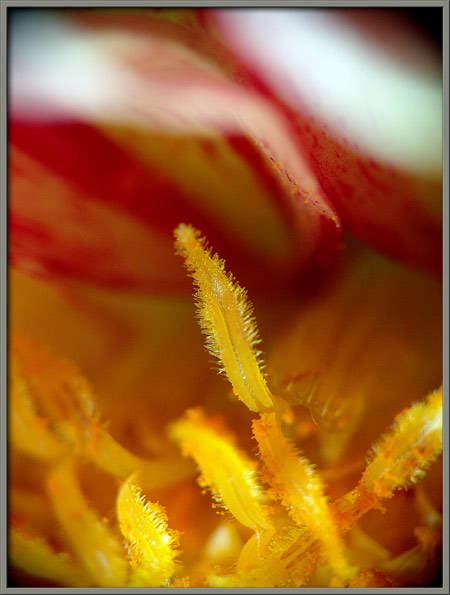
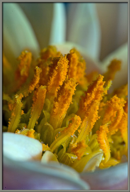
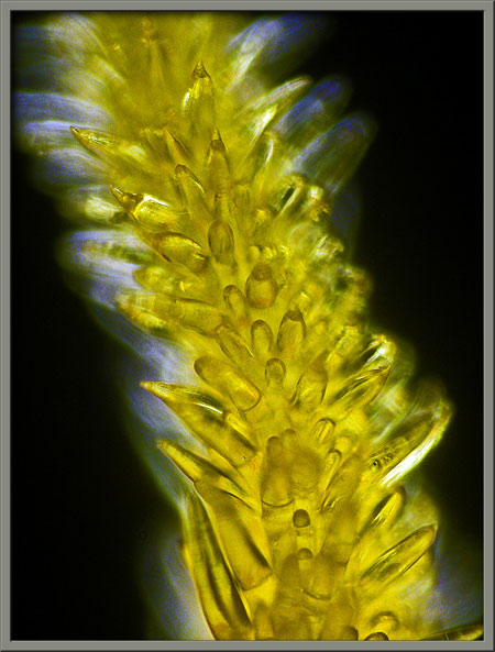
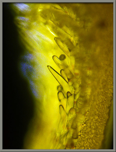
The two images that follow show the
bi-lobed stigmas of disk flowers. Notice the fringe of hair-like
protuberances on each lobe.
I must confess that I do not have a
green thumb. Many times, after photographing a flower, I have
attempted to keep the plant alive for a protracted period of
time. Usually the attempt is unsuccessful! In this case,
flowers continued to bloom, but they were smaller, and much less
spectacular than the original blooms. Here is an example of a
second generation flower to prove my point.
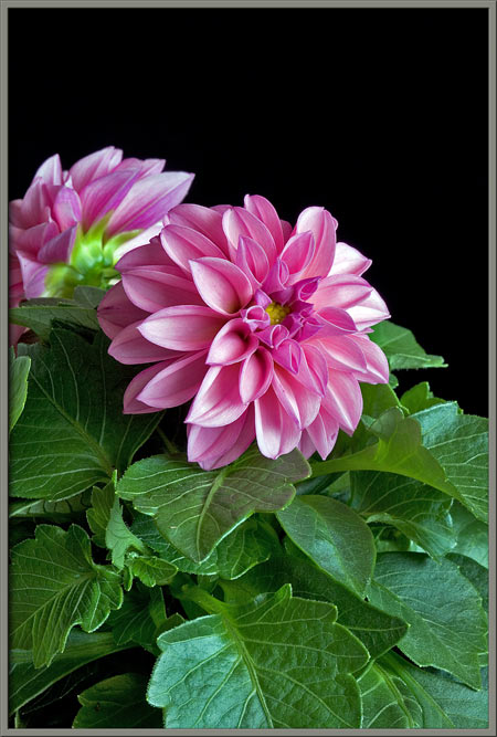
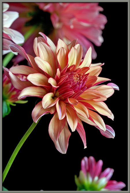
Dahlia
hortensis Apricot Sunrise
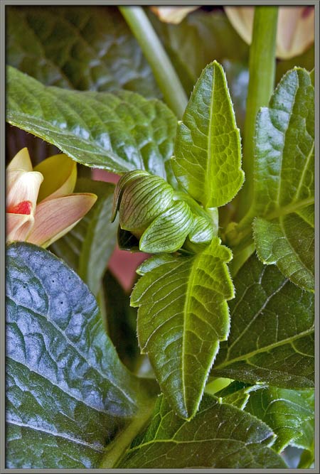
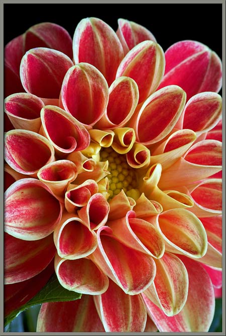
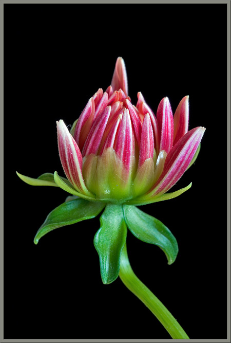
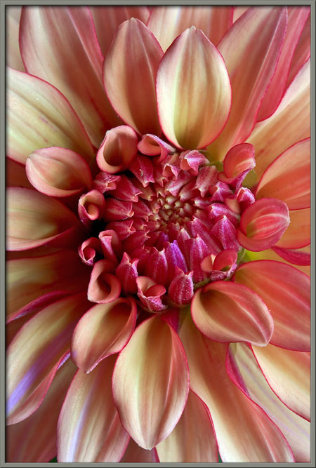
This second cultivar has a less
complicated petal decoration. Notice how the innermost petals
have a rolled-into-a-tube appearance. Also note that the bud
shown in the image on the right has two small leaflets beneath it, as
well as a ring of striated smaller leaflets at its base.
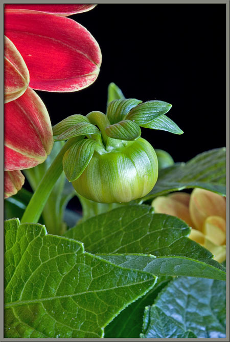
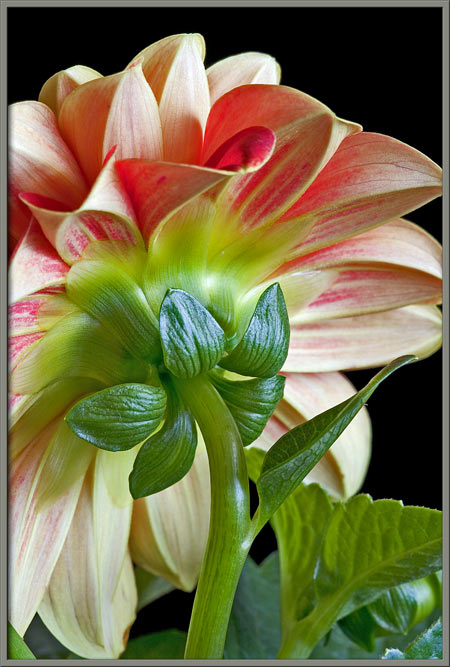
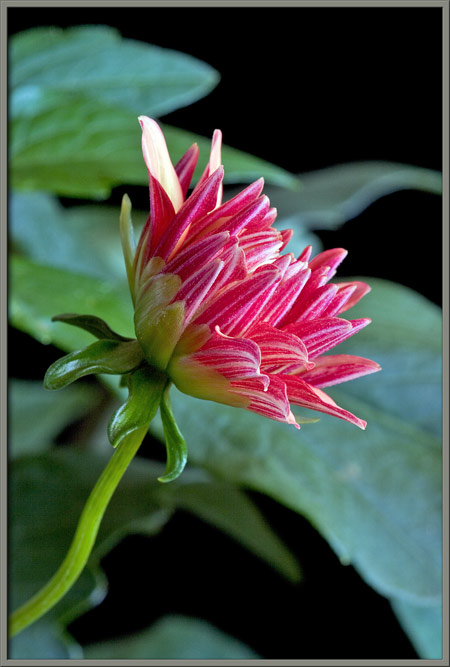
A short time later, the leaflets
have separated from the pumpkin-shaped bud.
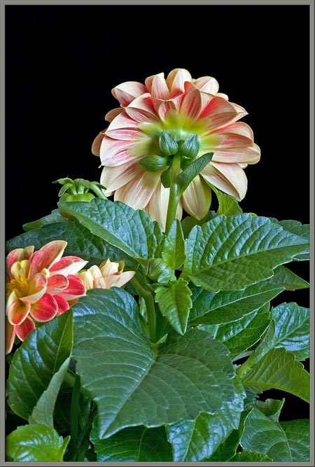
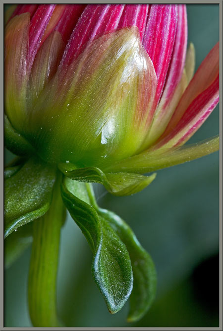
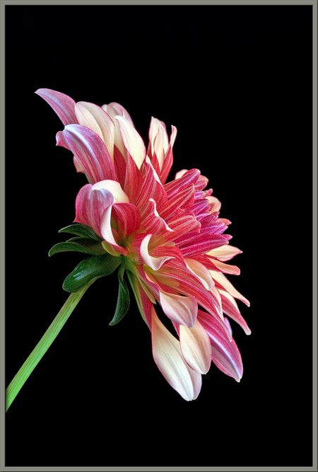
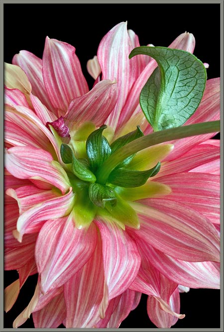
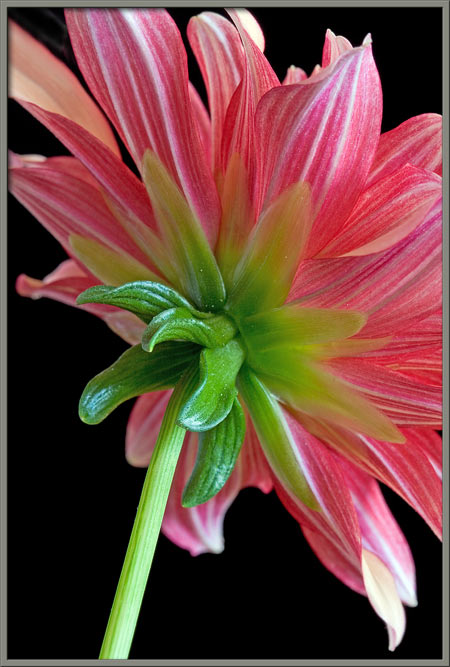
If a blooming flower is viewed from
behind, the ring of leaflets, and a second ring of larger, paler green
sepals (closest to the petals) can be seen clearly.
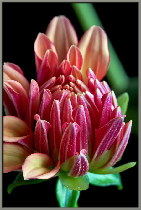
When the flowerhead first blooms,
it is the outer ray flowers that predominate. A dome of
yet-to-bloom disk flowers can be seen at the centre of the flowerhead.
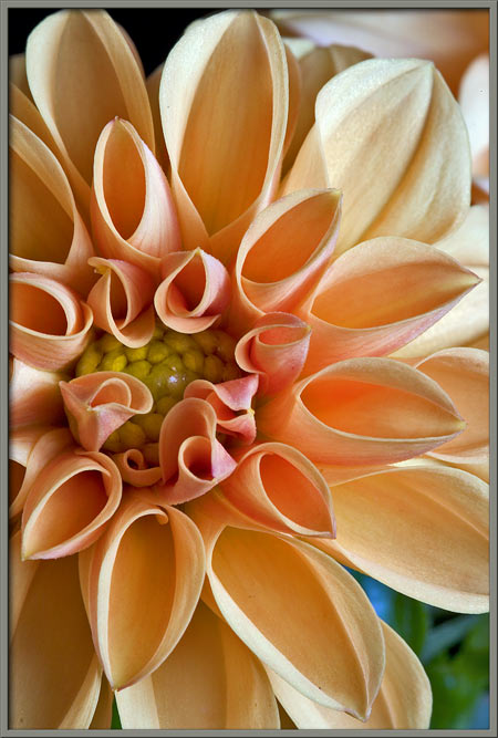
Somewhat later, the ray flowers
have opened up to reveal the central disk flowers with their
projecting, pollen covered pistils.
The higher magnification view below
shows that not all of the disk flowers have bloomed.
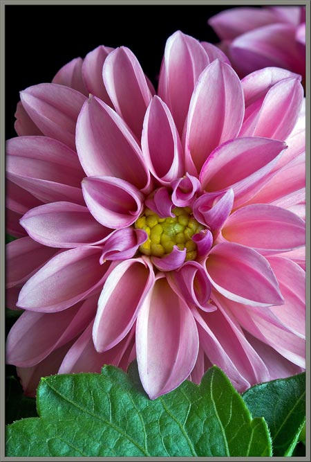
Dahlia hortensis Emily
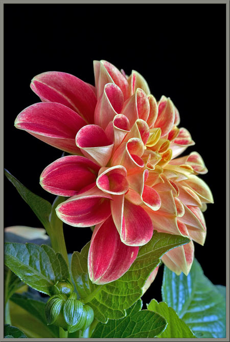
This third hybrid has solid pink
colouration. The bloom has just opened, and it too has ray
flowers with petals that are curled into tubes near the central
disk. Notice in the image at right, that none of the disk flowers
have opened.
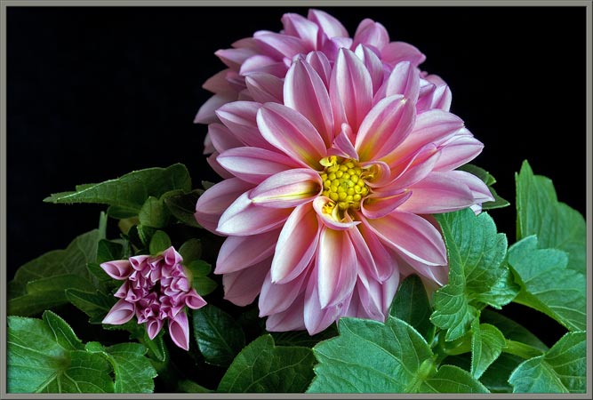
Notice the striking contrast
between the opening bud at left and the blooming one at right.
The same strangely angular bud is
shown twelve hours later in the image on the right below.
Dahlia
hortensis Linda
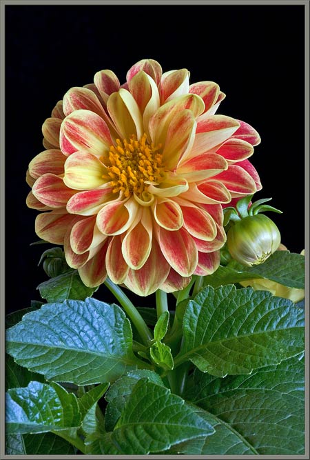
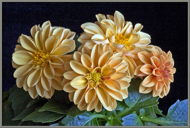
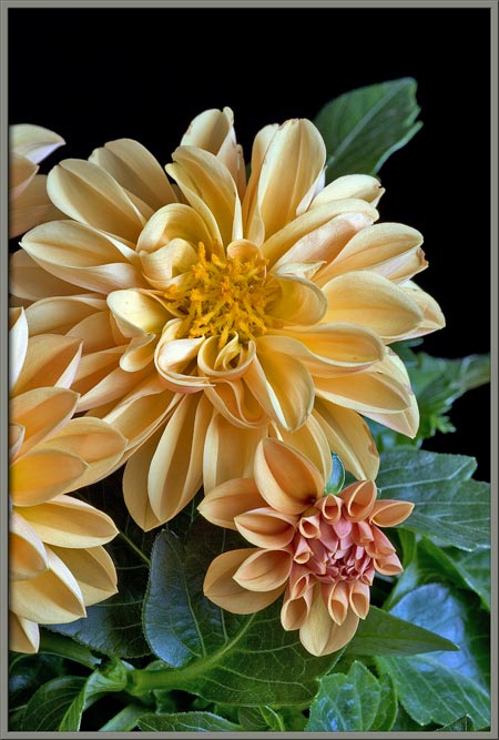
Here is another hybrid, this time
with a solid yellow-orange colouration. Early stage blooms tend
to be more orange than yellow (far right). The bloom in the
centre has the outermost disk flowers open, while the one on the left
is almost mature.
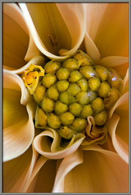
Notice the change in appearance of
the bud as it grows. Unopened yellow disk flowers are just
visible at the centre of the bud in the image at right.
As the ray flowers bend away from
the centre, the central disk flowers, in bud stage, become visible
. It appears that they are all temporarily covered by a thin
transparent membrane. In the image at right, if you look closely,
you can see that some of the outer disk flowers have bloomed, and the
pollen coated stigmas extend out from the distorted fused petal
assemblies.
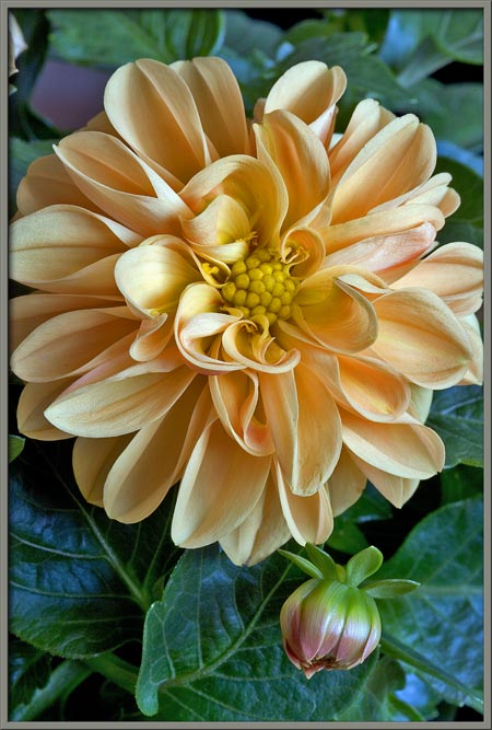
A mature dahlia flower of this
hybrid can be seen below. Several dozen stigmas extend an
appreciable distance out of the disk flowers.
If you look just below centre in
the right-hand image, you can see one of the disk flowers with its five
tiny, pointed, yellow petals fused together. Also note the very
stocky style supporting an elongated, orange stigma coated with pollen.
Dahlia
hortensis Unknown
Unfortunately, the supplier of this
dahlia hybrid neglected to insert the usual identification tags into
the pots. I have been unable to identify it.
Strangely,
this cultivar has ray flower petals with a solid pinkish-beige colour
on the upper surface, and brilliant red and white longitudinal stripes
on the under surface. In addition, even when a bloom is
completely mature, it displays no disk flowers.
The striped under-surfaces can be
seen perfectly in the opening bud. Here again, there is a lower
ring of five, bright green leaflets, and another ring of lighter green
sepals beneath the outer ray flowers.
An hour later, the ray flowers have
begun to open out towards their final positions.
Notice the extreme contrast between
the upper, and what will eventually be the lower surfaces of ray flower
petals.
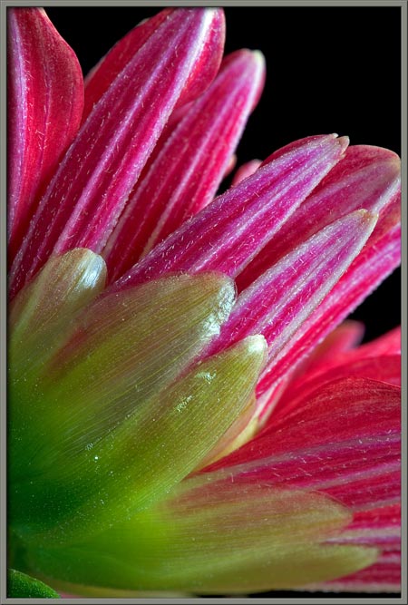
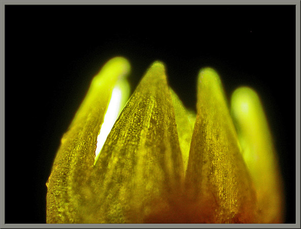
The pale, translucent, green sepals
are in contact with the ray flower petals at this stage. Notice
the fine, white, longitudinal striations on each of the sepals.
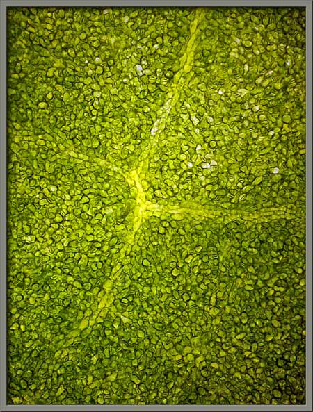
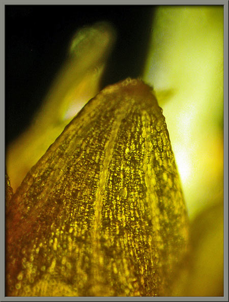
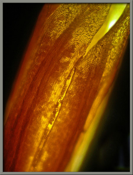
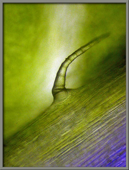
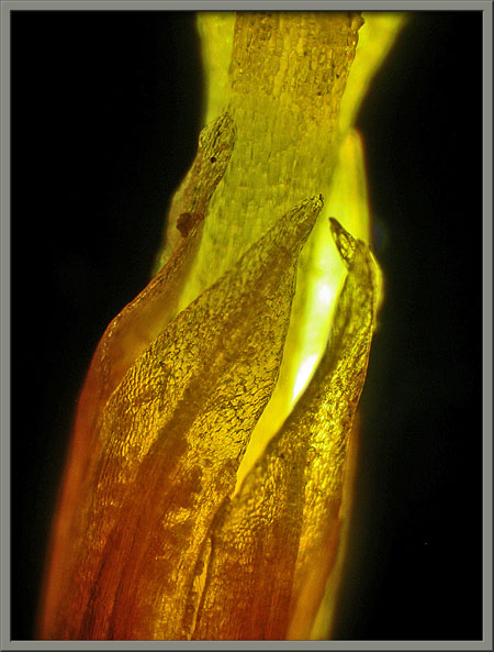
If one of these sepals is examined
under the microscope, the oval stoma
and guard cells that regulate
gas entry into and out of the sepal can be seen.
Here are front and side views of an
almost mature bloom.
Why look at the back-side of a
flower? In the case of this hybrid, the back is the best side!
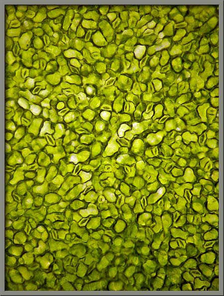
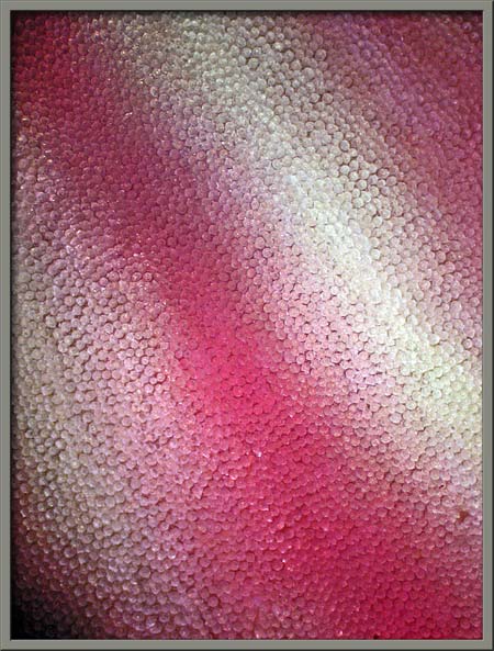
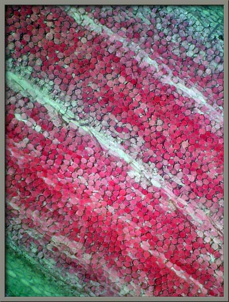
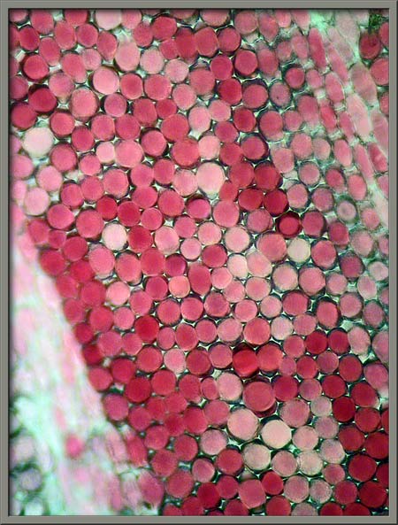
A low magnification photomicrograph
of the underside of a ray flower petal can be seen on the right below.
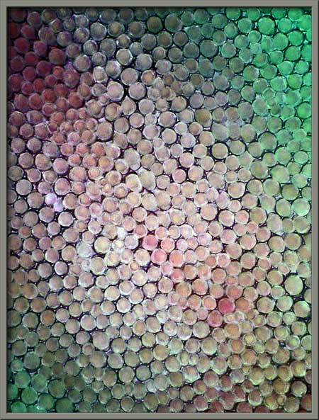
Higher magnifications reveal the
almost spherical cells that form the surface layer of a petal.
Note that the three images have had Photoshops
auto-levels applied in order
to increase contrast. The images are thus not true-colour.
The two photomicrographs below show
a typical dahlia disk flower. Notice the five, pointed petals,
that are fused together below their tips. The first image reveals
the style protruding out from the ring of petals. Where
are the stamens with their anthers, and supporting filaments?
They never protrude far enough out of the disk flower to be visible to
an observer. If you are sharp-eyed however, you can just make out
a darker shadow behind each petal, (the anther), and a number of
particles surrounding each shadow, (the pollen grains). As the
stigma is pushed out of the petal tube by the style, it comes into
contact with the anthers, and copious amounts of pollen become stuck to
its many hair-like protuberances. Self-pollination is discouraged
(but not entirely prevented), by having the stigma become receptive
only after its passage through its own ring of anthers.
A complete graphical index of all of my flower articles can be found here.
The Colourful World of Chemical Crystals
A complete graphical index
of all of my crystal articles can be found here.