|
Nineteenth Century British Microscopy and Natural History: Part 6 by Richard L. Howey, Wyoming, USA |
Visit the Micscape Library to read other parts in the series.
Good news for those of you who are rotifer fans. The key article in the February 1881 issue of the Journal of the Royal Microscopical Society is titled: “On Oecistes Janus and Floscularia trifolium, two new species of Rotifers”. The article was written by C.T. Hudson, M.A., LL.D., F.R.M.S. and is accompanied by two stunning plates.
For those of you not familiar with rotifers, you are in for a treat. These are microscopic organisms which exhibit a wide variety of shape, structure, and lifestyle. Wim van Egmond and I collaborated on a photo-essay called “Welcome to The Wonderfully Weird World of Rotifers” which will give you an idea of their variability. Wim did all the hard work and provided all of these splendid images while I just scribbled some words.
Some, like the two we’re going to be looking at, live in tubes, others like Asplanchna proceed through the water like an animated hand-blown Steuben lead crystal vase, and yet others, like Philodena, look like miniature inch-worms crawling along the substrate. Rotifers are also known as “wheel animalcules” since most species possess 2 hemispherical lobes which they can extend and retract. These are covered with cilia and when extended can produce powerful micro-currents which sweep food particles down through minute chambers to the mastax which are “jaws” which grind up the particles coming in. The mastax is an extraordinary structure and specialists sometimes utilize this feature to make species determinations. If you collect from ponds, lakes, and streams, you will almost certainly find rotifers. They live in many different sorts of habitats and for such small organisms, their morphology is surprisingly complex.
Let’s take a look at Plate I of Oecistes Janus, but before we do, let me remind myself of something, namely, that the taxonomy of rotifers is a super nightmare and what Hudson is calling Oecistes, I would have immediately said was Floscularia. So let’s all get down and Google (isn’t that the name of a cyberspace dance?).
In the meantime, however, let’s look at the plate and we’ll get back to the classification problem later.
Hudson kindly provides us with an explanation of the plates.
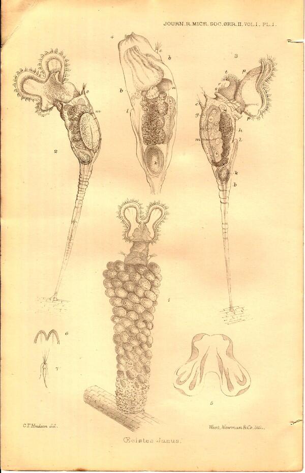
“Plate I Oecistes Janus
Fig. 1—Female in tube, antoral view, expanded.
Fig. 2—Female out of tube, nearly oral view, expanded.
Fig. 3—Female out of tube, side view, expanded.
Fig. 4—Female out of tube, side view, closed.
Fig. 5—Trochal disk; showing its thickenings.
Fig. 6—Extremity of chin.
Fig. 7—An antenna.
In all the figures:–a, horseshoe row of small cilia; b, longitudinal muscles; c, antenna; d, crop; e, tube from mouth into crop; f, mastax; g, ovary; h, stomach; k, its lower division; l, vent; m, transverse muscle; n, gastric gland; o, ganglion; p, thickening of trochal disk; r, curved bristles; s, knob-covering gland; t, ciliated chin.”
Now, of course, unless one knows a certain amount of specialized terminology, this explanation is not terrifically helpful and there is a further complication. In the last 127 years since Hudson’s article appeared, there have been significant terminological shifts with respect to the morphological features of rotifers such that a modern description of the structure of the same rotifer might seem to us radically different in some respects. Just to give you two examples: in modern terms rotifers have neither “chins” nor “antennae”.
So, what are we to make of this plate? Well, for one thing the drawings are splendid; they are so good that if you found one of these organisms under your microscope, you would be able to say–ah, yes, that’s what Hudson called Oecistes Janus. Furthermore, Hudson’s careful observations are so carefully recorded in these figures, that one would have an excellent idea of what structures to look for.
There is an intriguing aspect of this organism clearly demonstrated in Figure 1 and the nature of which Hudson was aware of and he mentions it briefly in his account. The tube in which this organism lives is constructed of what look to be tiny eggs, but which in reality are fecal pellets combined with detritus!
Let’s take a look now at the second plate which Hudson says is Floscularia trifolium.
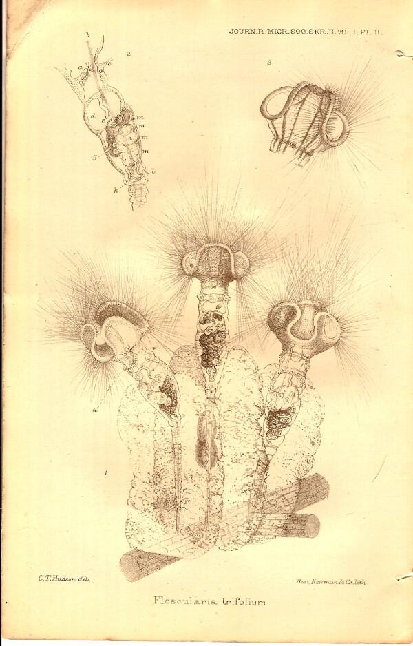
This too is clearly a remarkably strange organism in its appearance. As you can see, a large number of spines, setae, or acicular processes project from around the “head’. These are in fact very long cilia and form a loose, but very efficient, “net” for food capture. It is also evident from the plate that this rotifer secretes a substantial, gelatinous “house” into which it retreats instantaneously when disturbed.
Hudson provides the following explanation for this plate:
“Floscularia trifolium
Fig. 1—Three females, from different points of view.
Fig. 2—Side view of the body.
Fig. 3—Back view of the trochal dis; showing the two rows of setae down one side of a lobe.”
The references for the lettering of various parts are the same as for Plate I.
Now, let’s wrestle briefly with the taxonomy. For those of you who are not interested in the intricacies of classification, you may well want to skip this discussion. At this time, there was a family of rotifers called Melicertidae which now applies to thecate hydroids. In the taxonomy of rotifers, the genus has been replaced by Floscularia. In 1860 Philip Henry Gosse argued that the 5 different genera under the family Melicertidae should be collapsed into a single genus. Gosse you may remember for his popular book Evenings at the Microscope or his splendid treatise on sea anemones. So, a lot of classificatory juggling went on, and in recent literature, I can’t even find any reference to the genus Oecistes which Hudson says is the organism in Plate I. In fact, my first impression was correct; this is a specimen of Floscularia. So, what about the specimen in Plate II which Hudson calls Floscularia? As it turns out, this organism has now been assigned to the genus Collotheca. So, what difference does it make? When Shakespeare asks and then states: “What's in a name? That which we call a rose by any other name would smell as sweet," he is presenting us with a half truth. The psychology of language is a strange phenomenon and I have a vague feeling that if the name for “rose” “were “rotting rhinoceros”, we might not have quite the same affection for it. However, that’s not really the point with regard to these rather abstruse considerations regarding the classification of rotifers. We all know that taxonomy can be critical when it comes to organisms such as parasites or pathogens, but it doesn’t seem all that crucial with regard to rotifers. There are indeed times when the minute distinctions of taxonomists seem to be niggling nit-picking and I am tempted to regard them as the accountants of the biological sciences. Nonetheless, they are indispensable, for without them you and I might fight a duel over the morphology of a mastax, only later to discover that we were talking about completely different organisms.
This issue of the journal is chock full of interesting but, on the whole rather technical, reviews and discussions on a wide variety of subjects zoological and botanical. Because they are primarily of interest to specialists, I won’t consider them here with one brief exception. There is a 7 page examination of Mereschkowsky’s investigation of the two dominant theories at the time regarding the movement of diatoms. The first theory centers around protoplasmic streaming through the pores in the frustules, while the second focuses on osmotic processes to account for the movement. It is an interesting account with persuasive arguments on both sides and the author concludes that diatom movement is in all likelihood the result of a combination of both sets of processes.
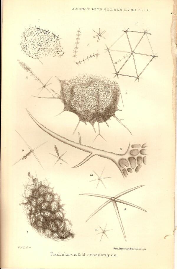
Now, let’s move up to the April 1881 issue which is one of exceptional interest containing as it does the presidential address of Lionel S. Beale, a paper by Ernst Abbe, who was an Honorary Fellow of the Royal Microscopical Society, and a long collection of “Notes on Aperture, Microscopical Vision, and the Value of wide-angled Immersion Objectives” which was a compilation derived from an extensive correspondence of Abbe with a number of fellows of the R.M.S. Some of this will very likely require a separate essay since there are a number of other wonderfully odd items to consider, beginning with the introductory paper by Professor P. Martin Duncan, M.B. (London), F.R.S.,&c, Vice-President F.R.S., titled “On a Radiolarian and some Microspongida from considerable depths in the Atlantic Ocean” accompanied, of course, by a splendid plate.
Here, visually, we have a lovely combination of the bizarre and the beautiful. I quite like Professor Duncan’s attitude toward the specimens he obtained and I wish to quote his first paragraph.
“In cleaning some specimens of recent corals, which had been dredged up during the expedition of H.M.S. ‘Porcupine,’ and others which had been given to me by the late Count de Pourtals, from the Caribbean Sea, I found a great number of minute sponge-like bodies. Some few were entangled in the consolidated ooze with which the cups of some dead corals were filled, and the rest were fixed to different parts of all kinds of living and dead corals, many being parasitic within.”
More than likely they were not parasites, but ectocommensals or accidental aggregations and Duncan simply assumed that they were parasitic because of their location in the dried state. He goes on to describe how there were some very minute, fragile bodies in the ooze of the cups and that he managed to disentangle 3 of them in a largely intact state. He then remarks: “On examining these little bodies, I was struck not only with their great beauty, but also with the Radiolarian appearance of two of them. The third was indubitably a true hexactinellid sponge of the Lyssakine division.”
I very much like the fact that he comments on the beauty of these structures–the symmetries are quite wonderful. Equally, however, I greatly appreciate Duncan’s interest in examining the dried bits and the ooze. I urge every amateur who manages to get such material to poke through all the bits and pieces; sometimes it will be disappointing, but from my experience, it is much more often highly rewarding. Figures 1,2, and 3 in the plate, he identifies as a radiolarian of the genus Aulosphaera from the radiolarian plages x and xi of Ernst Haeckel. Haeckel’s organism is A. elegantissima, but Duncan is convinced that his is a new species and generously names the species A. Pourtalesi after the late Count who provided him with the material in which Duncan found the specimen.
Professor Duncan is not only generous, but careful in his taxonomic assessments as well. Regarding Figures 4,5, and 6, he states: “If it is a sponge, it falls into the Dictyonine division of the Hexactinellids. But if the dermal spiculae are accidental, and if there is no oscule, the form will come under the Radiolaria. I propose to leave the question of the zoological position open.”
The real problem is the spicule presented in figure 5, the one that Duncan calls the brush spicule. It does indeed appear to be a spicule from a hexactinellid sponge but, if as Duncan conjectures, it accidentally got attached to the central mass represented in Figure 5, then it is not conclusive in deciding whether this is a micro-sponge or a radiolarian.
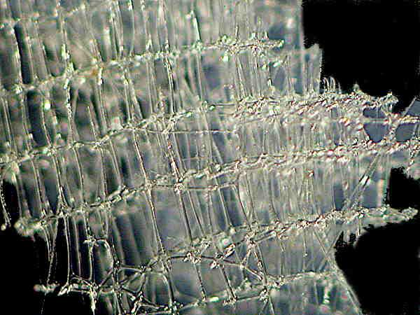
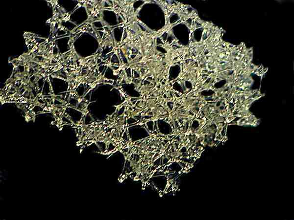
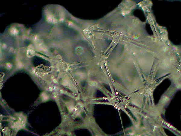
Figures 7-10 show what is clearly a hexactinellid micro-sponge. The hexactinellids have spicules with 6 prongs, hence the name. They are the “glass sponges”, the most famous and beautiful of which is the Venus Flower Basket (Euplectella aspergillum).
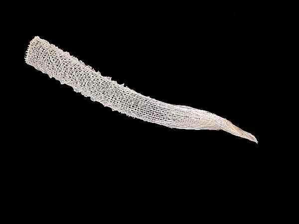
However, there are many other kinds. The main thing they have in common is the six–pronged, silica (glass) spicules. Here are a few images of some other aspects and types of hexactinellids.
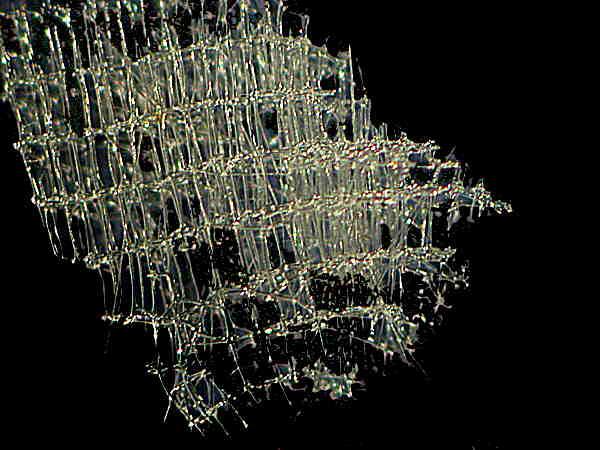
If you don’t know whether a sponge sample you have has silica spicules or not, take a small section, place it on a clean slide, and add a drop of 10% hydrochloric acid. [CAUTION: This is a corrosive and dangerous substance even when diluted. Handle with great care.] If the spicules dissolve, they were calcareous; if they didn’t, they’re almost certainly siliceous. However that doesn’t mean that you have a hexactinellid; siliceous spicules come in a wide variety of shapes and sizes, so you still have to determine if the spicules you are examining have six prongs.
Here we encounter another example of nature’s endless trickery—only some of the spicules will be hexactinellid; in fact, Duncan describes 6 different kinds of spicules from this sponge. As you can see from the plate, only those labeled #10 have 6 prongs. There are in addition quadriradiate and quinqueradiate spicules (such as the “feather” spicules shown in Figure 9.) These I find most intriguing and would very much like to have a sample to examine. Hint, hint, to all you sponge specialists in the region of Portugal.
One of the delights of being a naturalist is that there is no end of surprises to find all around one including the esoteric interests of other naturalists. In this issue Mr. A.D. Michael, F.L.S., F.R.M.S. has a paper titled: “On a Species of Acarus, believed to be Unrecorded”, in other words, an unidentified mite which as it turn out is a parasite on cormorants–now that’s fairly exotic. Parasites are, however, pretty eccentric in their lifestyles and have made some astonishing morphological alterations to adapt to their environments. The first thing that stuck me when I glanced at this plate was that this little mite looks like it has wheels.
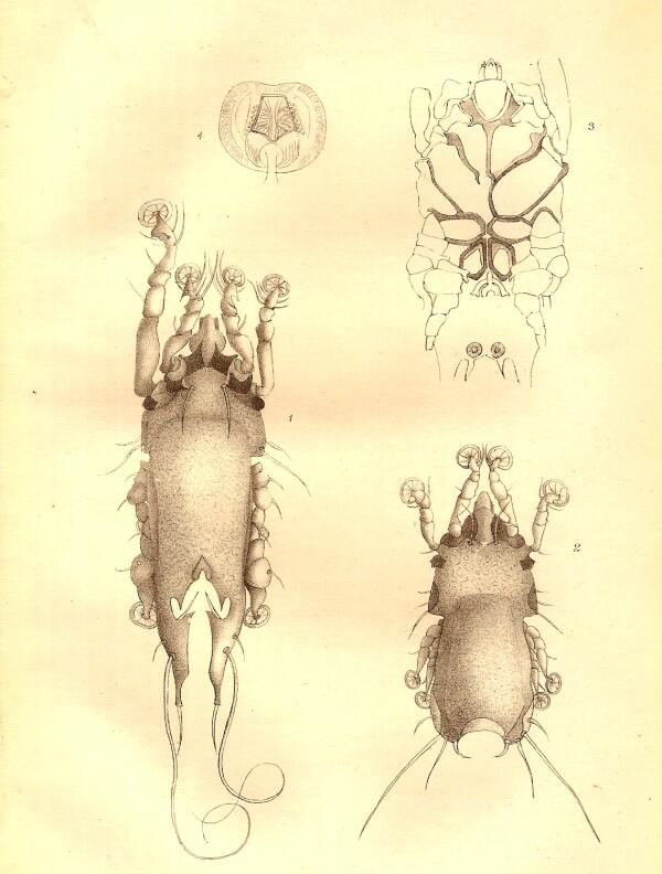
Sorry to disillusion you, but these extraordinary structures are suckers by means of which these mites attach to their hosts, in this case, cormorants.
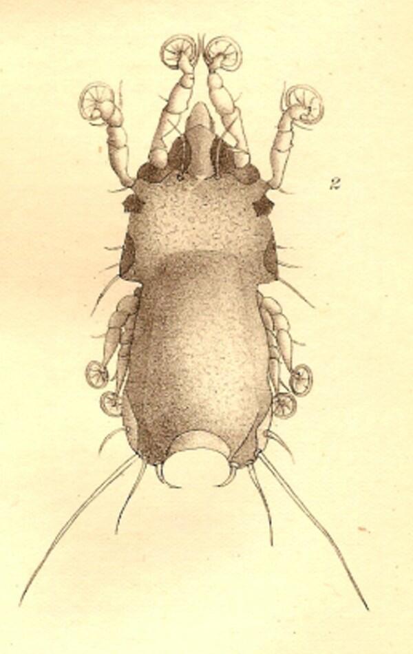
Mr. Michael proposes the species name heteropus if in fact it turns out to be unrecorded and places it in the genus Dermaleichus which contains a large number of avian parasites. Figure 1 shows the male and Figure 2, the female, each magnified 95 times and as you can readily see, the male is considerably larger. What struck Mr. Michael was the disproportionately large, left, second leg in the male. Figure 3 shows the underside of the female and Figure 4 one of the suckers, highly magnified.
WARNING: The next paragraph is rated “R” and contains language relating to sex and should not be read by anyone under the age of 40 unless accompanied by a parent or guardian.
The feature that struck Mr. Michael most was, as I mentioned, the exceptional length of the second, left leg in the male. He remarks:
“This occurs in the male only, and I have no doubt that the enlarged leg is for holding the female during coition, although I have not been able to find a pair in that act, as one usually may in most species of the genus.”
Good heavens!–randy mites and a voyeuristic naturalist; what will nature come up with next?
The reports and reviews of recent researches are, as usual, varied and rich, but many of them would be of interest only to a specialist, so I will just mention in passing or brief summary a few of those which caught my attention. I will save Beale’s presidential address for the next essay.
Let me begin with a somewhat technical review to make a very important point about a tendency which many of us have and which can lead to some rather fundamental mistakes. The report about an article by Mr. P. Geddes on “Perivisceral Fluid of the Echinoidea”. Sounds scary, doesn’t it? Echinoids are sea urchins which almost all of you are familiar with. Let’s examine the first brief paragraph.
“Mr. P. Geddes remarks that the close resemblance between uncoloured elements of the blood, the amoeboid corpuscles, and the true Amoebae is very remarkable in the class of animals he has selected for study; he is of the opinion that they ought to be able to lead us to a resolution of the important question, Does the term amoeboid express an accidental analogy, or a deep-seated resemblance?”
Just to make things more interesting, Geddes considers not just sea urchins, but some sea cucumbers as well and not just amoebae, but slime molds also. His conclusion? “The author is of the opinion that the theory which looks on the amoeboid character as being a fundamental character of the animal cell is fully justified.”
This is, of course, rubbish. We love to find analogous structures and indeed there are many significant examples in nature. The deadly conceptual error arises when people such as Geddes yield to the temptation to universalize such analogs. Indeed amoebocytes occur in animals from sponges to human beings, but this does not justify claiming that this is “a fundamental character of the animal cell.” It would be like claiming that because hemoglobin or hemoglobin-like pigments are found in the blood of humans, large clams and giant tube worms found near volcanic funes, and a wide variety of mammals, that therefore blood is always red. There are, for example, some tunicates which have green blood. In fact, this author points out that in the irregular sea urchin Spatangus purpureus, “purple, blue, green, olive, or yellow corpuscles may be found in the blood vessels.” Given that Mr. Geddes knows this, it is more remarkable that he makes the error regarding amoeboid-type cells.
There is a brief, interesting review titled: “Early Stages of Renilla.” Renilla is a colonial coral also known as the Sea Pansy.
You can find an image of one here, or just do a search on Google Images and type in “Renilla”.
It does indeed look like a deep reddish-purple, heart-shaped flower with tiny white blossoms across the surface when the polyps are extended and feeding. It is a remarkable and to me, very puzzling, creature, but then I find almost all colonial organisms puzzling. The “flower” contains tens of thousands of minute spicules which retain a deep burgundy color even after being subjected to bleach in the process of dissolving away the tissue to isolate the spicules for study.
A further oddity is that it possesses a peduncle or a kind of “tail” which is used to anchor the organism in the substrate. Furthermore it is bioluminescent and has been the object of considerable study recently regarding a green protein material that alters the usual pale blue light to fluorescent green.
Another review reports the work of Dr. W. Dybowsky on the sponges of Russia and, in this instance, those from that unique environment of Lake Baikal. If I were younger and healthier, I would love to spend a summer collecting in Lake Baikal. I’d even take my wife along since she knows Russian. Baikal contains 1/5th of the surface freshwater of the entire planet. It once had an open channel to the sea which then later closed and, as a consequence, there are species of organisms–from protozoa to freshwater seals–found nowhere else on Earth.
So, I’ll finish up this essay so that you can go call your travel agent and make the arrangements to go to Lake Baikal. Be sure to take the Trans-Siberian Express from Moscow to Irkutsk; it’s only a five or six day train journey. What an adventure!
ADDENDUM
I want to once again express my thanks to my wife for her patience, encouragement, and diligence in proofreading article after article. Without her input, these essays would be garbled, barely intelligible ramblings.
All comments to the author Richard Howey are welcomed.
Editor's note: Visit Richard Howey's new website at http://rhowey.googlepages.com/home where he plans to share aspects of his wide interests.
Microscopy UK Front
Page
Micscape
Magazine
Article
Library
Please report any Web problems or offer general comments to the Micscape Editor .
Micscape is the on-line monthly magazine of the Microscopy UK website at Microscopy-UK .