|
|
|
| From Brightfield to
'Smoke Rings' (Part 1) A Personal Odyssey By Paul James |
The properties of light that revealed the aerial images from the very first water drop lenses several hundred years ago hasn't changed at all. In fact, in principle once the compound microscope itself was envisaged little has changed with that, other than the lenses involved which are more perfectly shaped, and used in greater number. Of course the science of imaging and resolution was responsible for the general improvement of the instrument and yet in the last century nothing significant has changed in the way brightfield illumination is practiced. The generating of the illuminant, in principle, hasn't changed much either, just refined a little to eke out the best imagery from the optical train. Manufacturers for a long time have in their own particular ways modified the traditional illumination principles to suit their own philosophies concerning substage management.
But the basic brightfield image has only survived over the centuries because of the profundity of semi-transparent subject matter that litters the whole gamut of microscopic life forms. Opaque specimens are not flattered in brightfield for obvious reasons. Fortunately the bulk of microscopic forms of organic origins are richly endowed with structures that vary from the slightly opaque to the transparent with much in between. Had our prey been in general terms less obliging to the transmission of light, the microscope's evolution would have developed using vertical illumination, and our knowledge of the inner workings of the cell would have been all but closed off from view. However, in our brightfield scenario there is one particular property of light which is crucial in delineating fine structural detail: it is known as diffraction.
Thus when light skirts past the actual border of any opaque object its initial path is altered very slightly. So small is this deviation that it is not noticed in normal visual everyday occasions, but most certainly can be when images are amplified in the microscope and telescope. Strangely enough for the astronomer diffraction is a hindrance as it spoils the subtle details of imagery, which despite being self illuminating become tainted with diffraction generated rings around a star image caused by the inner edge of the main objective mounting. For the microscopist, it is brightfield's saving grace for without diffraction the more delicate cellular structures would not be visible or at least not so apparent. However this natural imaging enhancement phenomenon as it were comes at a price for it can so easily become too dominant in imagery when the ascending illumination pencil of light is narrow, almost parallel and of low na. Such imagery is easily induced by closing down the substage condenser iris as we only know too well. Diffraction dominated imagery is of no use to us as the subtle details are completely lost in the thickened silhouetting of the structure observed. The trick is to bring about a balance that satisfies the observer's eye for detail and tonality which is usually arrived at when the substage iris is about 3/4 open.......that's the traditional practice, and I have no doubts about that, BUT in actuality much depends on the opacity of the specimen in the first instance. In entomology for instance, diffraction only serves to exaggerate hard edges of imagery. In fact the small tonal range offered by these insect specimen parts on the whole is better served with a more diffusely charged illuminant which stretches the tonality and therefore making apparent more subtle variations in the structure observed by flooding out some of the diffractive artifacts which are not desired.
A very simple way therefore to reduce the diffractional component in brightfield is to introduce the diffusion or ground glass screen in the illumination train. In the past I used it frequently but placed below the condenser, but have also tried putting one just beneath the slide. In this situation the chaotic output from the diffusion screen offers higher na potential since the source is so much nearer the specimen and objective's front lens.
|
|
|
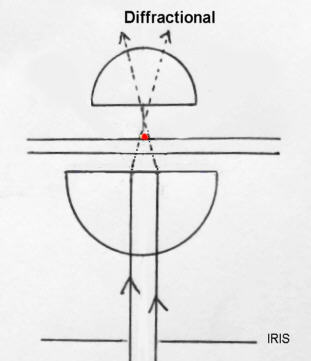
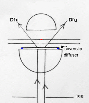
A very simple way of demonstrating the effects of diffraction and diffusion on the imaged specimen is symbolised in the diagrams above. At left we see the very narrow yet highly diffraction inducing pencil beam impinge on the specimen, and above right the simple effect of a coverslip diffusion screen atop of the condenser which 'shreds' the pencil beam into a very wide fan of light, albeit of diminished intensity. The result which you can easily demonstrate yourself is that the effects of diffraction are 'drowned' out. It is instructive that closing down the condenser's iris diaphragm with the diffuser in place has virtually no effect on image quality subject of course to choice of specimen. The virtue of observing with diffuser screens is however somewhat dependent on the specimen observed. The skill therefore in this scenario is knowing your subject matter with regard to its effects on light. In general terms, diffusion sourced illumination favours entomological studies and others also having robust or dense structures. Radiolarians etc. I think suit diffuse lighting too.
Solar lighting
|
|
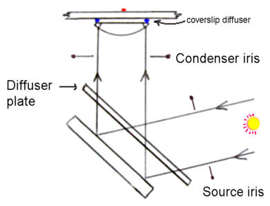
I thought at this point, before leaving brightfield I'd mention diffuse solar lighting because its phenomenal imaging clarity might interest some readers. BUT Do not attempt to observe before diffuser screen(s) are in place. Setup a microscope as shown above using a glass diffuser preferably taped temporarily to the substage mirror placed as shown below the stage. In this position the diffuser doubly subdues the intense sunlight for safety reasons, and the coverslip diffuser generates the widely scattered source of chaotic light onto the specimen. You can also control the sun's intensity by using a perforated piece of card/iris as shown, but I found this unnecessary. At less than x 100 this method of lighting is exceedingly good though admittedly inconvenient without a small heliostat to counter the sun's motion. At higher amplification the loss of the diffractive component in the image will be considered too 'soft' for some. Fortunately entomology can be enjoyed at these lower powers.
Observing a blow fly's proboscis with a Wild M20 through a x10 Fluotar produced such high quality imaging that it was all too easy to imagine no glassware was being used to image the specimen at all. The detail and tonality were in a league unknown in my standard brightfield microscopy. This was not too big a surprise since daylight is totally natural and is of course THE perfect source for natural imaging with the eyes and camera.
Some Final thoughts
There's more to the brightfield image than covered here of course, but essentially the dictum of using a slightly closed down substage iris diaphragm clearly has to be taken loosely since so much depends on the specimen's transparency and delicacy of structure. Though the concept of diffuse lighting may appear to be a vague term, lacking mathematical preciseness its overwhelming existence in the daylight of the real world must surely give it much more credence than does traditional microscopy practice. Diffused light exists in the illumination train of the average microscope to a greater degree than theorists would admit, so 'adding' a little more in certain circumstances to suit the nature of the specimen seems a sensible approach to low power microscopy.
To be continued.......
| All comments welcome by the author Paul James |
Microscopy UK Front Page
Micscape
Magazine
Article
Library
Please report any Web problems or offer general comments to the Micscape Editor.
Micscape is the on-line monthly
magazine of the Microscopy UK web
site at
Microscopy-UK