|
SUMMARY
The
habitat of Atractosteus tropicus,
the Mexican gar, captured in the
State of Tabasco, and 3 of its parasites are described and
illustrated. Pictures of them and graphs of their lifecycles are
included. Because of the shape of its head, and hard ganoid scales the
Mexican gar, also called tropical gar is locally known as
pejelagarto that translates as crocodile fish.
Key words: Atractosteus tropicus,
gar, pejelagarto, Clinostomum
complanatum, Cystoopsis atractostei, Proteocephalus singularis,
Perezitrema bychowsky, Centla Swamps
INTRODUCTION
On
October 22 of 2005, the Wilma Hurricane swept with
its winds of up to 300 km/hour over the city of Cancun and stayed over
it
for 36 distressing hours. When finally it slowly retired, there was
not left a single leaf on the trees that
still stayed up in the city and their environs, showing their skeletons
against the shady sky. The
city, with its destroyed buildings, seemed a bombed city, the population
was at the border of chaos, looters invaded the stores, and the
neighbors were organizing themselves for their defense and survival.
There was no electricity, and to illuminate the streets at night,
bonfires were ignited with the moist remnants of the fallen vegetation.
The black and pungent smoke covered the sky.
I
received an asylum offer in the city of
Villahermosa, Tabasco, and,
with my family, I fled from the city. There is no other word to describe
it. And we were amongst hundreds of those that fled.
Tabasco
has also been whipped by
hard hurricanes, before, and, mainly after, our visit. After a year
the city of Villahermosa itself was flooded almost totally by
overflowing waters of the rivers that surround it.
Those
used to employing GoogleEarth can see Villahermosa
City
and pictures
of its building, parks and floods!!.
But
at the moment of our visit it was flooded
by the sun, and the boisterousness of the busy life of the flourishing
oil
city that it is, and was really green because of its very abundant vegetation.
The
city is framed by the Grijalva River, and the
Carrizal River, and is seated
over a marshy plain that is always present because of the existence of
innumerable lagoons, pools and little swamps, scattered throughout the
city structure. For a microscopist interested in the aquatic
microfauna,
Villahermosa (and all Tabasco) is an Eden.
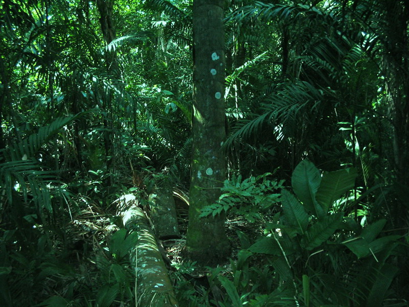
And,
around the city there is the
jungle, in which man has opened extensive prairies to cultivate
cattle. But which is always present, and is easy and extremely tempting
to walk under its sun speckled shadow.
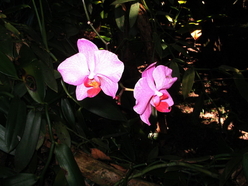
Surrounded
by a church-like silence it
is possible to see the bromeliads hanging from tree branches, and, from
time to time, some of the multiple species of tropical orchids that
decorate it.
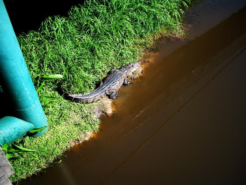
And
while you ramble across the
city and its environs, it is common to see along the shores of the
rivers, what Mexicans call a lagarto (lizard): the crocodile, of
which there are two species, Crocodylus moreletii (the swamp
crocodile), and Crocodylus acutus (the river crocodile) like
the one
whose picture I took while crossing a bridge.
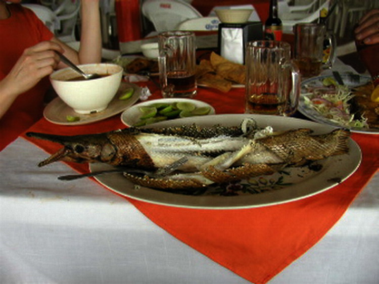
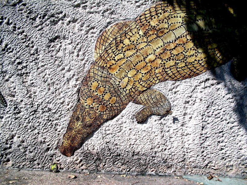
But
it is still more surprising to
see this scene when you enter a restaurant.
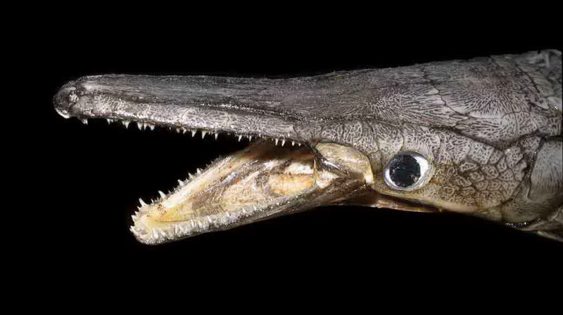
NO, it is not
really a small
crocodile (although people also eat the meat of these), it is a fish.
Not any fish of course. It is a survivor with a history of millions of
years, a living fossil: The PEJELAGARTO
(crocodile fish), the tropical
gar
Atractosteus
tropicus
This is the frightening aspect of
its head. The long
and narrow shape, and its sharp teeth, was worth its nickname by
comparison
with the head of a crocodile
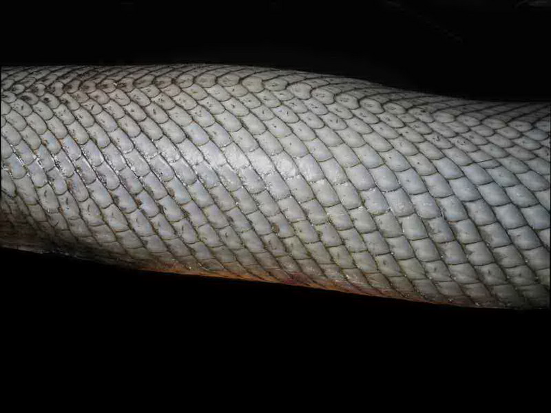
The Atractosteus belongs to an old group of fish,
the Lepisosteidae, with a history of more
than 140 million years, whose fossils are already found
even in the Permian. It
is characterized by its special ganoid scales, (see http://www.amonline.net.au/fishes/what/scales/index.htm) so different from those of the common fish. The Family is now
restricted to two genera, and is limited to the
south
of the United States (Texas and Louisiana - 5 species), Mexico (1
species),
Central America (1 species), and Cuba (1 species). When
adults these fishes can
reach a length of about 2 meters.
We can
summarize its taxonomic position according to these categories:
Kingdom:
Animalia
Phylum: Chordata
Class: Actinopterygiids
Order:
Lepisosteiformes
Family:
Lepisosteidae
The family has
two
genera Atractosteus and Lepisosteus, with
these species that I
list with their geographic distribution
Atractosteus
spatula United
States
Atractosteus tristoechus Cuba
("Pantanos
de Zapata", Zapata Swamp)
Atractosteus tropicus México
and Central América
Lepisosteus oculatus United
States
Lepisosteus osseus United States
Lepisosteus platostomus United
States
Lepisosteus
platyrhincus United States
Normally gars
vary
between 0.6 and almost 2.0 m in total length, but Atractosteus
spatula is the champion reaching more than 3.0
meters. In order to have a clear idea of its
dimensions it is sufficient to enter Atractosteus in a browser and
click images. Most of the pictures are examples of
the
enormous trophies captured in different fishing competitions in southern
USA.
All are
powerful
predators. In the United States the fishermen consider them a pest
because
they feed on the fish that they wish to fish, and they shoot
them or,
according to a journal article (dated June 2007), they even
use bows
and arrows (to classify this as a sport surely).
In Mexico
we eat them. I can attest that its meat,
cooked with due care, is really tasty. It is the
gastronomical
emblem of Tabasco and is offered in all type of restaurant. It is not
cheap,
because until recently, due to indiscriminate fishing, it was
considered a
species threatened with extinction. I must give notice here that this
fondness
for gars is partially based on the alleged aphrodisiac properties
of the
meat.
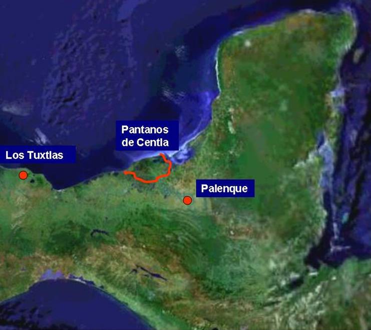
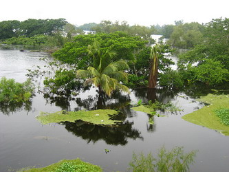
Natural
(and spectacular) habitats of tropical gars are the Centla
Swamps (los Pantanos de Centla) even if they are widely
distributed in Tabasco. Centla is a large expanse of
shallow waters,
with plentiful
aquatic vegetation and surrounded by tropical forests, one of whose
notable
characteristics is the high fish species richness, most of which are
edible and
of great quality.
The Pantanos
de Centla,
are located in the
neighborhood of the city of
Frontera, and their coordinates, for
whoever who
wishes to use Google Earth, are
Longitude: from 18º40 to 18º02 N
Latitude: from 92º16 to 93º05 W
From 1987
it has been a protected ecological area (declared
a Reserve of the Biosphere in 1992) that includes 3093 km2 .
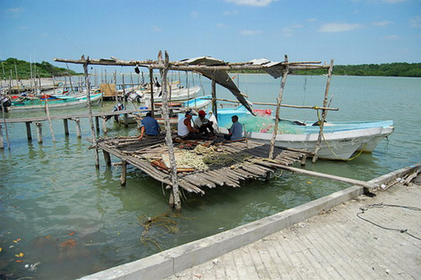
Theoretically, commercial fishing is forbidden
within
all its area. But in defined areas, and through the innumerable
marshes included
around the Centla, the pejelagarto lives and is fished intensely in
artisan
form.
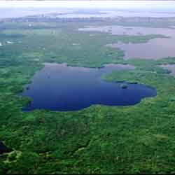
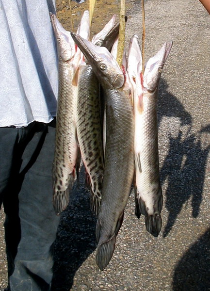
This is a picture
from a dedicated
photographer (ventolinmono) that has grasped the atmosphere of the area
in his
beautiful collection of pictures published in Flickr http://www.flickr.com/photos/dystopiatv/sets/72157605278930063/
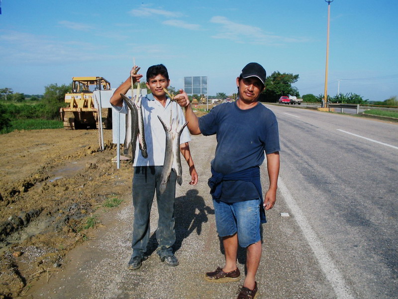
You can see the fishermen offering their merchandise
throughout the highways of the area. The offered examples are small, as
the
following photos demonstrate, by comparison with the size an adult
reaches. It was
considered a species threatened with extinction. And its capture was
prohibited
(without great success).
|
The
culture of gars
The only way to defend this species from
man, the only
enemy who is able to threaten their millions of years
existence, has been to establish
its culture, with aims to repopulate the swamps, or even to try
commercial aquaculture.
To do this it required not only a deep study of its own anatomy,
physiology
and ecology, but of its parasites also.
Lacking my microscope, and interested by my first
gastronomical contact with such an attractive living fossil, I
approached the
University Juárez Autónoma of Tabasco and its departments of
Aquaculture and Parasitology.
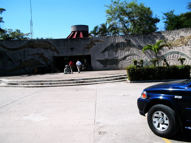
The
admiration the people of Tabasco have for their crocodiles (the true
ones, of
course) is demonstrated by the entrance to its buildings where these
have been
beautifully sketched in their admission portal.
In the aquaculture area I could observe the large
(and
small) tanks in which the gars are grown step by step, from the alevine
stage
(the youngest fish, which is born from the egg) until the breeding age.
There is even a local market, for the local
aquaculturists to buy and care for the little animals, with the
understanding to
return them to their natural habitat when they are uncomfortable in
their house
aquariums. The rest of the country is excluded from this hobby. With a
good
ecological criterion the exportation of living gars is banned out of
Tabasco.
Breeding examples are impressive animals, being
almost
two meters in length and large in diameter. Unfortunately due to
lighting
conditions, I did not obtain any decent photo of the breeding tanks or
the
nursery.
But the hatchery is successful at the species
reproduction level and is able to produce juveniles in high quantities
(300,000
a year it is said) to be released at the Tabasco marshes to aid with
the
recuperation of the wild population of Atractosteus.
Experimental farms, with relatively rustic,
extensive
aquaculture conditions, have been also installed to grow the gars to an
appropriate
harvest size. (2007)
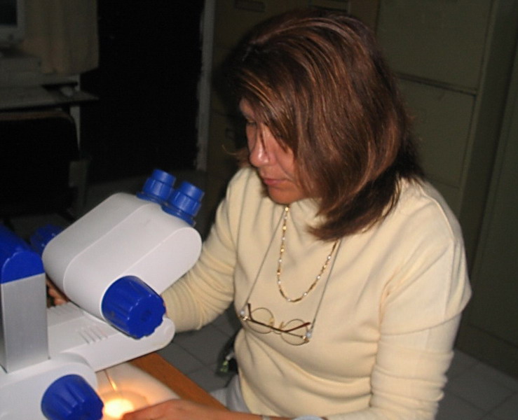
I was more fortunate in Parasitology, where Dr.
Leticia
García Magaña not only kindly gave me copies of their
papers on the parasites
of atractosteus, but also put at my disposal some microscopes, and mounted
preparations of several of them.
Dra
Leticia García Magaña
A
first
note for those who are not familiar with fish
parasites, is that these can be classified in three great groups: the
Nematoda,
the Trematoda, and the Cestoda.
The
reader interested not only in the images, but in
the organisms which they represent, if they do not have enough
knowledge of Zoology,
would have to read at least some of the following articles on the
Internet,
where
in addition they will learn the technical terminology applicable to the
anatomy of
each group:
http://en.wikipedia.org/wiki/Trematodes,
follow up the
references for aspidogastrea, digenea and monogenea
http://cas.bellarmine.edu/tietjen/images/platyhelminthes.htm
http://en.wikipedia.org/wiki/Nematoda
http://www.1911encyclopedia.org/Nematoda
http://www.itg.be/itg/DistanceLearning/LectureNotesVandenEndenE/imagehtml/ppages/CD_1071_086c.htm A good sketch of digenean anatomy
http://home.austarnet.com.au/wormman/wltape.htm focused on human
parasitology (Cestoda)
http://www.aber.ac.uk/parasitology/Edu/Cestodes/CestTxt.html another
interesting site
The reader must be warned
that as human beings host several parasites, and human parasitology
has great sociological
and clinical importance, most of the general parasitological
information on the
Internet refers especially to human parasites.
Although I worked with a
Leitz Photomicroscope, my lack of familiarity with it and my hurried
visit made
that, finally, the better pictures I took with my Canon Powershot A300,
by the
simple
artifice of applying the lens to the exit pupil of the eyepiece
of a
normal binocular microscope.
In
the State of Tabasco (Salgado-Maldonado,
et al, 2005, Salgado-Maldonado, 2006)
there are detected on Atractosteus
Adult
trematodes. Perezitrema bychowskyi
Trematode Metacercariae: Clinostomum
complanatum, Diplostomum sp., Posthodiplostomum mínimum;
Adult
cestodes: Proteocephalus
singularis
Adult
Nematodes: Cistoopsis
atractostei, Procamallanus (S.) rebecae
Nematodes
larvae: Contracaecum sp. tipe 1, Contracaeucum sp. 2,
Spiroxys sp.
The species I could observe are written in blue.
Aside
from the pictures that I could obtain, and in order to complete the
information
and make more useful this article, I have added complementary images
whose
origin is detailed in each one.
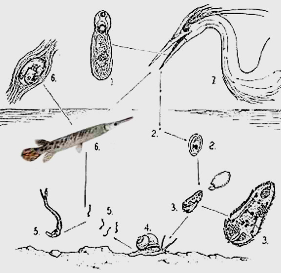
Trematodes
Clinostomum complanatum (Rudolphi, 1819)
Clinostomum complanatum
is
a parasite that in its adult stage (1) is very common in aquatic
birds. Its
normal habitat is their mouth and sophagus. Their
eggs (2) fall into the water, where they release
one first larva, the miracidium
(3), that invades a snail (4), where it develops through two additional
stages (Sporocyst
and Redia) until the cercaria (5) which is freed and finally infects
the Atractosteus, producing a cyst under its
skin, that lodges a larva denominated metacercaria (6), already very
similar
to the adult. By their color, the cyst is called a
yellow grub, and is a
very common disease in the fishes of the area. If the fish is eaten by
the host
bird, the metacercaria frees and methamorph into an adult (1), starting
a new cycle.
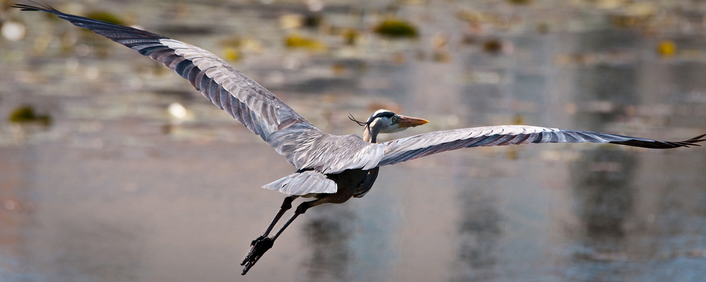
"Flying
blue heron" Picture by Joe Orban (see http://www.flickr.com/photos/vidular/3556033031/)
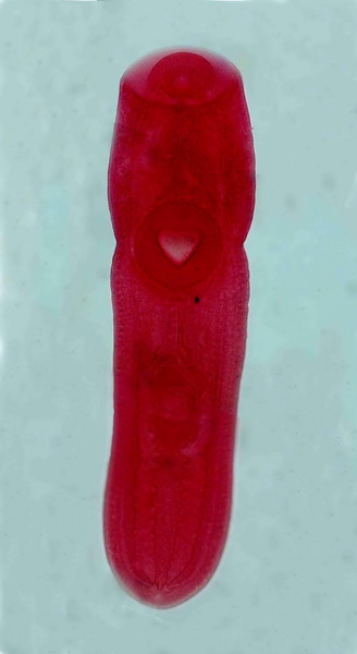
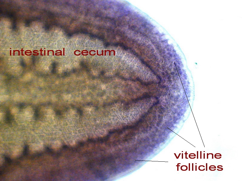
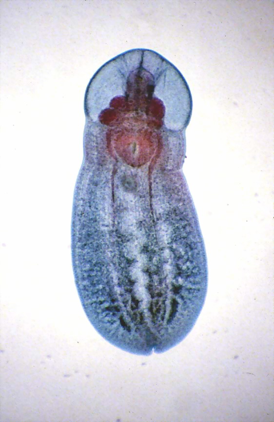
This is an image of a metacercaria, pulled out of
its
cyst, stained with Carmine and mounted in Canada Balsam. It is a four
image mosaic.
Although it occupies much more space, I present it in
a vertical format so
that its morphology is better appraised. The complete original image is
a 6 Mpx
one.
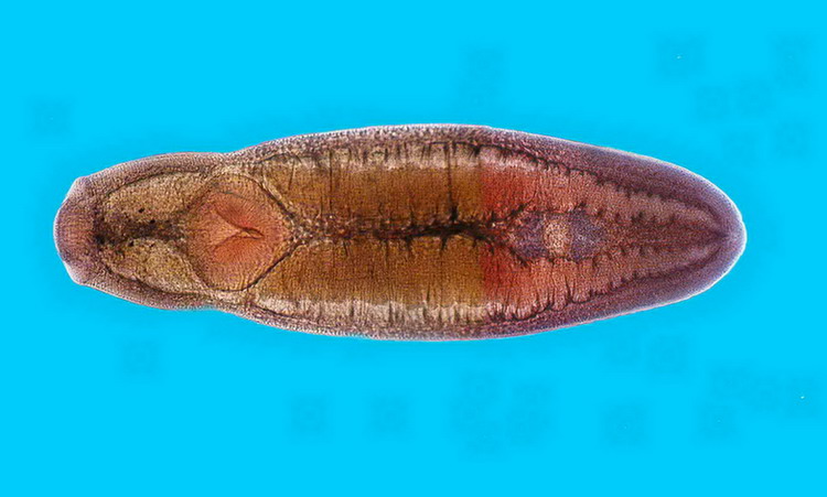
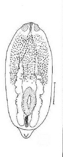
This
is also a mosaic of which I think is a pre-adult, probably stained with
Hematoxylin.
Adults have their uterus full of eggs, which is not
the case here as you see in the third picture. Above it has been
reduced, to show its general
anatomy,
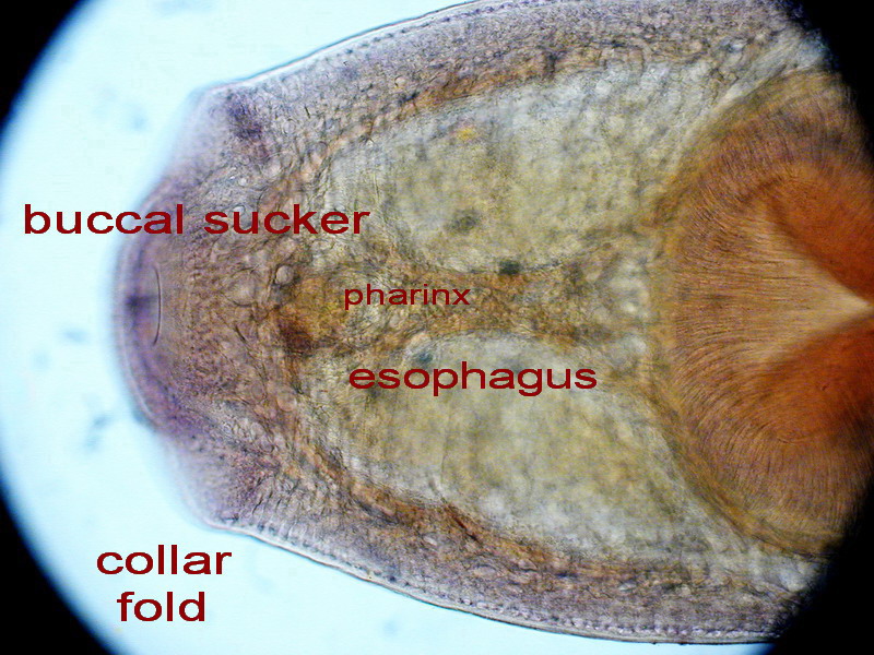
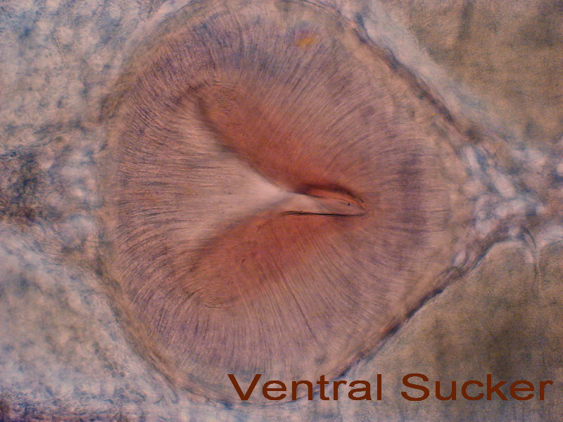
but in the following images I show it through 4 images at a bigger
magnification. Each original picture was 2 Mpx.
For Perezitrema
, Posthodiplostomum,
and Diplostomum
I present the following images only with the intention to better
complete the
panorama of this fish's parasites.
|
|
|
| Perezitrema bychowsky
|
Posthodiplostomum
minimum
|
Diplostomum sp. |
NEMATODES
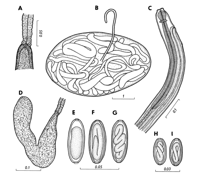
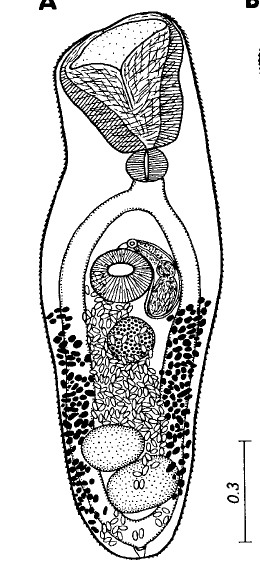
Cystoopsis
atractostei Moravec and
Salgado Maldonado. 2003
It
is a relatively large nematode, that lodges under the Atractosteus
skin. Its description is at http://www.ibiologia.unam.mx/pdf/directorio/s/salgado/cystoopsis.pdf
from where I reproduce the identifying drawings, and
where you can investigate
the meaning of the
different drawings. For
those not familiar with the group, this could be a good occasion to
read a complete
scientific description of a new species.
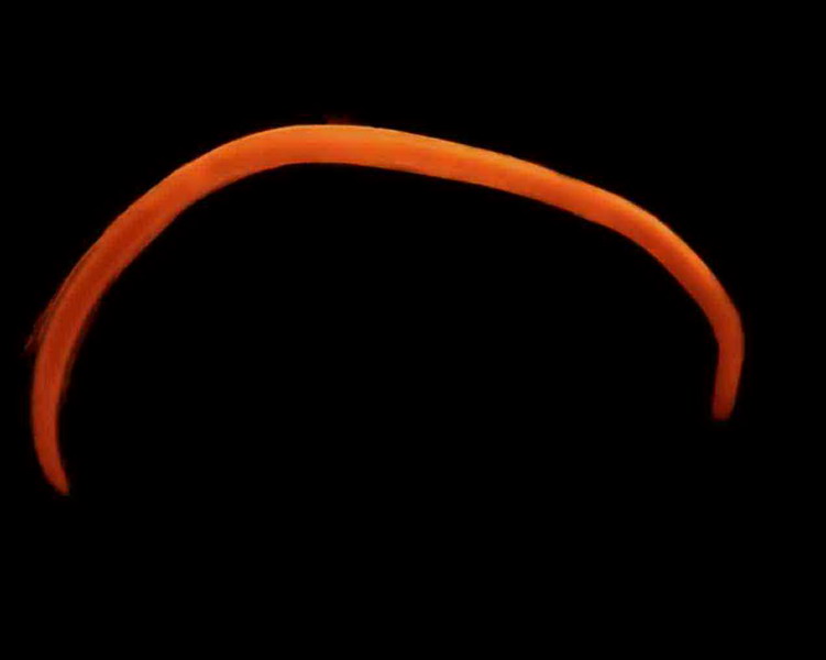
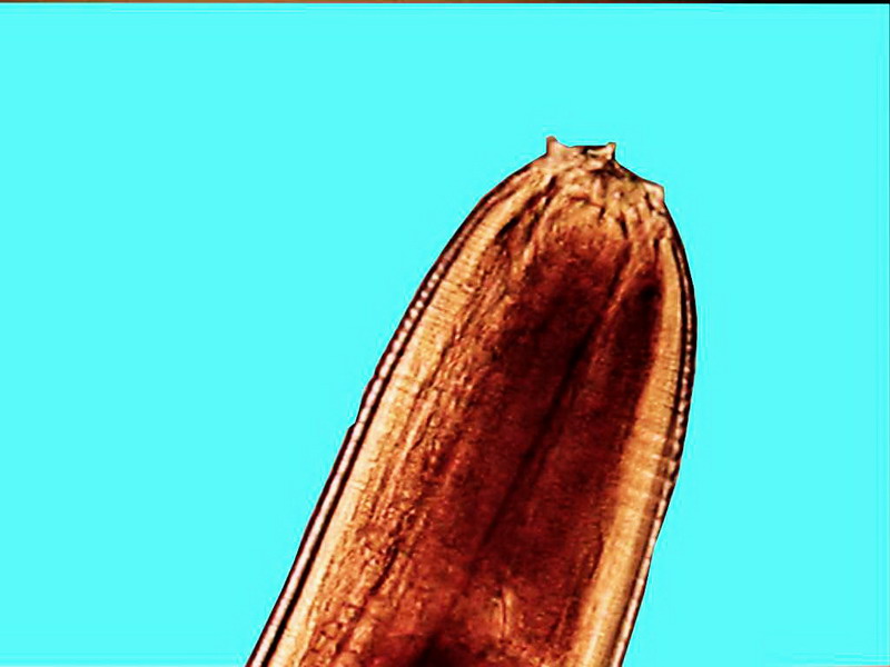
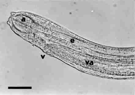
This is the worm seen under the stereomicroscope
And
this is its head.
Clearing
the nematode with chloralphenol you can see the structure of the head.
a
buccal bulb, e sophagus, v
vulva, va vagina. The bar is 40µm long
reproduced from Moravec &
Salgado-Maldonado
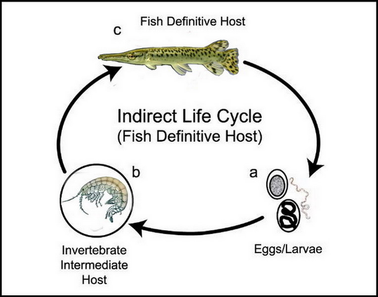
Life cycle is unknown, but we can hypothesize that
it is
similar to the cycle of Cystoopsis acipenseri, a
better known species. It
must be
relatively simple and similar to the one show below.
|
CESTODES
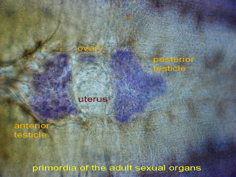
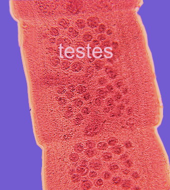
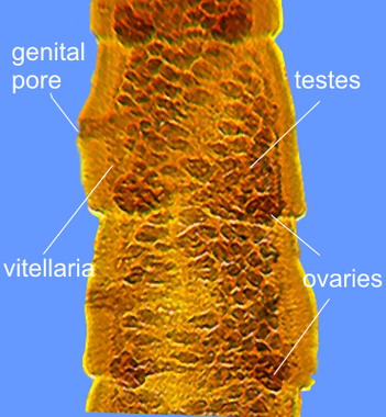
Proteocephalus singularis La Rue, 1911
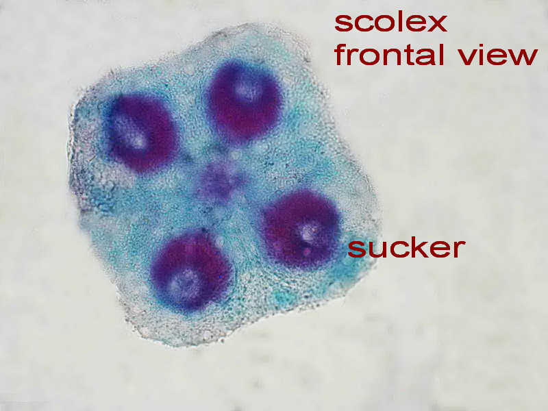
Cestodes
(tapeworms) are basically characterized by the anatomy of their
constituent
proglottids (segments). This is the site of the sexual organs
(whose
morphology is essential for species differentiation). Nevertheless,
the scolex (the first segment, or head, the holdfast organ) has
generally also
a distinctive structure, based on the suckers, grooves, hooks or spines
that
ornate some species. Both structures serve to fix the tapeworm to the
wall of
the intestine of their host. The genus Proteocephalus
has a scolex with a very simple structure as it is seen in the
following
frontal figure. The preparation has been stained with Hematoxylin and
mounted in
Canada Balsam.
It is similar
enough to
the one of another genus named Ophiotenia.
Differentiation between both genera is based on the distribution of
the
testicles in the proglottid, in Ophiotenia
they are distributed in two
distinct
lateral fields, whereas those of Proteocephalus
form an ample, central, and unique field, as the following image (left)
shows,
the right image is a preparation of one mature proglottid.
Salgado-Maldonado et al.,2003, states
that The life
cycle of this cestode in Mexico is unknown, but we assume that a
copepod serves
as an intermediate host. Plerocercoids of proteocephalids have been
found in
the viscera of several freshwater fishes in Tabasco. This is
consistent
with
what is known about the life cycles of the cestoda, that can be so
summarized:
The adult
of
Cestoda (1) lives in the
intestine of the definitive
host fish (in
this case the Atractosteus), where it
sheds free proglottids which free its eggs that enter the water
with the feces (2). The eggs are
generally eaten
by a copepod (still now unknown) (3) and
develop into a first larva named procercoid. If the infected copepod is
eaten by a small
fish (4) (also
unknown
but there are a lot that are food for the tropical gar) the parasite penetrates the gut
and develops in the internal
organs or the mesentery into a second
larval stage (plerocercoid).
Finally, the definitive
host (5) eats an
infected small fish and the adult
cestode develops in its intestine.
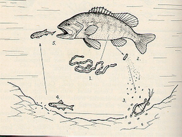
So they think the cycle is similar to the one of the
proteocephalid of the black bass shown above and reproduced from an
original drawing in
https://www.msu.edu/~gillilla/basstapeworm.html
I want to offer
thanks here for the
corrections that Prof. Leticia García Magaña made to my
draft, which improved the article.
NOTE:
if you like
to know better the State of
Tabasco, I recommend that you visit this incredible collection of
pictures, most
of them in HD: http://www.skyscrapercity.com/showthread.php?t=518975
Comments to the author,
Walter
Dioni
, are welcomed.
© Microscopy UK or their
contributors.
Published in the June 2009 edition of
Micscape.
Please report any Web problems or offer general
comments to the
Micscape
Editor.
Micscape is the on-line monthly magazine of
the Microscopy UK web
site at
Microscopy-UK
© Onview.net Ltd, Microscopy-UK, and all
contributors 1995 onwards. All rights reserved.
Main site is at
www.microscopy-uk.org.uk
with full mirror at
www.microscopy-uk.net
.
|
|