An Overview of Human Cells for Light Microscopists
Part III - Human Eyes and Insect Eyes
A 3D modelling article
by Mol Smith 2010
Please Donate to our Appeal to Fund the Creation of 3D Models for Microscopic Entities!
Please give the pages in this article time to load: medium size video files involved.
You will need a minimum version 6.1 of
Apple Quicktime Player Plug-in to view the movies here.

Page:      1  |  2  |   3   |  4  |  5 |  6                       For Part 1 The Human Cell - go here!    Part 2 Human Skin - go here!

 Insect Eyes    Insect's View: Simulation1   Simulation2   Simulation3   Human Eyes1   Human Eyes2   Resources and external links
   

Human Eyes
Humans and most mammals have Complex Eyes which use a very sophisticated lens system to focus light onto receiving nerve sensors at the back of the eye. This type of eye cannot sample the outside world at the same fast speed of the insect's compound eye, and is a lot slower. However, the information gathered is highly detailed.

On the right is a diagram of a human eye.
The Retina has two distinct types of light-sensitive nerve cells: rods and cones.

Rods detect monochrome (black-and-white) imagery and are especially well suited to low light levels. There are no rods at the Fovea or blind spot, and more exist for peripheral vision than central vision. The Fovea region is highly important in the human eye, as it contains more cones than in other parts of the eye. This allows much greater detail in our central vision (see A in diagram) where light waves are gathered and focused onto the Fovea through the centre of the pupil. Thus we receive a sharper picture when we look directly at an object.

Cones enable color vision but they need brighter light to function than rods. Cones come in three types:
long-wavelength sensitive (red colour detection), medium-wavelength sensitive (green colour detection), and short-wavelength sensitive light (blue colour detection).

Cone and rod cells connect to nerve fibres of the optic nerve through intermediary cells in the retina. When light falls on the rods and cones, nerves send electrical impulses into the brain.


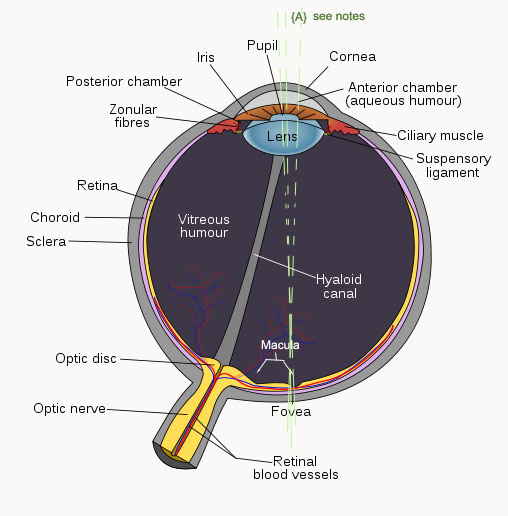
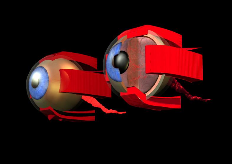
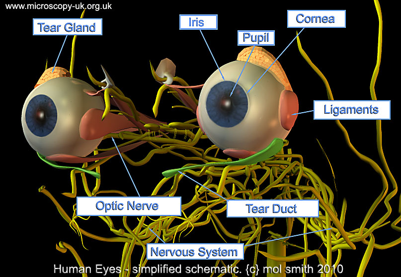
The astounding things about human eyes is that they auto-focus and they also adjust their irises to control light input. They are self-cleaning, and because we have two of them placed side-by side, they provide us with two separate views of the external world - an attribute which the brain uses by combining the two images into a single stereoscopic 3D image. This aids us in determining depth and the relative distances of objects in our field of view.

Our eyes have a blind spot. This is the area at the back of the eye which is occupied by the optical disc. the point at which the nerve cells leave the eye to connect to the brain. There can be no rods or cones in this area to sense light. The alternate 3D model rendered below may also help to illuminate the parts of the human eye.

Watch a 3d model rotation of human eyes in situ and relative to the human brain here.


Comments to the author
Mol Smith are welcomed.

Microscopy UK Front Page
Micscape Magazine
Article Library

all material © Mol Smith except where indicated


Published in Jun 2010 Micscape Magazine.
Please report any Web problems or offer general comments to the
Micscape Editor.
Micscape is the on-line monthly magazine of the Microscopy UK web site at
Microscopy-UK


© Onview.net Ltd, Microscopy-UK, and all contributors 1995 onwards. All rights reserved. Main site is at www.microscopy-uk.org.uk