Her
mist of primroses within her breast
Twilight hath folded up, and
oer
the west,
Seeking remoter valleys long
hath
gone,
Not yet hath come her sister
of the
dawn
George William Russell
(1867 1935)
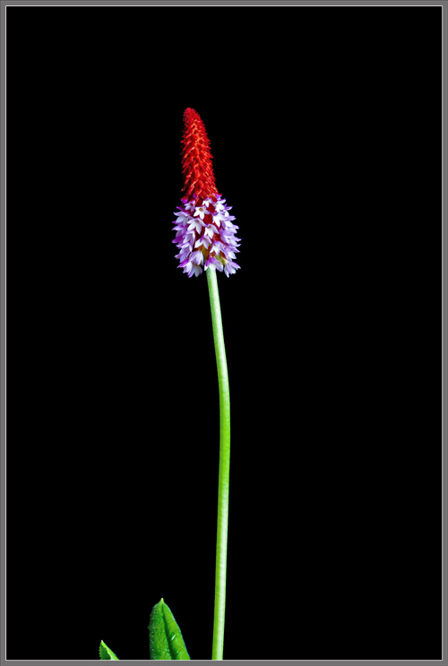
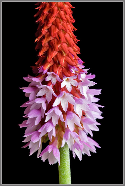
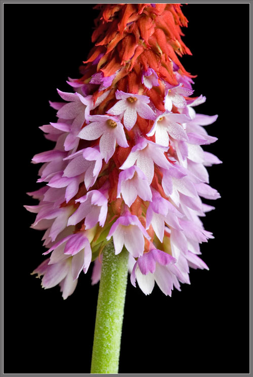
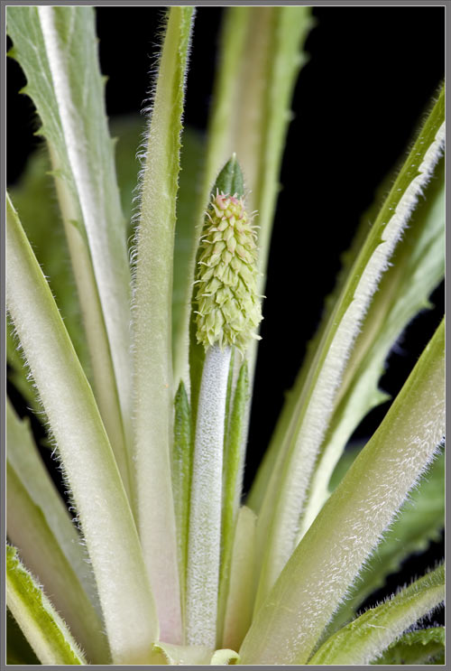
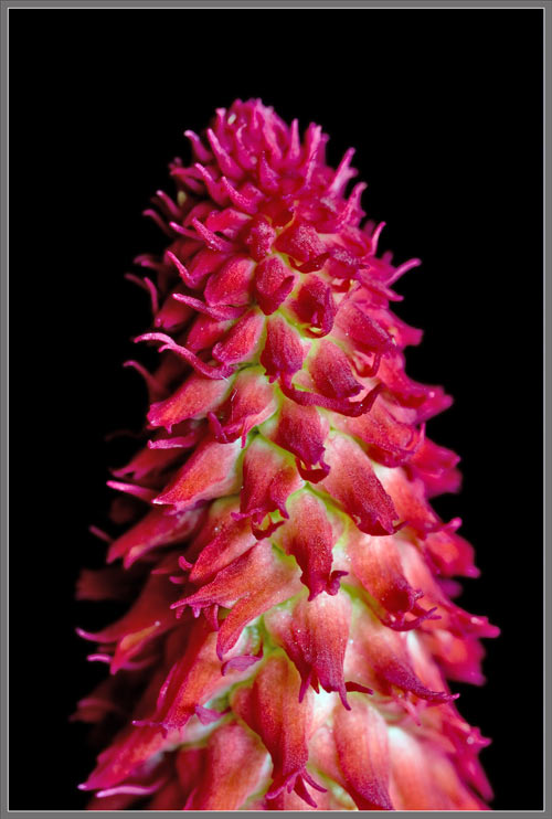
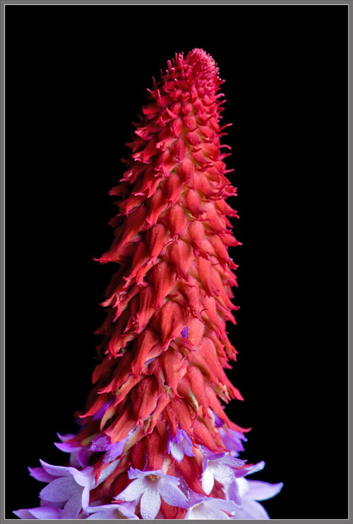
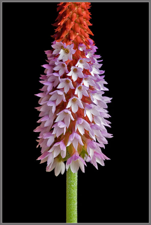
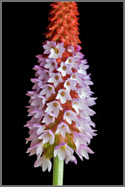
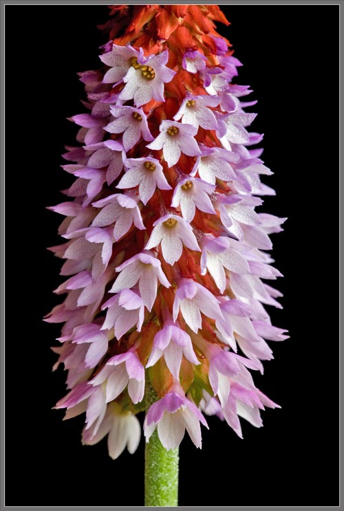
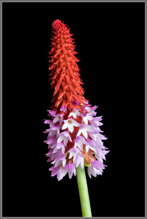
This Primula vialii hybrid certainly
looks different than other Primroses! Perched atop a long
cylindrical stem, its two-toned, rocket-shaped flower-head has
brilliant red bracts, and orchid pink flowers. The plant
grows to
about 40 centimetres in height, and has a rosette of short,
unusually
shaped basal leaves. Other common names for this species
include
Foxtail Primrose, Poker Primrose, Orchid Primrose, and Wayside
Primrose.
The Primula genus of perennials
contains around 400 species, most of which are found in the
Northern
Hemisphere temperate regions. China and the Himalayas
possess the
greatest number of species, and Primula
vialii is in fact native to Chinas Yunnan Province.
Images follow that show the
plants
long stem, and two-toned flower-head.
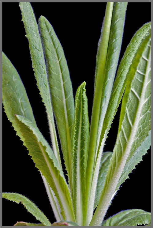
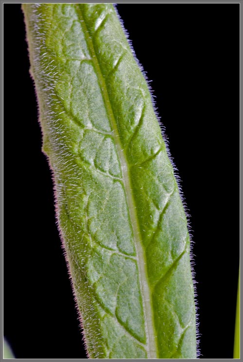
Most leaves in the basal
rosette
are positioned vertically, and thus show their back surface to
the
observer.
The front of a leaf is
intensely
hairy and possesses a central longitudinal vein with irregularly
positioned offshoots.
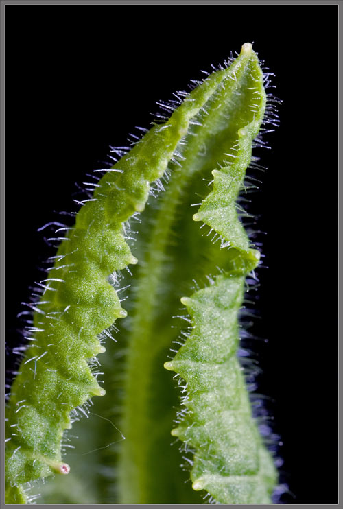
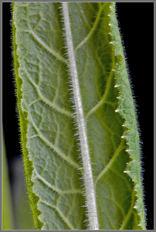
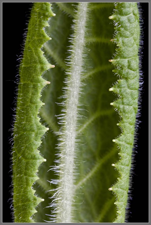
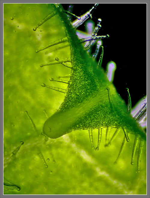
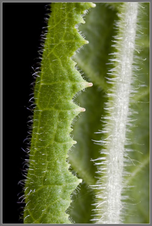
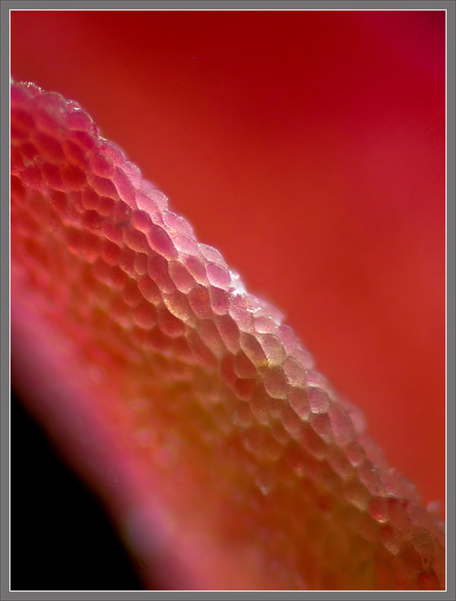
As can be seen below, each
leaf has
an extremely concave back surface, and strikingly toothed
margins.
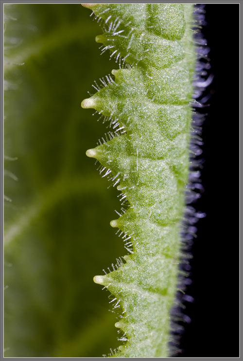
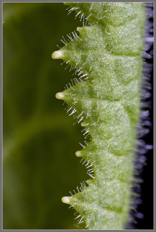
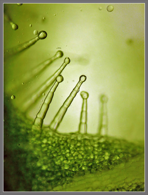
The two images that follow
show the
white peg that forms the tip of each tooth along a leafs
margin.
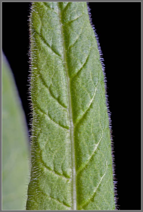
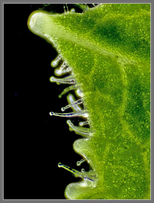
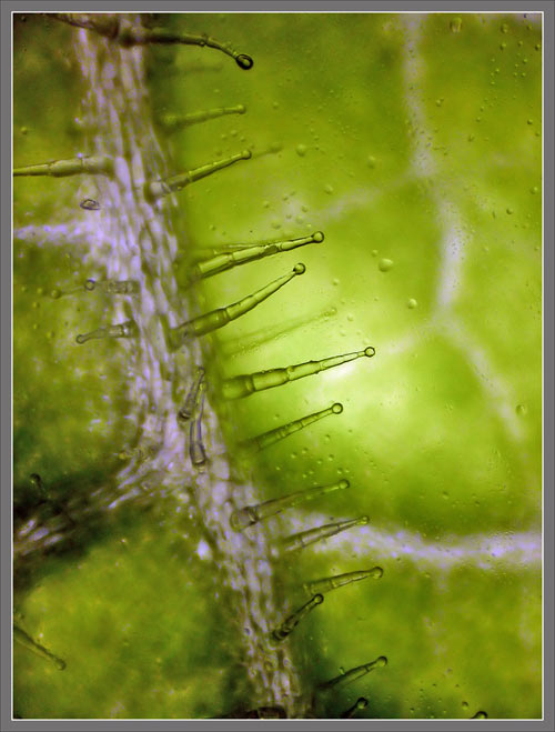
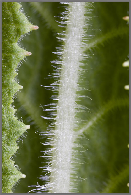
Notice in the image at left
below,
how prominent is the main vein on a leafs underside. It
is also
extremely hairy. The photomicrograph on the right shows
one of
the teeth along the leafs margin, and the bulbous-tipped
glandular
hairs that grow from its surface.
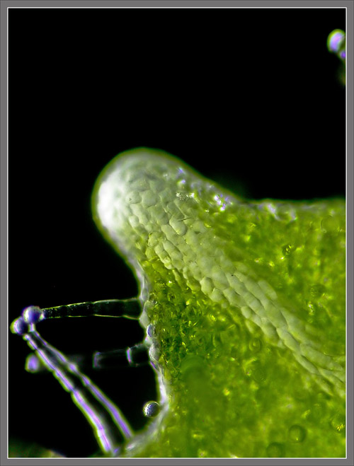
The cellular structure of one
of
the pegs at the tip of a tooth can be seen in the image on the
right
below.
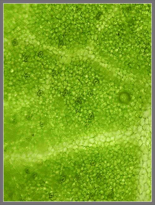
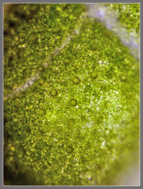
Stomata and guard cells, which
control gas entry and exit from a leafs underside, are visible
in the
photomicrograph below.
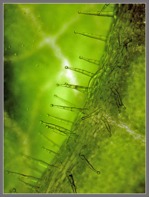
Photomicrographs follow that
show
the segmented glandular hairs that grow from the veins on the
underside
of a leaf.
Viewed from directly overhead,
the
glandular hairs appear as shaded spheres.
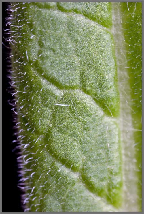
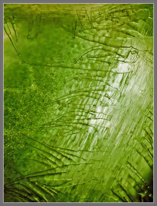
A leafs main vein is not
imbedded
deeply in the undersurface of the leaf, but is very prominently
raised,
with minimum connection between vein and leaf.
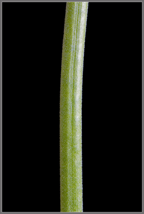
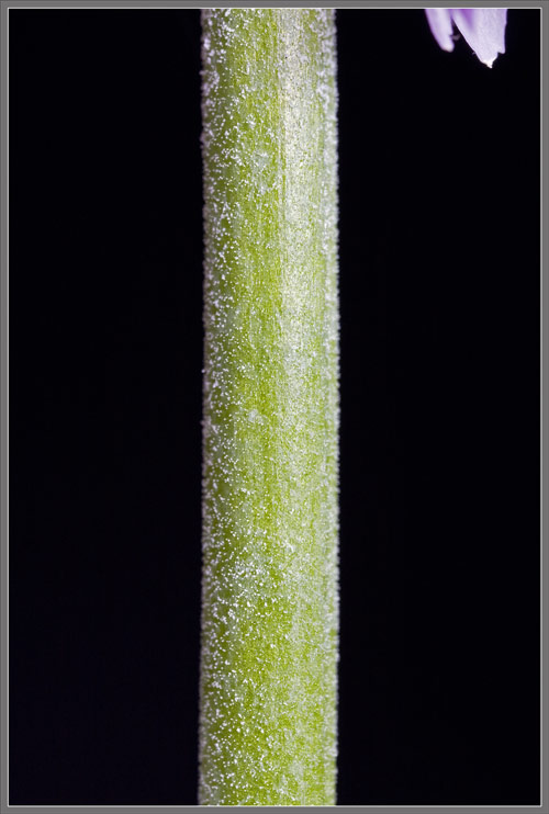
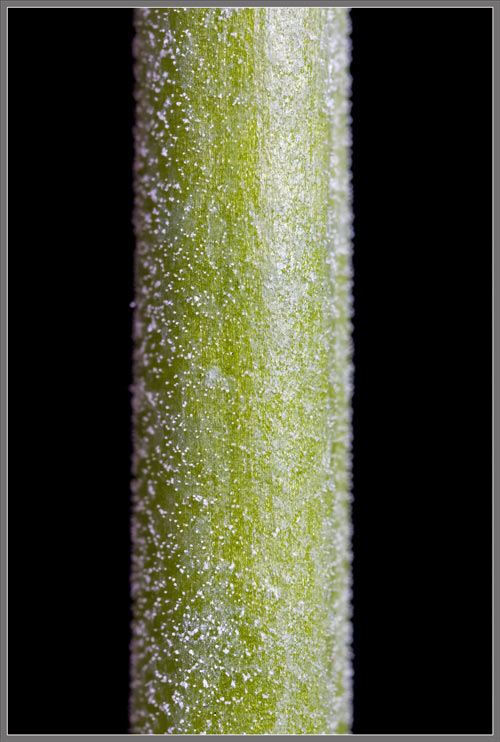
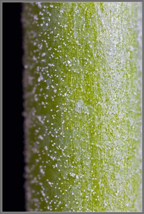
The plants main stem has an
approximately circular cross-section, with a single, shallow
longitudinal groove on its surface.
Closer views reveal that the
stem
is also liberally covered with short glandular hairs.
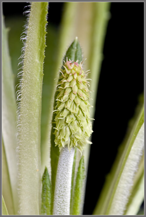
The following sequence of
images,
taken with increasing magnification, shows a very early
bud-stage
flower-head. At this point the buds are still completely
protected by pale green bracts, (modified leaves). Near
the tip
of the flower-head, hints indicating the eventual red colour of
the
bracts have begun to appear.
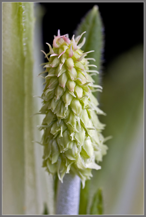
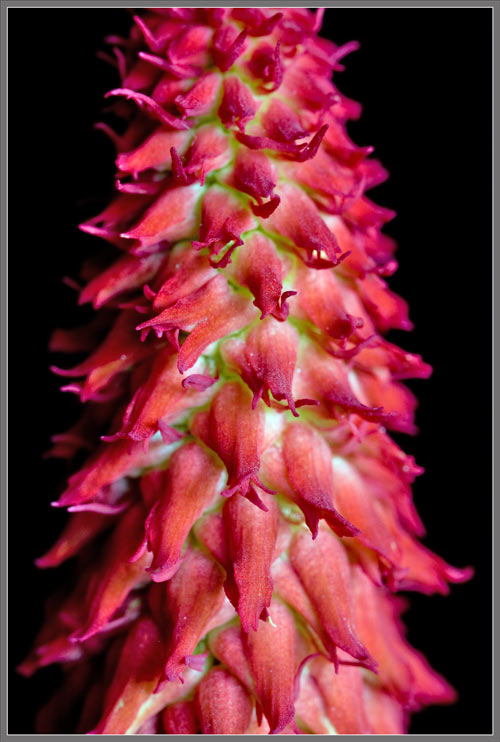
A week later, these same
bracts are
brilliantly red coloured on an almost white background. At
this
stage, no signs of the underlying flower buds are visible.
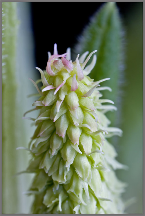
At the middle of both images
below,
you can see the purple tips of bud petals peeking out from
between the
bright red bracts.
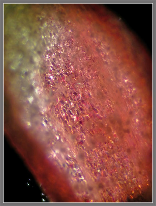
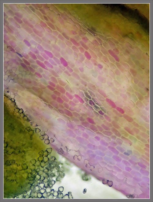
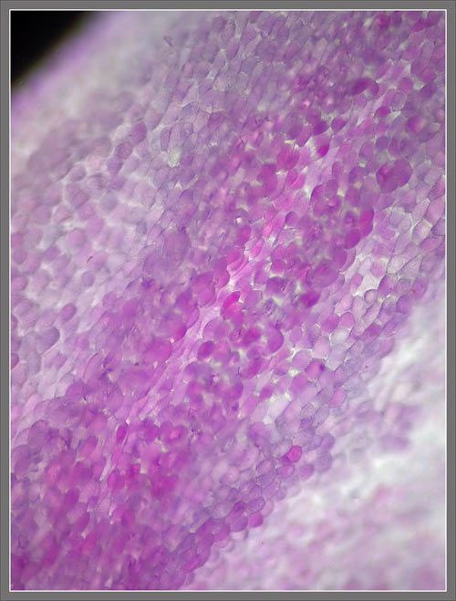
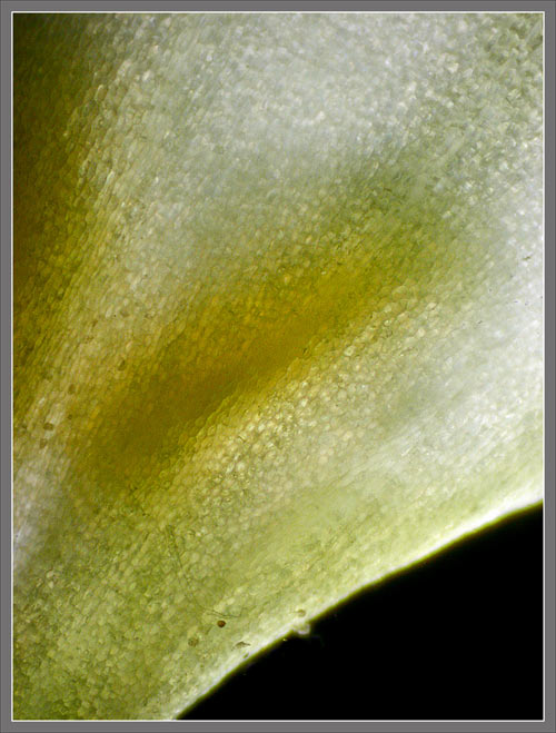
If the surface of one of the
bracts
is examined under the microscope, its cellular structure becomes
visible.
Higher magnification reveals
more
detail.
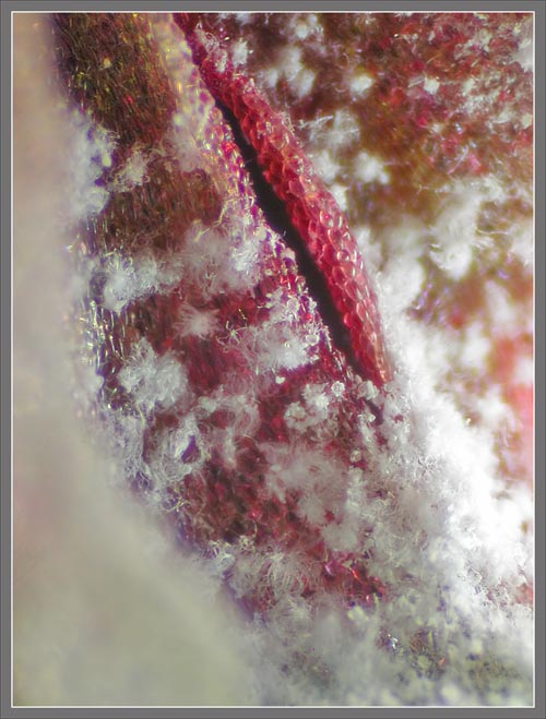
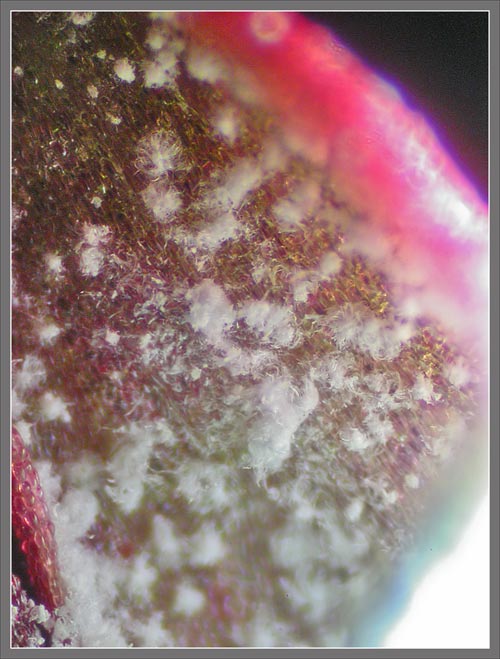
While working with the plant,
I
noticed that most of the structures comprising the flower-head
were
covered with what appeared to be microscopic snow
flakes. Under
the microscope, these appear to be tufts of white fibrous
material
whose purpose is unknown.
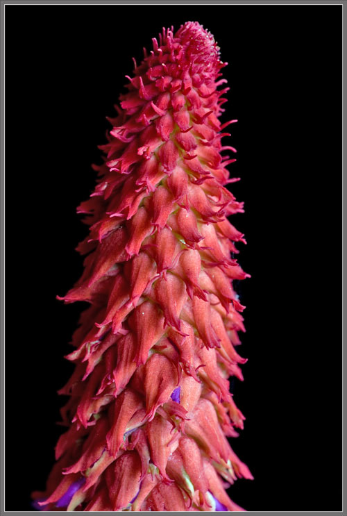
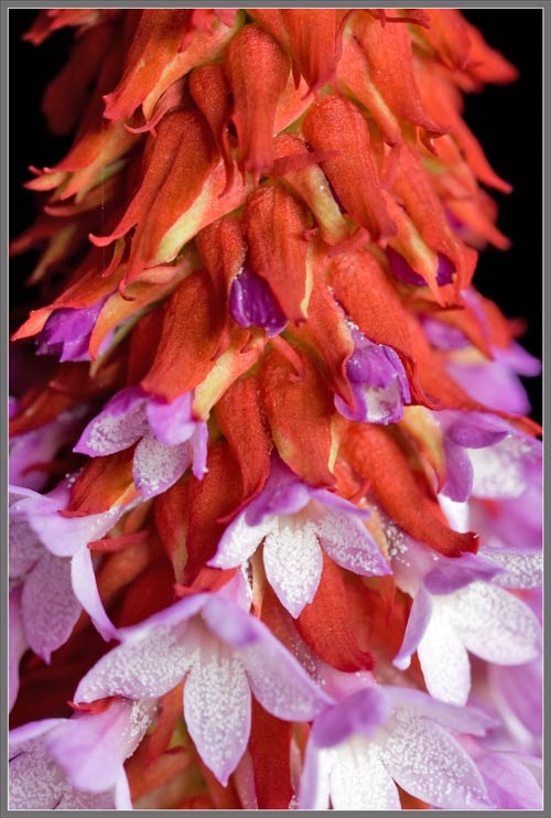
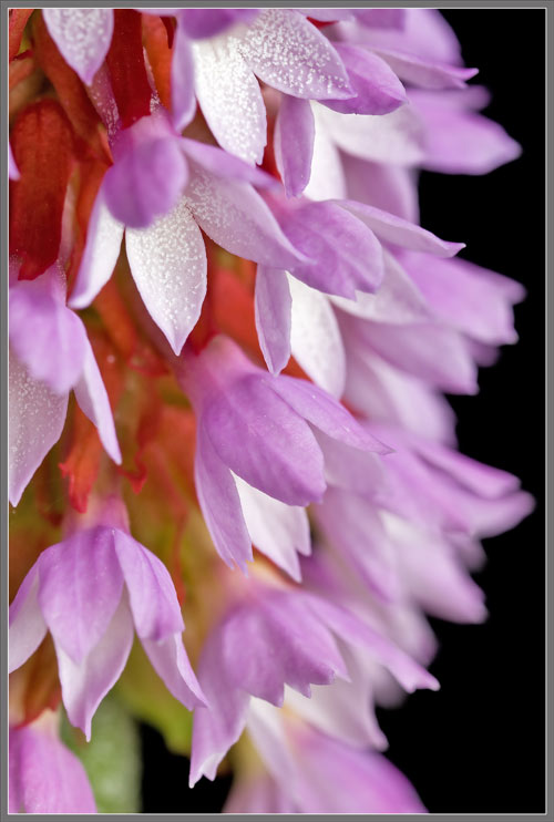
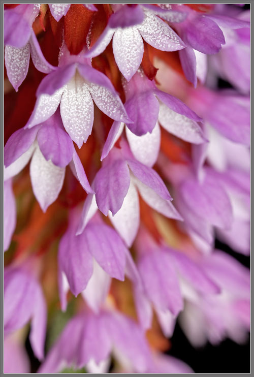
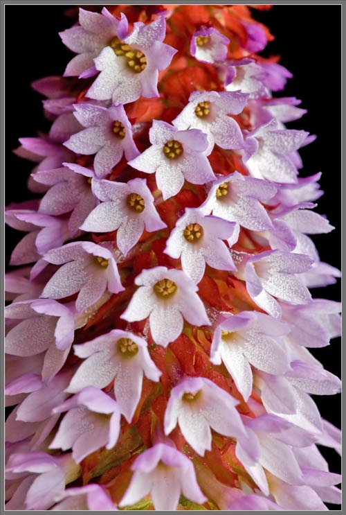
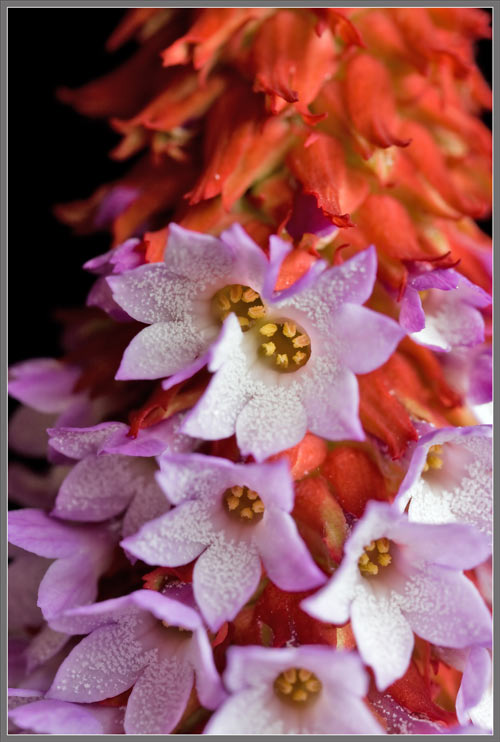
The three images that follow
show
the flower-head in bloom. Flowers have five pink, pointed
petals
fused at the base to form the corolla. At the centre of
each
flower, there is a group of yellow anthers. Note that the
top of
the stem has been angled away from the viewer in order that he
or she
can see into the recesses of the corollas. Normally, the
flowers
are angled downwards, preventing the reproductive structures
from being
seen.
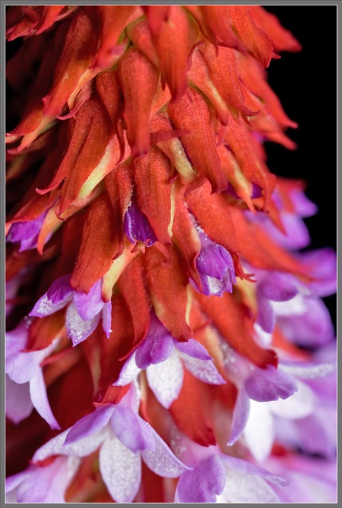
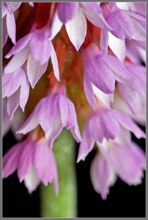
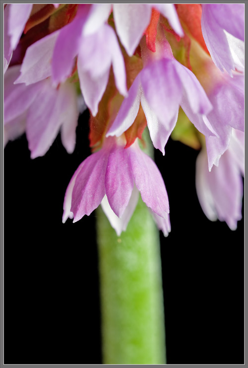
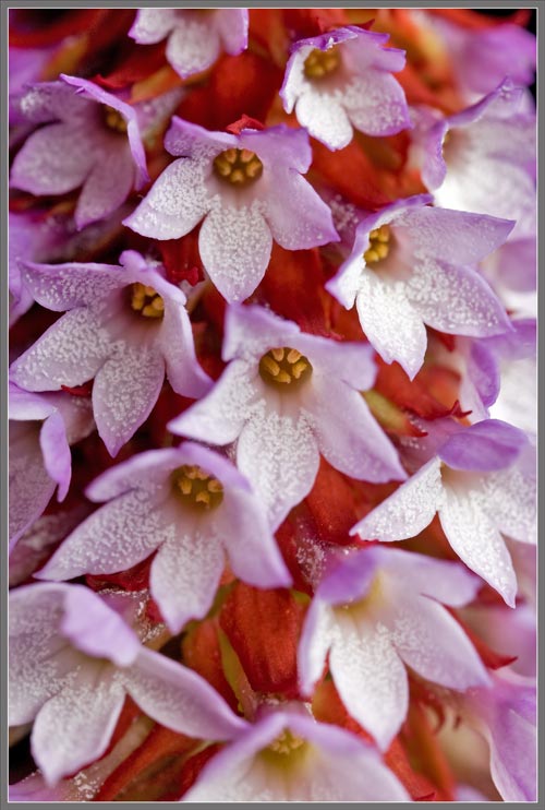
This normal orientation can be
seen
in the images below. Note that each flower is attached to
its
stalk through the narrow tubular base of the flared
corolla. In
many images, the pure white tufts of fibrous material mentioned
earlier
can be seen coating the inner surfaces of corollas.
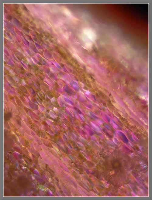
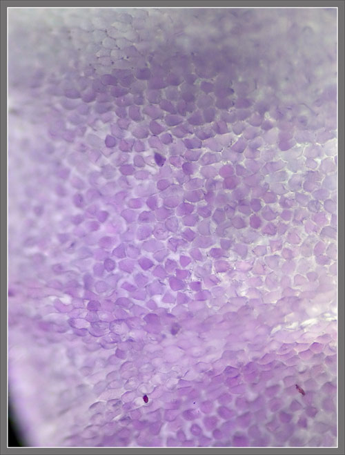
The cellular structure of the
outer
surface of a petal is visible in the photomicrographs
below. Note
the variable shape of pollen grains in the second image.
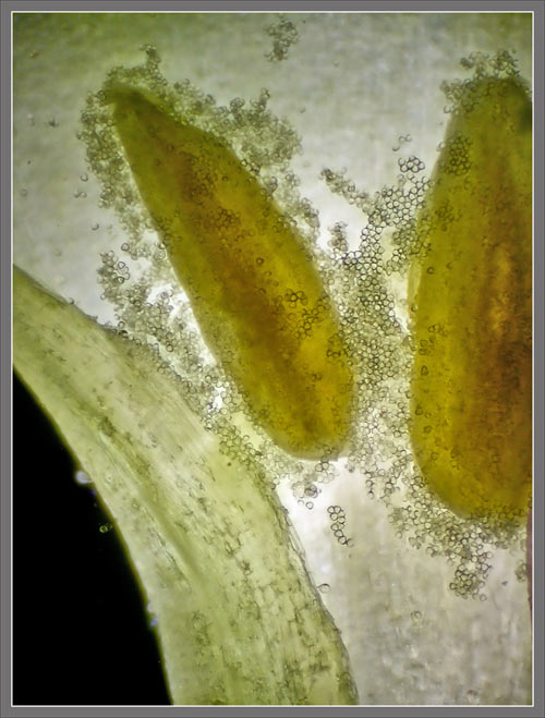
Closer views of the ring of
anthers
at the centre of each flowers corolla can be seen in the images
that
follow. Some flowers possess six anthers, while others
appear to
have only five.
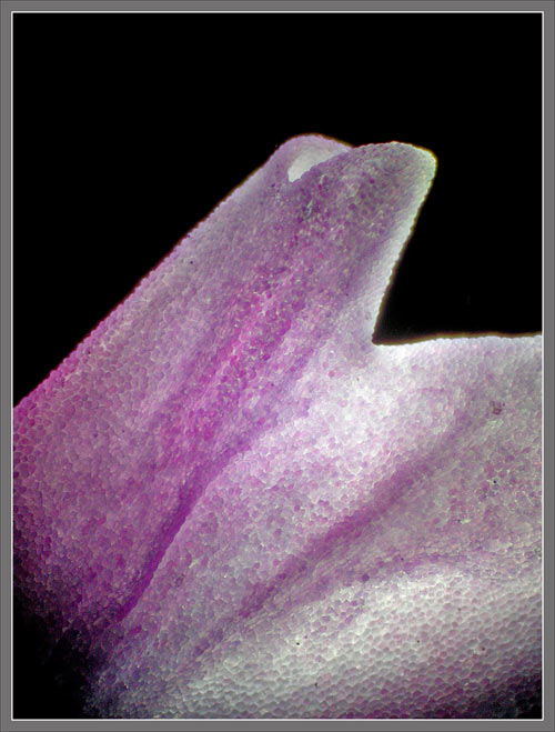
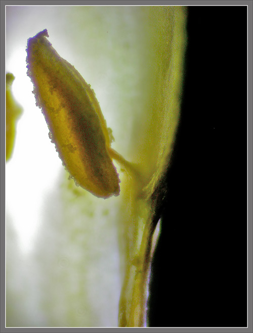
The image that follows on the
left
shows a portion of the tubular base of a flowers corolla.
A dark
shadow is being cast by some internal structure. The image
on the
right shows this structure an anther joined to the wall of the
corolla by a short, remarkably small diameter filament.
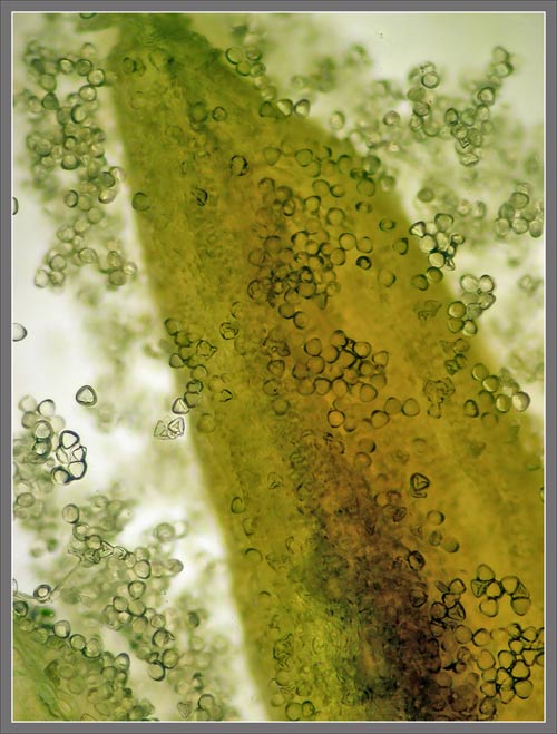
The final views show more
highly
magnified anthers with their variably shaped pollen grains.
The hybrid Primrose studied in
this
article, Primula vialii
Chinese
Pagoda, was awarded the Royal Botanical Society Award
of
Garden Merit in 1993. It is indeed a spectacular plant!
Photographic
Equipment
The low magnification, (to
1:1),
macro-photographs were taken using a 13 megapixel Canon 5D full
frame
DSLR, using a Canon EF 180 mm 1:3.5 L Macro lens.
An 10 megapixel Canon 40D
DSLR,
equipped with a specialized high magnification (1x to 5x) Canon
macro
lens, the MP-E 65 mm 1:2.8, was used to take the remainder of
the
images.
The photomicrographs were
taken
using a Leitz SM-Pol microscope (using a dark ground condenser),
and
the Coolpix 4500.
A Flower Garden of
Macroscopic Delights
A complete graphical index of
all
of my flower articles can be found here.
The Colourful World
of
Chemical Crystals
A complete graphical index of
all
of my crystal articles can be found here.