Exploring the cultivated silk moth Bombyx mori
Part 1: The forms and fine structure of the adult wing scales, a subject studied by Leeuwenhoek.
by David Walker, UK
Image right: From Wardle 1881 (14).
|
Exploring the cultivated silk moth Bombyx mori Part 1: The forms and fine structure of the adult wing scales, a subject studied by Leeuwenhoek. by David Walker, UK
Image right: From Wardle 1881 (14). |
|
In Explorer browser, setting 60% in 'Print Preview' prints out on 8 sheets of A4 paper.
Part 2 (September 2012 Micscape) - other aspects of adult silk moth.
Part 3 (October 2012 Micscape) - compound eyes of adult, facet count.
The cultivated silk moth (Bombyx mori) was one of the first insects whose structure and life cycle was investigated in detail using optical aids. The insect would have been well known to the first microscopists in the 17th century given that sericulture was by then well established in mainland Europe (1, 3b, 15). Some of the earliest studies are associated with leading names in the history of microscopy and include Malpighi, Swammerdam and Leeuwenhoek (13, 18).
These workers' practical and observational skills using some of the earliest microscopes, despite the inherent limitations of the instruments, can often be better appreciated if complemented by studying the subjects using a modern microscope. My own interest in the early silk moth studies was sparked when reading Hoole's 'The Select Works of Antony van Leeuwenhoek' (2). Leeuwenhoek studied and illustrated in detail the many forms of the scales, their insertion and spacing in the wing and also counted the maximum number of fine ribs he'd observed on the wing scales. I became particularly intrigued as to how accurate this latter measurement was as hadn't seen the wing scale micrometric studies discussed in references seen to date. The first part of this article summarises Leeuwenhoek's reported studies and is followed by modern studies presenting both published SEM images and my own hobby level 'explorations' with an optical microscope.
Leeuwenhoek's published studies of Bombyx mori
Cole in 1937 published a 96 page paper in two parts entitled 'Leeuwenhoek's Zoological Researches' (6,7). Part 2 meticulously tabulates where each of Leeuwenhoek's letters have been published in Dutch, Latin and English editions and an index listing which letters discussed a given zoological group (listing to species where known). This identified letter 146 as the main letter discussing his studies of the various stages of the silk moth life cycle. It was written on April 20th 1702 and addressed
to Karl [Charles I], Landgrave of Hessen-Kasel (3) who Dobell describes as an 'amateur of science' (4b) and who Ratcliff describes as 'an important patron of science and prince' (8). This letter therefore does not feature in those addressed to the Royal Society and published in their Philosophical Transactions.
The most accessible English translation of a selection of Leeuwenhoek's letters remains Hoole's (in 2 vols. 1798, 1807) and is available online and in modern budget reprints. Dobell remarks that they are 'an excellent and scholarly translation' (with provisos). Letters on the Bombyx studies are compiled into the chapter 'On the Silkworm' (2). The Editors of the 'Collected Letters' note that Hoole's translation of letter 146 is:
'An all but complete English translation of the letter, the last part on the procreation excepted'. (3)
The definitive modern English/Dutch translation of Leeuwenhoek's letters with extensive footnotes by scholars are the 'The Collected Letters of Antoni van Leeuwenhoek', an ongoing project which has published 15 of the planned 19 volumes to date (see ref. 3 for details). Volume 14 contains letter 146 ('Collected Letters' number 236), a facsimile of the original drawing by Leeuwenhoek's draughtsman and a splendid reproduction of a plate engraving.
Leeuwenhoek's studies of the adult wings and scales of Bombyx mori
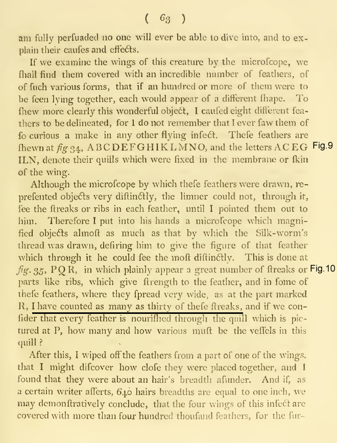
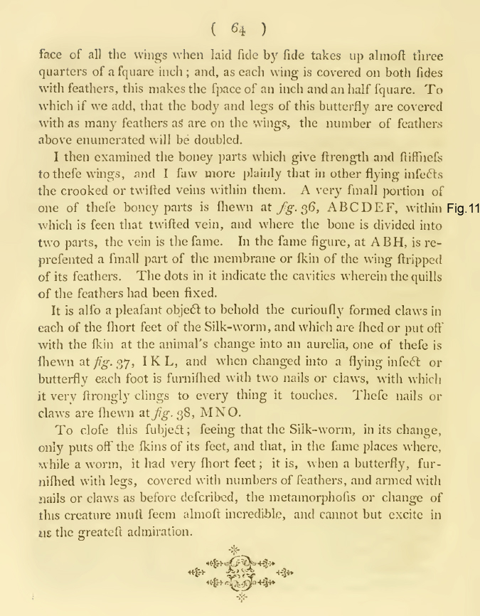
Hoole's translation of the wing scale studies reads very well and is presented below from www.archive.org (2). The fig. nos. in the margin are mine and refer to the published plate below (figs. were renumbered and laid out in Hoole). An 'f' is an old form of an 's'. Key points are the number of varied forms of scales he found (his 'feathers') and the number of ribs (his 'streaks') which he counted on
the larger scales at their widest point. These are discussed below in the context of modern microscopy studies with both an SEM and optical microscope.
Page scans from Hoole's chapter 'On the Silkworm' discussing the adult wing form and scales from his
'The Select Works of Antoni van Leeuwenhoek, vol I, 1800'. Source public domain books on www.archive.org .
Hoole's translation includes a plate showing Leeuwenhoek's silk moth scale studies but detail is inevitably lost; inspection of a contemporary published edition of Leeuwenhoek's letters with engravings, better shows the splendid original detail e.g. the 'Opera Omnia' (Latin) in four volumes which have been carefully scanned for the European Cultural Heritage Online (ECHO) website, plate presented below.
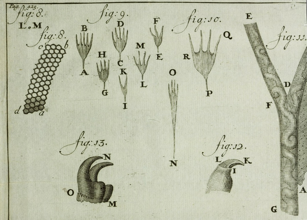
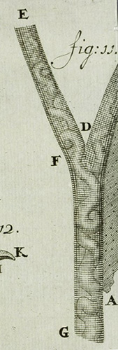
Plate LM from Leeuwenhoek's published letters in 'Opera Omnia', vol. 4, 1719. Figs. 9-11 inclusive refer to the adult silk moth and accompany his letter no. 146. These are engravings for the book from the original red chalk drawings by Leeuwenhoek's draughtsman which is shown on Plate VIII in the 'Collected Letters' vol. 14 (ref. 2).
ECHO website catalogue entry: 'Leeuwenhoek, Antoni van, Opera
omnia seu arcana naturae ope exactissimorum microscopiorum detecta, experimentis variis comprobata : epistolis ad varios viros, ut et ad integram, quae Londini floret, sapientem
Societatem, cujus membrum est, datis, comprehensa, & quatuor tomis distincta; vol. 4: [Continuatio arcanorum naturae], 1719.'
ECHO 'source information': Digitised by and images © Max Planck Institute for the History of Science. The ECHO venture has an 'Open Access Policy' where public display of works including derivative works is permitted with proper attribution. Link to plate.
Note on Leeuwenhoek's micrometric studies
Leeuwenhoek's wide ranging microscopy studies included subjects where measurements and/or counts were taken and were reported in his letters. Both Dobell (3) and Schierbeek (4) provide summaries of selected studies and his micrometry techniques. Schierbeek also notes that:
"Leeuwenhoek was not the first microscopist, but he was the first to measure microscopic objects." (4).
Leeuwenhoek took linear measurements by comparing with one of his standard subjects of appropriate size. In decreasing size these standards included an inch defined by his five inch measuring rod, three grades of sand grains, hairs from known sources, silkworm thread, red blood corpuscle and 'the smallest animals in pepper water' (bacteria). These have been tabulated with typical actual size ranges from modern measurements in each volume of the 'Collected Letters' (2) and were also discussed by Dobell (3).
Exploring the Bombyx mori wing and scale fine structure with a modern microscope
SEM
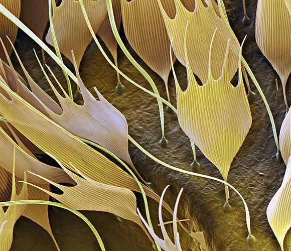
The scanning electron microscope's ability to image subjects with both high resolution and extended depth of field can give striking images of insect wing scale layout and structure; two splendid examples for Bombyx mori are shared below from the website www.eyeofscience.com courtesy of Oliver Meckes.
|
Right: False colour SEM of Bombyx mori wing scales in situ showing both the fine structure and how the scales are inserted in the wing. Magnification is 400x at 10 cm wide image. © www.eyeofscience.com and shared with permission. Contact the website for any image use. |
|
|
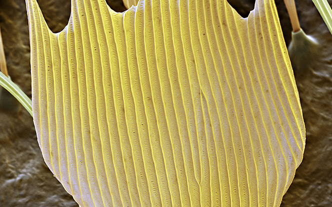
The scale on the right in the above image is presented in the screen plane and allows an accurate count of rib number. The image right is a crop from the master image of a close-up of this scale to show both the ribbing more clearly and the fine structure between ribs. The scale width at widest point is ca. 63 µm. Crop of a master image supplied by and © www.eyeofscience.com and shared with permission. |
|
Leeuwenhoek's micrometric studies of scale fine structure and size, and scale studies by later workers:
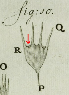
As highlighted in the Hoole translation above, Leeuwenhoek inspected many scales noting the variety of forms, the fine ribs and that he 'counted as many as thirty of these streaks' at the widest point of the largest scales, point R on fig. 10 on plate above. The modern 'Collected Letters' translation reads
that he 'counted at least thirty'; i.e. depending on interpretation, thirty ribs was either the upper limit which Leeuwenhoek observed or greater. The SEM image above of the particular single scale facing forward was exactly thirty give or take one! Counting finely spaced features in free space without a frame of reference, as Leeuwenhoek would have had to do, can also be tricky.
To resolve the scale ribs measured at ca. 2.1 µm apart from the above SEM image, requires a lens with numerical aperture of at least 0.14; studies of Leeuwenhoek's remaining nine lenses by van Zuylen (9) show that two were capable of this, the best extant 'Utrecht' lens has a magnification of 266X, an NA of 0.31 and a measured resolving power of 1.36 µm. Although the scale fine structure is not a demanding subject to resolve, Leeuwenhoek comments that although he could see the ribs, a better lens had to be given to his draughtsman ('limner') to show him the ribs to draw (Boltjes remarks on this, 10).
Boltjes in 1941 (10) prepared a range of 'blown' single lenses and presented an excellent series of photographs taken with them of a selection of subjects studied by Leeuwenhoek. This included Bombyx mori scales and shows the ribs crisply resolved in Plate IV using a lens 1.2 mm diameter and magnification of 250X (but didn't comment on Leeuwenhoek's micrometric studies of the scales). [Permission to share plate pending].
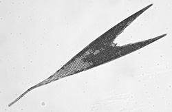
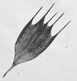
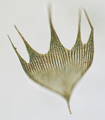
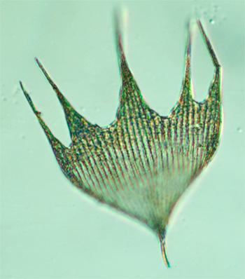
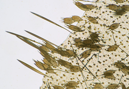
Wardle in his 'Handbook of the Collection Illustrative of the Wild Silks of India ...' 1881, presented drawings of 24 species of silk moth to show the variety of scales from different parts of the wing as well as selected scale detail. The variation of scale forms shown for Bombyx mori are not dissimilar to some other species i.e. showing shield-like and hair-like forms. Two plates are shown below.
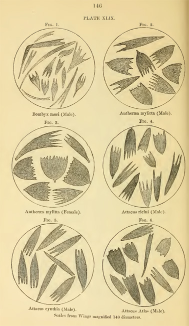
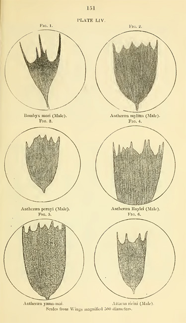
Two plates from Wardle's 'Handbook of the Collection Illustrative of the Wild Silks of India, in the Indian Section of the South Kensington Museum, with a Catalogue of the Collection and Numerous Illustrations', 1881. (14)
Optical microscope studies
Obtaining adult specimens of Bombyx mori
There are a number of entomology suppliers who source specimens legally and responsibly, but although many striking and exotic species can be readily obtained, the adult Bombyx mori is a small, relatively drab looking moth, presumably unappealing to the collector and have yet to find a supplier. Occasionally, an eBay entomology dealer does offer examples. The silk
moth's life cycle does feature in a variety of educational materials and a local silk farm may be a more ready supply. I am indebted to William Hyett of www.silkfarm.co.uk for supplying by post two types of adult specimen at a small price just to cover his costs: 1) a number of dead adults normally discarded, these can be rather tatty for wing scale studies but offers a ready supply for trials at dissection for studying
the more robust parts such as the legs and head; 2) viable cocoons which potentially allowed emergence to be followed as well as obtaining pristine adults for wing scale and other studies.
Incubating cocoons to emergence
The viable cocoons were incubated in a small part-covered tray under the recommended conditions of 20-25ºC with some humidity. The cocoons emerged a week after receipt. I set up a tripod and camera in the hope of filming the emerging adults, but they didn't read the film script; they usually emerge on the onset of daylight, but the tray was transferred to an airing cupboard each night to maintain the temperature and the adults were already fully emerged while still in
the dark.
I also hoped to film the mating of the adults but those emerging were the same sex, so after some still image photography, they were quickly killed in a covered Petri dish* with a small wad of cotton wool present dampened with ethyl acetate (one of the recommended humane killing methods for insects). (* For small insects a covered dish seemed to be better than the advised formal 'insect killing bottle' as very little ethyl acetate is required to build up a high vapour level and the adults died rapidly and easily removed.)
|
|
|
Left: The simple incubator used to keep the cocoons at 20-25ºC. The small square box contained water to maintain a local humidity. The fluorescent light panel (for viewing 35mm film slides) on which the tray sits gave trickle heat when required. Tray lid removed for clarity. The white tissue gives a hold to aid the emerging adults.
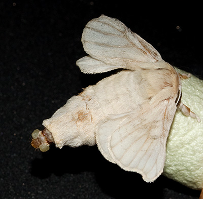
Right: A newly emerged adult, the gland at posterior is typical of a female, it releases pheromones to attract a male (16). (The race emerged has a much paler cocoon than for the race shown left.)
Wings and scale structure at low power
For many moths and butterflies, the wing scales are arranged like a neat 'tiled roof' with most scales laying near flat, although the colours and forms of scales can vary greatly to build up the wing patterns e.g. see Nijhout (11). Such arrangements are readily seen under the stereo or hand lens but SEM photography shows it particularly well (e.g. see the splendid SEM studies on www.psmicrographs.co.uk
by Syred and Power and by Dennis Kunkel).
Low power images of the wings are shown below. Interpreting the scale layout of the silk moth was not as straightforward as for some Lepidoptera because the smaller scales are overlain extensively in many areas with long thread like scales. The small shield-like scales have a more vertical presentation on the wing than typical tile-like wing scales (as the SEM above shows).
Note on low power imaging below: A stereo microscope was used to provide a wide field and greater depth but does not have the NA for crisp fine detail. The compound microscope offers the NA but stitching and stacking is required for creating a 'bigger picture' (which I have yet to master). The magnification for each image in brackets is the optical mag of the stereo before projection into a Nikon D5000 DSLR body.
|
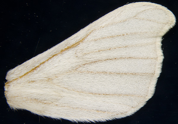
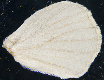
Right forewing from freshly hatched Bombyx mori. Max. width 17 mm. There are no colour derived wing scale patterns (in this example); the partly scaled veins providing the only markings. (4X with 0.4X accessory lens on stereo.) |
Right hindwing from same source (to scale with forewing). Max. width 13.5 mm. |
|
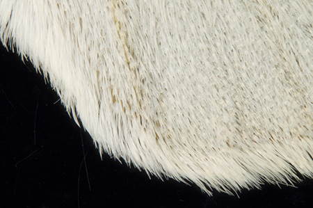
Detail from forewing along lower edge showing the hairlike scales. (20X) |
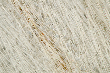
Close up of scales in centre of forewing. The hair-like scales overlay in many areas the small shield-like scales. (50X) |
|
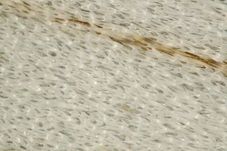
Close up of shield-like scales in an area less populated with hair-like scales. (50X) The scale insertion pattern is not readily determined cf the typical 'tiled-roof' pattern seen on many Lepidoptera, e.g the forewing of a large white butterfly shown right. (32X) |
|
Wings and scale structure at high power under the compound microscope
When exploring the tiny scales under a binocular microscope with modern optics and mechanics, it's worth remembering that Leeuwenhoek made his observations with his typical single lens microscope held very close to the eye with their inherent limitations. But as modern studies by scientists have shown, notably the work of Brian Ford in using the finest remaining extant Leeuwenhoek lens and modern counterparts (12), the carefully made single lens was capable of excellent work in the right hands and superior to
the compound microscopes of the time.
The wing scales as in many insects, readily rub off with a light brush and these can be transferred to a microscope slide and covered dry with a slip. Leeuwenhoek mounted larger subjects directly on the microscope pin for study but scale strews were likely have been first supported e.g. on a thin glass plate or cleaved talc both of which he is documented as using (5b).
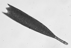
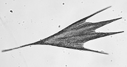
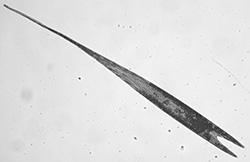
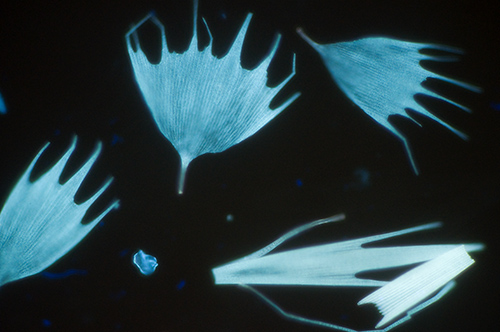
Scanning a strew at low power shows the variety of both the shield-like scales and the longer thread-like forms, as Leeuwenhoek noted and illustrated. The modern worker has the benefit of a mechanical stage to scan wide fields with subjects remaining in focus. Leeuwenhoek would have had to persevere with the close focus lenses, tiny field of view, crude focussing and awkward fine manipulation of e.g. glass plates with likely repeated studies to report the many scale forms, eight of which are carefully drawn by his draughtsman in the plate accompanying his letter (shown earlier). As described earlier, Wardle showed that other silk moths have a similar variety of scale form.
A selection of scales at various magnifications are shown below. Many scales on the moths studied to date have some sort of deposit which masked a clear presentation of the ribs under the modern microscope. This would have added additional difficulty in counting ribs on scales if Leeuwenhoek encountered similar scales.
Images below were taken using a Zeiss Photomicroscope III with Zeiss optics and a Nikon D5000 consumer DSLR on photo tube (Zeiss 10x Kpl W eyepiece on short collar as projection eyepiece.) HFW= horizontal field width.
|
|
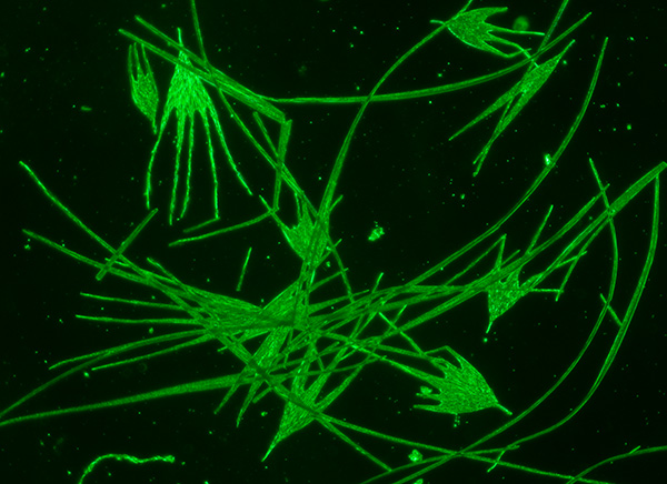
Darkfield image of a dry scale strew at low power (Zeiss 4x objective) shows the scale variety and size of the small shield-like scales with respect to the much longer hair-like scales which were typically 900 µm long. The large shield-like scale upper left is 425 x 230 µm
|
||||||
|
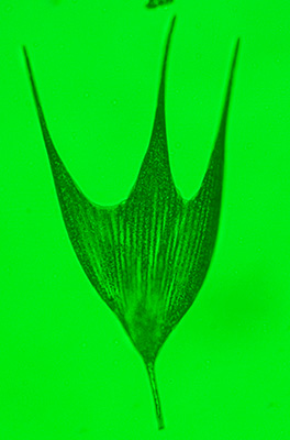
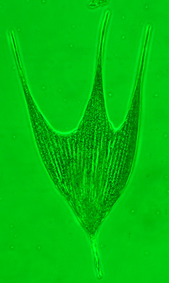
Left above: A typical shield-like dry scale with the 16x objective. The left-hand image is in brightfield, the right-hand with phase but the latter does not really aid the clear presentation of the dry scale ribs. |
Note that there is a double-edge appearance in the sculpted curve bases of the scales, a feature which Leeuwenhoek clearly depicted in his drawing reproduced below (arrowed) from earlier 'Opera Omnia' plate.
|
||||||
|
Left and above: A selection of scales from the wing showing the wide variety of forms present. |
||||||
|
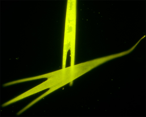
Some Lepidopteran scales can exhibit autofluorescence (17). The above image shows this with UV (400 nm LED) excitation using a Leitz 6.3 NPL Fluotar objective (epi III RS head on Photomicroscope, 10 sec exposure at ISO 400). The image right is with the Zeiss 16X objective with deep blue (455 nm LED) which also shows the deposits that can make study of the ribs trickier in brightfield. |
|
||||||
|
Left and right: One of the wider scales selected to count the ribs. Brightfield, Zeiss 16X objective. There are 30 ribs present as Leeuwenhoek also noted and have yet to find a scale with more. |
Above: DIC on same scale doesn't really aid study. Try counting the ribs in free space as Leeuwenhoek did. It isn't that easy without a frame of reference. The preference is to use a pen and touch the screen or print out and count. |
||||||
|
|
|
||||||
|
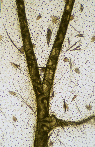
Image above left: Leeuwenhoek's study (drawn by his draughtsman) of the wing with scales removed to show the scale insertion points, veins and interior detail. |
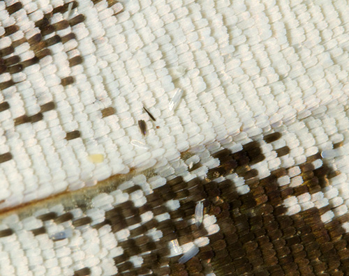
Above: Detail of the wing edge to show insertion of the residual scales on the 'brushed off' wing. |
In Leeuwenhoek's letter no. 146 shown in part earlier, he studies the wing with scales removed ('wiped off' in Hoole, 'rubbed off ... with a brush' in 'Collected Letters'). As his Fig. 11 in the plate shows, this presents well the scale insertion points, the veins and internal detail (of the trachea remains, 3c). A repeat of this study is shown above using a paintbrush size '00' brushed back and forth which readily removes most scales and illustrates how keen his observations were; the modern microscope shows little extra detail at this magnification. There seems no obvious pattern to the scale insertion points or their distance apart. Leeuwenhoek observes that the insertion points were 'one hair's breadth' apart which a footnote in 'Collected Letters' notes is approximately 40 µm (3c); this is within the range measured for the wing above using a modern micrometer where the insertions were typically 15 - 47 µm apart.
Comments
The cultivated silk moth Bombyx mori is neither a striking looking moth nor possesses microscopic features of the wing scales markedly different from other moths (as Wardle shows, 14), but its long history in sericulture and being one of the earliest insects studied in detail gives it a particular appeal to the enthusiast to study under the microscope. By manipulating and studying the same subjects, an additional insight and
appreciation can be gained of Leeuwenhoek's remarkable skills, who was accurately describing, measuring and illustrating the features with single lens microscopes without any of the benefits which the modern microscopist enjoys!
Comments to the author David Walker are welcomed.
Acknowledgements: I would like to thank the following people, but any errors in the above article are solely mine.
- William Hyett of www.silkfarm.co.uk for supplying the specimens and for providing valuable advice on incubating the supplied cocoons.
- The Science Museum Library (London) staff for the copy of Leeuwenhoek's Letter in reference 3 (via their excellent postal photocopying service).
- Oliver Meckes of www.eyeofscience.com for permission to share the striking SEM images of Bombyx mori.
- Research Library staff of the Max Planck Institute for the History of Science for clarifying 'Open Access' image use from the ECHO website (Leeuwenhoek's 'Opera Omnia' bookplate).
- Jan Parmentier for the generous gift some years ago of the book in ref. 9 and which helped inspire my hobby level interest in Leeuwenhoek's work.
Notes and references
1. Sir T. Wardle, 'Silk: Its Entomology, History and Manufacture', as Exemplified at the Royal Jubilee Exhibition Manchester 1887. Book available to read online/download at www.archive.org .
2. S. Hoole, 'The Select Works of Antony van Leeuwenhoek, Containing His Microscopical Discoveries in Many of the Works of Nature.' 2 Vols, 1798, 1807. The chapter 'On the Silk Worm' is in volume 1 pp. 49-64 and Plate II. www.archive.org has both vols. but plates incomplete. Also available in various modern reprints.
3. "The Collected letters of Antoni van Leeuwenhoek = Alle de Brieven
van Antoni van Leeuwenhoek", Leeuwenhoek, Antoni van, 1632-1723. Volume XIV, 1996 Swets and Zeitlinger-Lisse. Letter no. 236 [146], 20 April 1702, pp.101-133
(odd numbered pages are in English), and plates VII and VIII.
The website www.lensonleeuwenhoek.com summarises this 'monumental' publishing project; vol. 1 was published in 1939, 15 of the planned 19 vols. have now been published (with vols.
16 and 17 due in Dec. 2012 from the current publishers CRC Press). The webpage of the Editor L.C. Palm of the publishing project
tabulates the letters published in each volume to date.
3b. ibid., page 103, footnote 2. Notes that silkworm cultivation in Europe was occurring 'from the fifteenth century onwards'.
3c. ibid., page 129, footnote 39.
4. C. Dobell, "Antony van Leeuwenhoek and his "Little Animals"", first published 1932. Dover Edition 1960, p. 200ff. and p.333 'Leeuwenhoek's Micrometry'.
4b. ibid, page 60, footnote 3.
5. A. Schierbeek, "Measuring the Invisible World", Abelard-Schuman, 1959, p. 55 'Leeuwenhoek and His Microscopes - Micrometry'.
5b. ibid, page 47. Quote from Martin Folkes who examined the microscopes bequeathed to the Royal Society in 1723.
6. F. J. Cole, 'Leeuwenhoek's Zoological researches.—Part I', Annals of Science, 1937, vol. 2, no. 1, pp. 1-46.
7. F. J. Cole, 'Leeuwenhoek's Zoological researches.—Part II. Bibliography and Analytical Index', Annals of Science, 1937, vol. 2, no. 2, pp. 185-235.
8. M. Ratcliff, 'The Quest for the Invisible: Microscopy in the Enlightenment', p.60.
9. J. van Zuylen, 'The Microscopes of Antoni van Leeuwenhoek', J. of Microscopy, 1981, 121/3, pp. 309-328. (Reprinted in 'Antoni van Leeuwenhoek 1632-1723', Eds. L. C. Palm and H.A.M. Snelders, Rodopi, Amsterdam, 1982, pp.29-55.) Table 1 summarises the properties of nine remaining Leeuwenhoek microscopes.
10. T. Y. Kingma Boltjes, 'Some experiments with blown glasses', Antonie van Leeuwenhoek, 1941, vol. 7 (1), pp. 61-76. Bombyx mori scales shown on p.67, plate IV, image 6.
11. H. F. Nijhout, 'The development and evolution of butterfly wing patterns', 1991, Smithsonian Institution Press.
12. B. J. Ford, 'The Leeuwenhoek Legacy', 1991, Biopress, London.
13. M. Cobb, 'Malpighi, Swammerdam and the Colourful Silkworm: Replication and Visual Representation in Early Modern Science', Annals of Science, 2002, 59, pp.111-147.
14. T. Wardle, 'Handbook of the Collection Illustrative of the Wild Silks of India, in the Indian Section of the South Kensington Museum, with a Catalogue of the Collection and Numerous Illustrations', 1881. Available to read online/download on www.archive.org
15. Sir F. Warner, 'The Silk Industry of the United Kingdom: Its Origin and Development', 1921. Includes history of its development in mainland Europe. Available to read online/download on www.archive.org .
16. S. A. Johnson, 'Silkworms', Lerner Publications, 1982.
17. F. W. D. Rost, 'Fluorescence Microscopy', volume II, p.13.
18. W. A. Locy, Malpighi, Swammerdam and Leeuwenhoek', 'The Popular Science Monthly', 1901, vol. LVIII, April, pp. 561-584.
Published in the June 2012 edition of Micscape.
Please report any Web problems or offer general comments to the Micscape Editor .
Micscape is the on-line monthly magazine of the Microscopy UK web site at Microscopy-UK
©
Onview.net Ltd, Microscopy-UK, and all contributors 1995
onwards. All rights reserved.
Main site is at
www.microscopy-uk.org.uk.