|
Chiton Teeth by Richard L. Howey, Wyoming, USA |
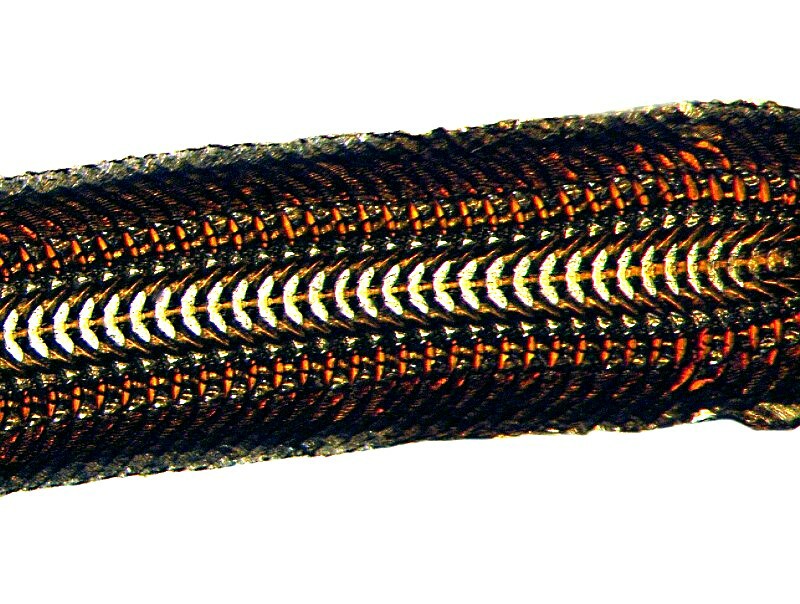
Looking for the hardest teeth on the planet? Look no further; chitons and limpets have them. It may be hard to believe and, in fact, this was discovered only relatively recently. Most animal teeth are composed, among other things, of calcium, as is bone, and we know that these can be very hard indeed. However, the teeth of chitons are composed of magnetite or goethite which is iron oxide combined with some complex proteins. They are arranged in long rows, are often morphologically elaborate, and they arise out of bands of powerful muscles that can move the strips or rows back and forth allowing them to abrade a surface to attain food. As Alex Hamilton, one of my e-mail correspondents, describes them: they look rather like unzipped zippers. This structure is called a radula and they are excellent objects for examination not only by brightfield, but also Near Infra-Red, Rheinberg, Polarizing, and Nomarski Differential Interference Contrast using the prism to do “optical staining”.
Radulae occur in a variety of molluscs, so let’s begin by looking at a few of the different types and examples. Let’s begin with the so-called “sea hare” which is a large sea slug or nudibranch. These can grow to over 2 and ½ feet in length and weigh in excess of 15 pounds! They are capable of expelling a cloud of toxic ink to discourage predators. The ink is a deep purple color which in the ancient world was known as Imperial Purple and was at times reserved for royalty and used to dye their garments; it was extremely expensive because of its relative rarity. This radula is rather different from those of chitons and limpets; it is flatter and the teeth are spread over a wider area. Below are 2 views; the second being a closeup to show the teeth in a bit more detail.
Next, I’ll show you a radula from another nudibranch, but this time a quite small one–only 1 and ½ inches long and its radula is more typical of those of other gastropods.
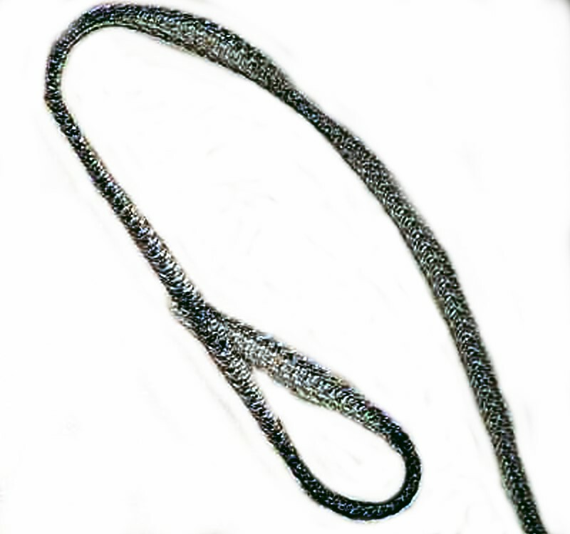
Radulae also are to be found in freshwater snails and below is a view of one which I extracted from a small pond snail. It’s length is greater than that of the snail itself! It is bowed in such a way that this creates a long apparatus for the scraping surfaces for algae.
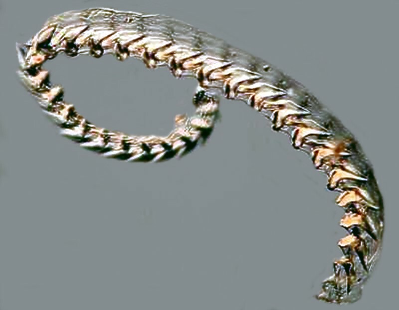
A very impressive radula is to be found in the whelk and, in general, more variation is seen in marine gastropods than in freshwater ones.
And here is a closeup image that shows the arrangement of some of the whelk teeth. Imagine what these teeth would be like if whelks were the size of Great White sharks!
Next up, we have a radula from an unidentified gastropod which gives a very clear idea of the complexity of this structure. This image was taken of a 19th Century slide.
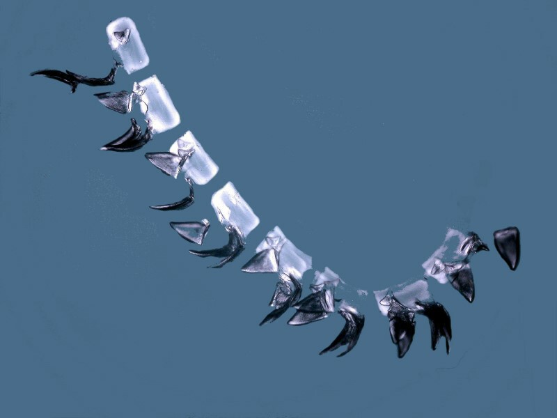
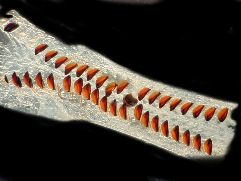
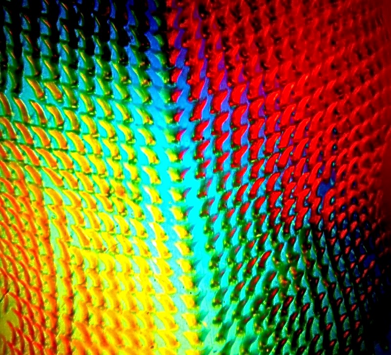
The mounter was fortunately able to extract a nice, rather complete section. In some molluscs the teeth are interlocked in such a way that they make it very difficult to extract the radula intact and fragments or even isolated teeth are the best one can hope for ordinarily. This is largely true of the radulae and teeth that I have of limpets and chitons. So, let’s move on to looking at a few teeth on a strip of the hyaline tissue of a portion of the radula. I say a few teeth, since the common limpet Patella vulgata has 1,920 teeth in 160 rows of 12 teeth each according to Wikipedia, because I certainly wasn’t going to try to count them. The radula of this species is longer than the organism itself as is the case with the freshwater snail which we looked at earlier.
So, what we are going to be looking at is a tiny fragment using Near InfraRed and Nomarksi Differential Interference Contrast.
We’ll begin with the Near InfraRed.
As you can see, the teeth stand out and appear both sharp and strong. In the living organisms, they are connected by bands of tissue creating a highly complex structure.
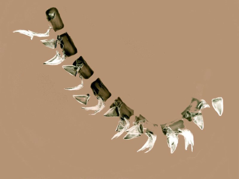
This next view is a result of applying the computer graphics “invert” function to the image immediately above.
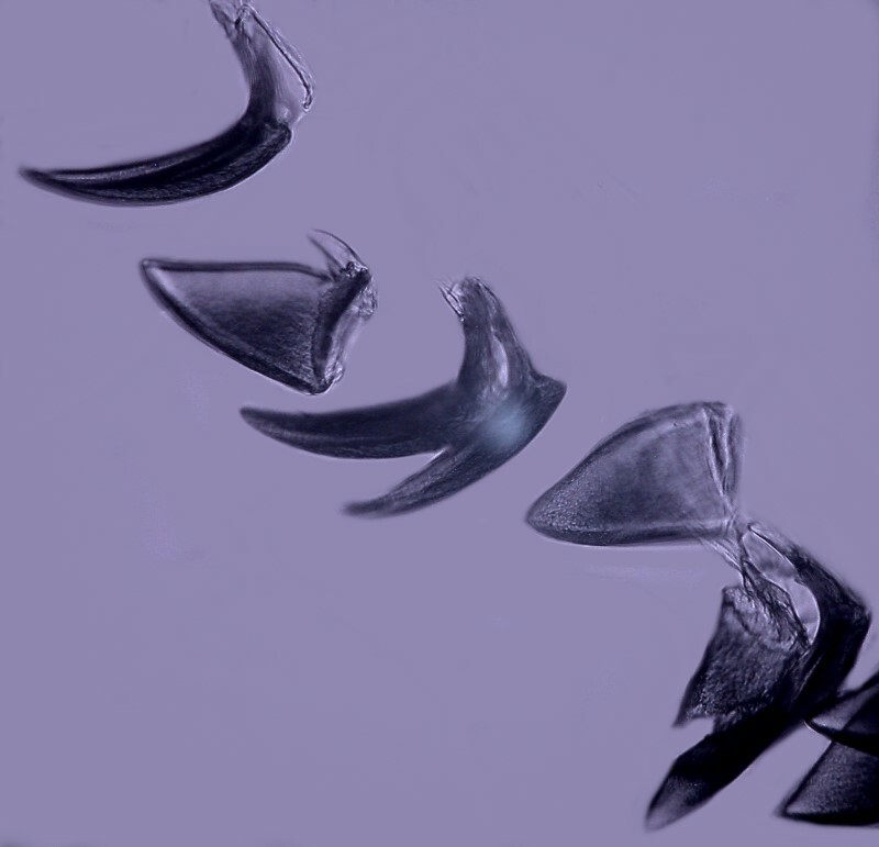
And now a closeup view of a few of the teeth.
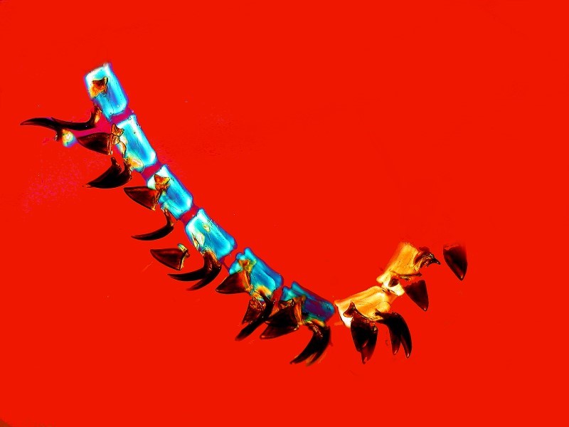
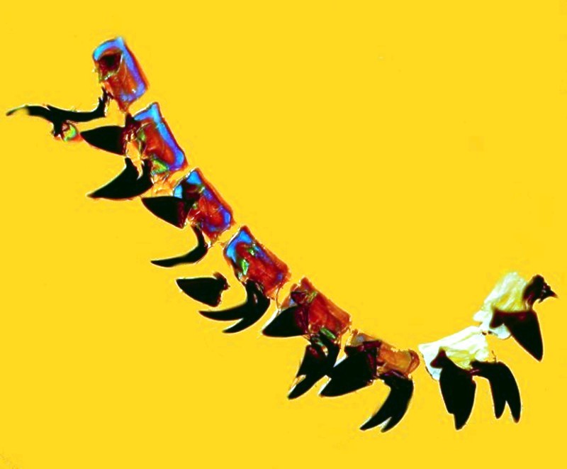
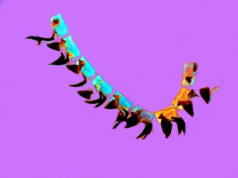
Earlier I mentioned using the “optical staining” capacity of Nomarski Differential Interference Contrast to observe the radulae. I chose one particular specimen and took a series of shots all of this single fragment. I rotated the prism to change the contrast and I will first show you a page of thumbnails with 12 views of the radula section.
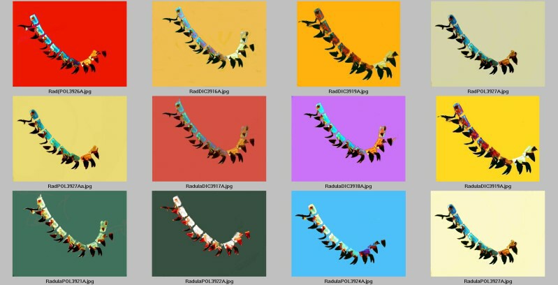
Next, we’ll look at four full images and you can see quite clearly how the contrast shifts from image to image.
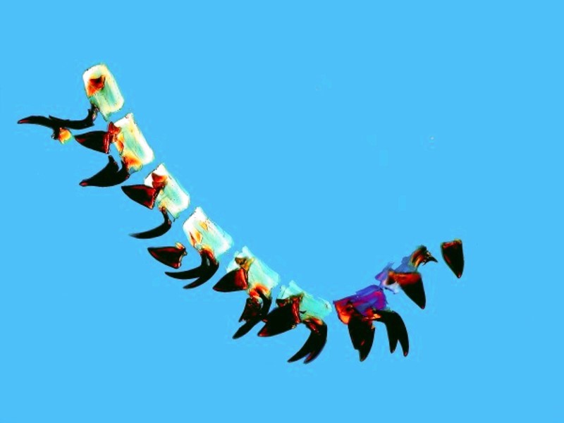
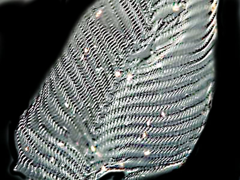
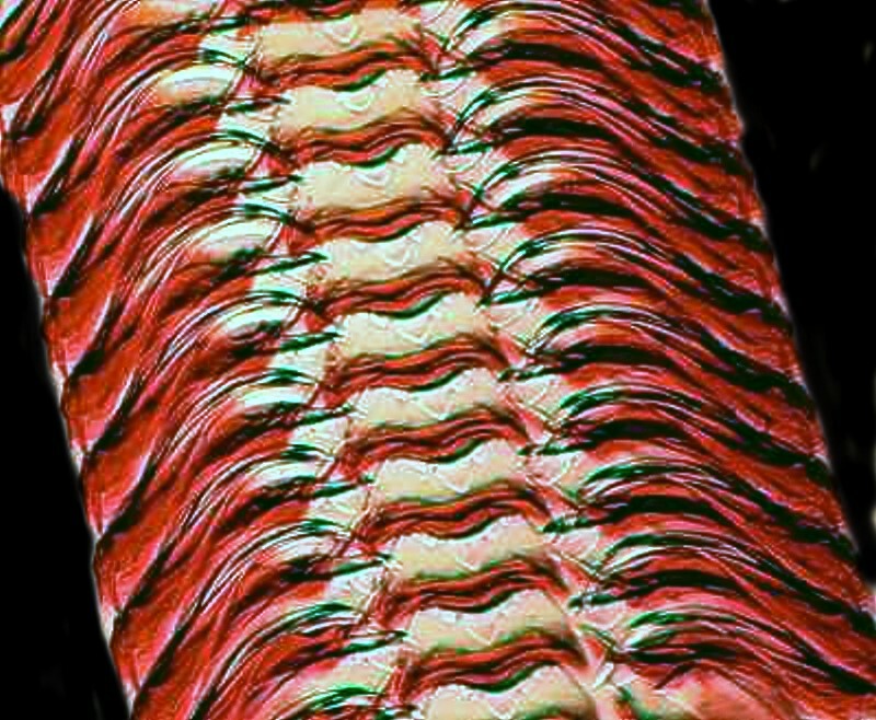
For several months I was examining chitons (May and July Micscape articles in 2016) and recently began looking at their radulae and so, I would like to share some images of parts of these structures with you.
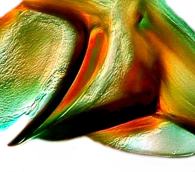
There is a significant variation in the size of teeth on a given radula and the large ones are clearly the ones that do the major scraping of larger bits of algae and detritus and the smaller ones, which are much more numerous, scoop up small particles and may also function to grind some of the larger pieces into a more manageable size.
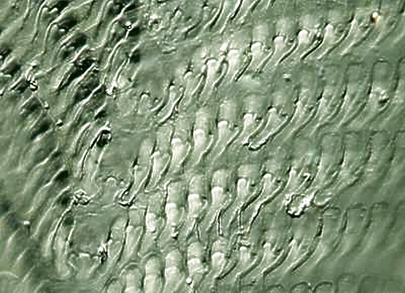
Here you can see that the major teeth are curved at ends like cutting blades.
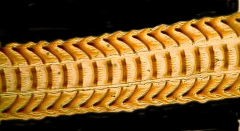
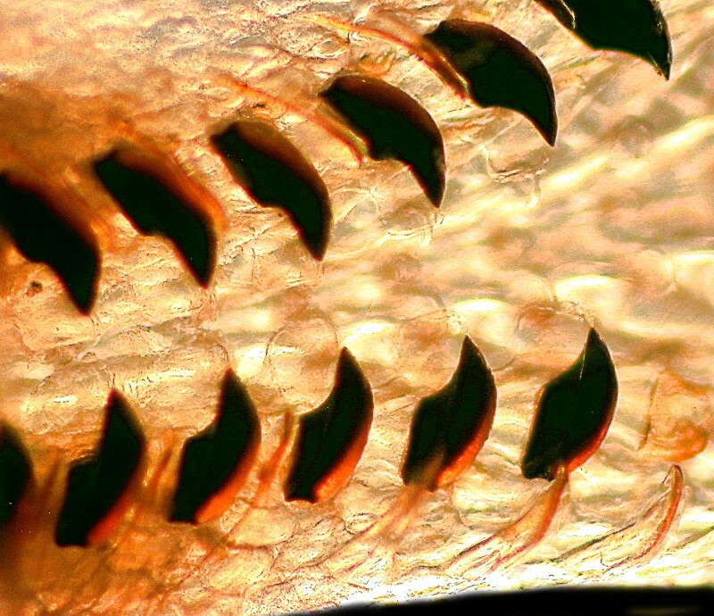
And next a view of a longer section
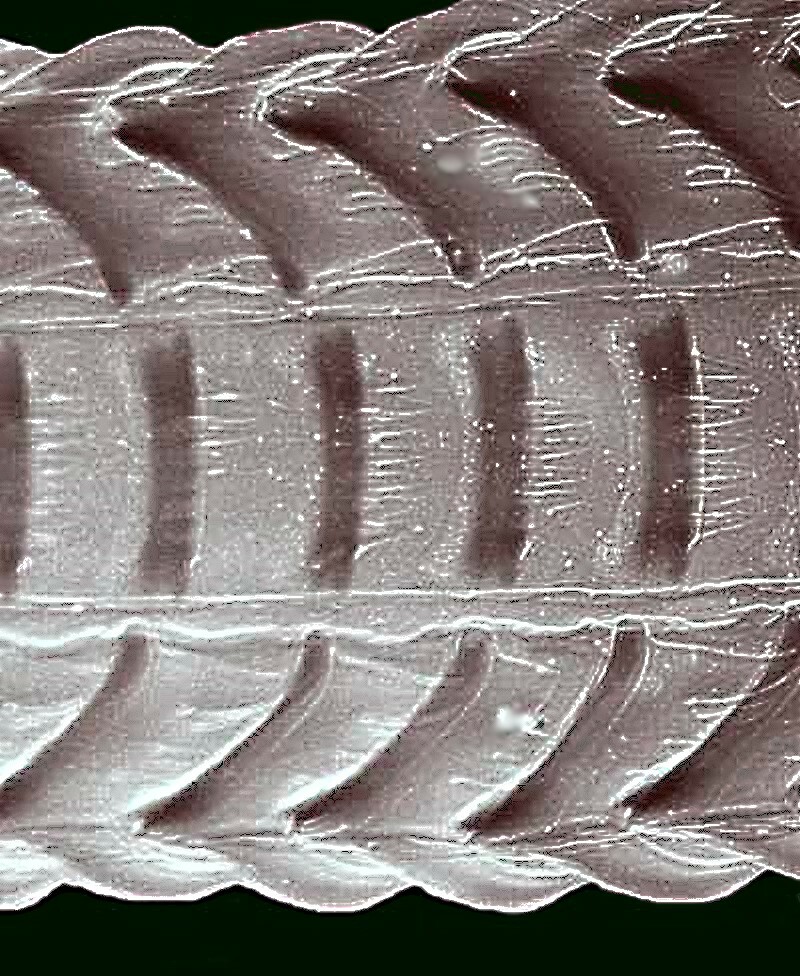
And yet another closeup view from a slightly different angle.
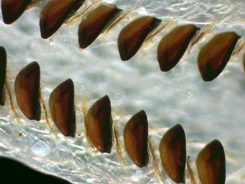
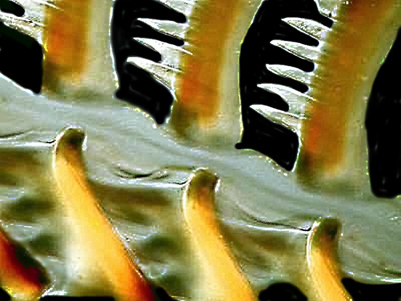
And finally, 2 very different views of sections from 2 other types of radulae. First, a closeup of a section with numerous small teeth and then a very close up view of formidable-appearing teeth giving us a glimpse of their complex layering.
In conclusion, I would add that I don’t think I would want to be a dentist for mollusks.
Addendum
For purists, I guess we will now have to do a bit of revision of
Shakespeare's King Lear. Where in Act 1, Scene 4, it reads: 'How sharper than a
serpent's tooth it is to have a thankless child.' should now read 'How sharper
than a chiton's tooth it is to have a thankless child.' I think it would have
to be chiton's tooth rather than limpet's tooth, because, well frankly,
'limpet's tooth' is just too limpid."
All comments to the author Richard Howey are welcomed.
Editor's note: Visit Richard Howey's new website at http://rhowey.googlepages.com/home where he plans to share aspects of his wide interests.
Microscopy UK Front
Page
Micscape
Magazine
Article
Library
Published in the June 2017 edition of Micscape Magazine.
Please report any Web problems or offer general comments to the Micscape Editor .
Micscape is the on-line monthly magazine of the Microscopy UK website at Microscopy-UK .
©
Onview.net Ltd, Microscopy-UK, and all contributors 1995
onwards. All rights reserved.
Main site is at
www.microscopy-uk.org.uk .